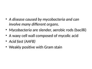
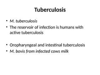
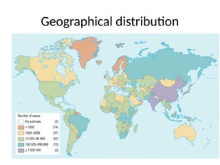
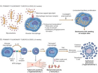
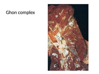
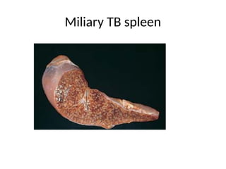
Tuberculosis
Dr. Mutaz Ibrahim M. Nur
• A disease caused by mycobacteria and can
involve many different organs.
• Mycobacteria are slender, aerobic rods (bacilli)
• A waxy cell wall composed of mycolic acid
• Acid fast (AAFB)
• Weakly positive with Gram stain
Tuberculosis
• M. tuberculosis
• The reservoir of infection is humans with
active tuberculosis
• Oropharyngeal and intestinal tuberculosis
• M. bovis from infected cows milk
Geographical distribution
Epidemiology
• After HIV, tuberculosis is the leading infectious cause
of death in the world
• Poverty, crowding, and chronic debilitating illness
– Diabetes mellitus,
– Hodgkin's lymphoma,
– Chronic lung disease (particularly silicosis),
– Chronic renal failure,
– Malnutrition,
• Alcoholism, and Immunosuppression
• HIV and M. tuberculosis
• Infection Vs Disease
• Person-to-person transmission
• Airborne droplets of organisms from an active case
to a susceptible host
• Primary tuberculosis: usually asymptomatic
• Fibrosis and calcification.
• Organisms may remain dormant for decades.
• Reactivation to produce communicable and
potentially life-threatening disease (secondary)
• Infection is followed by delayed
hypersensitivity to M. tuberculosis antigens.
• The Tuberculin test.
• 2 to 4 weeks after infection, intracutaneous
injection of purified protein derivative of M.
tuberculosis (PPD) induces a visible and
palpable induration that peaks in 48 to 72
hours.
• A positive test does not differentiate between infection and
disease.
• False-negative
– Viral infections,
– Sarcoidosis,
– Malnutrition,
– Hodgkin disease,
– Immunosuppression, and
– Overwhelming active tuberculous disease.
• False-positive reactions may result from infection by
atypical mycobacteria.
Pathogenesis
• Immunocompetent Vs immunocompromised
host.
• Sensitisation Vs 1st
encounter
• Cell-mediated immunity
• The effector cells that mediate immunity also
mediate hypersensitivity and tissue
destruction
• Caseating granulomas and cavitation
• The issue of hypersensitivity and immunity to
the organism!!
• Loss of hypersensitivity (tuberculin negativity
in a previously tuberculin-positive individual)
may be a sign that resistance to the organism
is lost.
Pathogenesis
• Macrophages
• Early in infection, the bacilli replicate
unchecked
• Later, the T-helper response stimulates
macrophages to contain the proliferation of
the bacteria.
Pathogenesis
• M. tuberculosis enters macrophages
• Replicates within them
• Early in primary tuberculosis (<3 weeks) in the
nonsensitized individual there is proliferation of
bacteria in the alveolar macrophages and
airspaces
• Bacteremia and seeding of multiple sites.
• However, most patients at this stage are
asymptomatic or have a mild flulike illness.
Pathogenesis
• About 3 weeks after infection, a TH1 response
against M. tuberculosis is mounted
• Macrophages become bactericidal
• Mature TH1 cells, in lymph nodes and in the
lung, produce IFN-γ
• The TH1 response also leads to the formation
of granulomas and caseous necrosis
Pathogenesis
• This response contains the bacteria
• In some people, the infection progresses and
the ongoing immune response results in tissue
destruction due to caseation and cavitation
To summarize
• Immunity to M. tuberculosis is mediated by TH1 cells,
which stimulate macrophages to kill the bacteria.
• This immune response comes at the cost of
hypersensitivity and the accompanying tissue
destruction.
• Reactivation or re-exposure in a previously sensitized
host results in rapid mobilization of a defensive
reaction but also increased tissue necrosis.
Clinical Features
Primary tuberculosis
• Unexposed, and therefore unsensitized, person
• Clinically significant in only 5%
• Loss of immunity (elderly and profoundly
immunosuppressed ) may result in the
development of primary TB more than once
(i.e. Loss of sensitization)
• The source of the organism is exogenous
Primary tuberculosis
• Primary pulmonary TB.
• Primary intestinal TB.
Primary tuberculosis
• Latent disease in most of the cases.
• Some progress (progressive primary
tuberculosis ):
• Resembles an acute bacterial pneumonia
• Lower and middle lobe consolidation (not
apical), hilar adenopathy, and pleural effusion
• Cavitation is rare
Clinical Features
Primary tuberculosis
• Lymphohematogenous dissemination
• Tuberculous meningitis and miliary
tuberculosis (may also follow pregression of
secondary TB)
Secondary tuberculosis
• In a previously sensitized host
• Reactivation
• Usually many decades after initial infection
• Weakened host resistance
• Exogenous reinfection
Secondary tuberculosis
• Reactivation is more common in low-
prevalence areas, while reinfection plays an
important role in regions of high prevalence
Secondary tuberculosis
• Apex of the upper lobes of one or both lungs
• High oxygen tension promotes growth of the
bacteria
• Pre-existence of hypersensitivity
• Prompt and marked tissue response
• Localisation of infection
Secondary tuberculosis
• Lymph nodes are less prominently involved
• Cavitation; results in
• Dissemination of mycobacteria along the
airways
• Source of infection when the patient coughs
up
Secondary tuberculosis
• Localized secondary tuberculosis may be
asymptomatic, or
• Insidious onset
• Malaise, anorexia, weight loss,
• Fever: low grade and remittent (appearing late
each afternoon and then subsiding), and
• Night sweats
• Sputum, at first mucoid and later purulent
• Hemoptysis
• Pleuritic pain
• Differential diagnosis??!!
Extrapulmonary TB
• Reactivation of dormant bacilli
• The GIT: ileocaecal, ascites
• The kidneys
• The epididymis
• Salpingitis, tubal abscesses and infertility in
females.
• Tuberculous meningitis and tuberculomas in
the CNS
Extrapulmonary TB
• Septic arthritis and osteomyelitis
• Potts disease of the spine and cold abscesses.
• Lupus vulgaris (TB of the skin)
• Constrictive pericarditis
• Adrenal TB, causing Addison’s disease.
Extrapulmonary TB
Tuberculous lymphadentitis
• Is common
• Hilar and paratracheal lymph nodes are the most
common.
• Initially the nodes are firm and discrete
• Later they become matted and can suppurate with
sinus formation.
• Scrofula =massive cervical lymph node
enlargement with discharging sinuses.
TB and HIV infection
• Stage of HIV infection
• Usual secondary TB
• More advanced immunosuppression (CD4+
<200 cells/mm3
): a clinical picture that
resembles progressive primary tuberculosis
(lower and middle lobe consolidation, hilar
lymphadenopathy, and noncavitary disease).
• Extrapulmonary involvement
TB and HIV infection
• Sputum-smear negativity for acid-fast bacilli
• False-negative PPD
• Lack of characteristic granulomas in tissues
Morphology
Primary Tuberculosis
• Pulmonary
• Intestinal; now rare
• Midzonal sub-pleural
• Ghon focus (inflammatory consolidation)
• Central caseous necrosis
• Drain to the regional nodes, which also often
caseate
• Ghon complex (the primary complex)
Ghon complex
Primary Tuberculosis
• In 95% of cases the Ghon complex undergoes
fibrosis and calcification; and despite seeding
of other organs, no lesions develop.
Primary Tuberculosis
• Granulomas
• Caseating and noncaseating.
• Tubercles (microscopic)
• Coalese to become macroscopically visible
• Immunocompromised people do not form the
characteristic granulomas
A granuloma with central necrosis
A higher magnification of the previous
photomicrograph
Epithelioid macrophages and a Langhan’s
giant cell
Numerous bacilli within and outside
macrophages (ZN stain)
Secondary Tuberculosis
• Initially, a small focus of consolidation close to
the apical pleura
• Central caseation and peripheral fibrosis
• Healing with fibrosis either spontaneously or
after therapy, or
• Progression and extension along several
different pathways
Secondary Tuberculosis
Progressive pulmonary tuberculosis
• The elderly and immunosuppressed
• Erosion into a bronchus (=cavity)
• Erosion of blood vessels (=hemoptysis)
• Healing by fibrosis distorts the pulmonary
architecture
• Spread by direct expansion, dissemination through
airways, lymphatic channels, or the vascular
system
Progressive pulmonary tuberculosis
TB with cavitation
Secondary Tuberculosis
• Miliary pulmonary disease
– Through lymphatics into venous return to the right
side of the heart and thence into the pulmonary
arteries
• Pleural effusions, tuberculous empyema, or
pleural fibrosis may develop
• Systemic miliary tuberculosis
– Liver, bone marrow, spleen, adrenals, meninges,
kidneys, fallopian tubes, and epididymis
Miliary TB spleen
Diagnosis
• History
• Physical examination
• Radiographic findings of consolidation or
cavitation in the apices of the lungs
Diagnosis
• Acid-fast smears (ZN) and cultures of the sputum
• Conventional cultures Vs liquid media-based culture
• Testing of drug susceptibility
• PCR is more rapid
• Multidrug resistance and implications on therapy
and prognosis
• Bacilli are easy to find early (exudative and caseous
phases)
• Become sparse later (fibrocalcific stages)
• Treatment
• Prevention and control
• Contact tracing
• Prophylaxis
• BCG vaccination

tuberculosis mycobacterium disease (3).pptx

  • 1.
  • 2.
    • A diseasecaused by mycobacteria and can involve many different organs. • Mycobacteria are slender, aerobic rods (bacilli) • A waxy cell wall composed of mycolic acid • Acid fast (AAFB) • Weakly positive with Gram stain
  • 3.
    Tuberculosis • M. tuberculosis •The reservoir of infection is humans with active tuberculosis • Oropharyngeal and intestinal tuberculosis • M. bovis from infected cows milk
  • 4.
  • 5.
    Epidemiology • After HIV,tuberculosis is the leading infectious cause of death in the world • Poverty, crowding, and chronic debilitating illness – Diabetes mellitus, – Hodgkin's lymphoma, – Chronic lung disease (particularly silicosis), – Chronic renal failure, – Malnutrition, • Alcoholism, and Immunosuppression • HIV and M. tuberculosis
  • 6.
    • Infection VsDisease • Person-to-person transmission • Airborne droplets of organisms from an active case to a susceptible host • Primary tuberculosis: usually asymptomatic • Fibrosis and calcification. • Organisms may remain dormant for decades. • Reactivation to produce communicable and potentially life-threatening disease (secondary)
  • 7.
    • Infection isfollowed by delayed hypersensitivity to M. tuberculosis antigens. • The Tuberculin test. • 2 to 4 weeks after infection, intracutaneous injection of purified protein derivative of M. tuberculosis (PPD) induces a visible and palpable induration that peaks in 48 to 72 hours.
  • 8.
    • A positivetest does not differentiate between infection and disease. • False-negative – Viral infections, – Sarcoidosis, – Malnutrition, – Hodgkin disease, – Immunosuppression, and – Overwhelming active tuberculous disease. • False-positive reactions may result from infection by atypical mycobacteria.
  • 9.
    Pathogenesis • Immunocompetent Vsimmunocompromised host. • Sensitisation Vs 1st encounter • Cell-mediated immunity • The effector cells that mediate immunity also mediate hypersensitivity and tissue destruction • Caseating granulomas and cavitation
  • 11.
    • The issueof hypersensitivity and immunity to the organism!! • Loss of hypersensitivity (tuberculin negativity in a previously tuberculin-positive individual) may be a sign that resistance to the organism is lost.
  • 12.
    Pathogenesis • Macrophages • Earlyin infection, the bacilli replicate unchecked • Later, the T-helper response stimulates macrophages to contain the proliferation of the bacteria.
  • 13.
    Pathogenesis • M. tuberculosisenters macrophages • Replicates within them • Early in primary tuberculosis (<3 weeks) in the nonsensitized individual there is proliferation of bacteria in the alveolar macrophages and airspaces • Bacteremia and seeding of multiple sites. • However, most patients at this stage are asymptomatic or have a mild flulike illness.
  • 14.
    Pathogenesis • About 3weeks after infection, a TH1 response against M. tuberculosis is mounted • Macrophages become bactericidal • Mature TH1 cells, in lymph nodes and in the lung, produce IFN-γ • The TH1 response also leads to the formation of granulomas and caseous necrosis
  • 15.
    Pathogenesis • This responsecontains the bacteria • In some people, the infection progresses and the ongoing immune response results in tissue destruction due to caseation and cavitation
  • 16.
    To summarize • Immunityto M. tuberculosis is mediated by TH1 cells, which stimulate macrophages to kill the bacteria. • This immune response comes at the cost of hypersensitivity and the accompanying tissue destruction. • Reactivation or re-exposure in a previously sensitized host results in rapid mobilization of a defensive reaction but also increased tissue necrosis.
  • 17.
  • 18.
    Primary tuberculosis • Unexposed,and therefore unsensitized, person • Clinically significant in only 5% • Loss of immunity (elderly and profoundly immunosuppressed ) may result in the development of primary TB more than once (i.e. Loss of sensitization) • The source of the organism is exogenous
  • 19.
    Primary tuberculosis • Primarypulmonary TB. • Primary intestinal TB.
  • 20.
    Primary tuberculosis • Latentdisease in most of the cases. • Some progress (progressive primary tuberculosis ): • Resembles an acute bacterial pneumonia • Lower and middle lobe consolidation (not apical), hilar adenopathy, and pleural effusion • Cavitation is rare
  • 21.
  • 22.
    Primary tuberculosis • Lymphohematogenousdissemination • Tuberculous meningitis and miliary tuberculosis (may also follow pregression of secondary TB)
  • 23.
    Secondary tuberculosis • Ina previously sensitized host • Reactivation • Usually many decades after initial infection • Weakened host resistance • Exogenous reinfection
  • 24.
    Secondary tuberculosis • Reactivationis more common in low- prevalence areas, while reinfection plays an important role in regions of high prevalence
  • 25.
    Secondary tuberculosis • Apexof the upper lobes of one or both lungs • High oxygen tension promotes growth of the bacteria • Pre-existence of hypersensitivity • Prompt and marked tissue response • Localisation of infection
  • 26.
    Secondary tuberculosis • Lymphnodes are less prominently involved • Cavitation; results in • Dissemination of mycobacteria along the airways • Source of infection when the patient coughs up
  • 27.
    Secondary tuberculosis • Localizedsecondary tuberculosis may be asymptomatic, or • Insidious onset • Malaise, anorexia, weight loss, • Fever: low grade and remittent (appearing late each afternoon and then subsiding), and • Night sweats • Sputum, at first mucoid and later purulent
  • 28.
    • Hemoptysis • Pleuriticpain • Differential diagnosis??!!
  • 29.
    Extrapulmonary TB • Reactivationof dormant bacilli • The GIT: ileocaecal, ascites • The kidneys • The epididymis • Salpingitis, tubal abscesses and infertility in females. • Tuberculous meningitis and tuberculomas in the CNS
  • 30.
    Extrapulmonary TB • Septicarthritis and osteomyelitis • Potts disease of the spine and cold abscesses. • Lupus vulgaris (TB of the skin) • Constrictive pericarditis • Adrenal TB, causing Addison’s disease.
  • 31.
    Extrapulmonary TB Tuberculous lymphadentitis •Is common • Hilar and paratracheal lymph nodes are the most common. • Initially the nodes are firm and discrete • Later they become matted and can suppurate with sinus formation. • Scrofula =massive cervical lymph node enlargement with discharging sinuses.
  • 32.
    TB and HIVinfection • Stage of HIV infection • Usual secondary TB • More advanced immunosuppression (CD4+ <200 cells/mm3 ): a clinical picture that resembles progressive primary tuberculosis (lower and middle lobe consolidation, hilar lymphadenopathy, and noncavitary disease). • Extrapulmonary involvement
  • 33.
    TB and HIVinfection • Sputum-smear negativity for acid-fast bacilli • False-negative PPD • Lack of characteristic granulomas in tissues
  • 34.
  • 35.
    Primary Tuberculosis • Pulmonary •Intestinal; now rare • Midzonal sub-pleural • Ghon focus (inflammatory consolidation) • Central caseous necrosis • Drain to the regional nodes, which also often caseate • Ghon complex (the primary complex)
  • 36.
  • 37.
    Primary Tuberculosis • In95% of cases the Ghon complex undergoes fibrosis and calcification; and despite seeding of other organs, no lesions develop.
  • 38.
    Primary Tuberculosis • Granulomas •Caseating and noncaseating. • Tubercles (microscopic) • Coalese to become macroscopically visible • Immunocompromised people do not form the characteristic granulomas
  • 39.
    A granuloma withcentral necrosis
  • 40.
    A higher magnificationof the previous photomicrograph
  • 41.
    Epithelioid macrophages anda Langhan’s giant cell
  • 42.
    Numerous bacilli withinand outside macrophages (ZN stain)
  • 43.
    Secondary Tuberculosis • Initially,a small focus of consolidation close to the apical pleura • Central caseation and peripheral fibrosis • Healing with fibrosis either spontaneously or after therapy, or • Progression and extension along several different pathways
  • 44.
    Secondary Tuberculosis Progressive pulmonarytuberculosis • The elderly and immunosuppressed • Erosion into a bronchus (=cavity) • Erosion of blood vessels (=hemoptysis) • Healing by fibrosis distorts the pulmonary architecture • Spread by direct expansion, dissemination through airways, lymphatic channels, or the vascular system
  • 45.
  • 46.
  • 47.
    Secondary Tuberculosis • Miliarypulmonary disease – Through lymphatics into venous return to the right side of the heart and thence into the pulmonary arteries • Pleural effusions, tuberculous empyema, or pleural fibrosis may develop • Systemic miliary tuberculosis – Liver, bone marrow, spleen, adrenals, meninges, kidneys, fallopian tubes, and epididymis
  • 48.
  • 49.
    Diagnosis • History • Physicalexamination • Radiographic findings of consolidation or cavitation in the apices of the lungs
  • 50.
    Diagnosis • Acid-fast smears(ZN) and cultures of the sputum • Conventional cultures Vs liquid media-based culture • Testing of drug susceptibility • PCR is more rapid • Multidrug resistance and implications on therapy and prognosis • Bacilli are easy to find early (exudative and caseous phases) • Become sparse later (fibrocalcific stages)
  • 51.
    • Treatment • Preventionand control • Contact tracing • Prophylaxis • BCG vaccination