Contents
• Introduction
• TrigeminalNuclei
• Functional Components
• Course & Distribution
• Trigeminal Ganglion
• Divisions of Trigeminal Nerve
• Clinical Examination of V Nerve
• Applied Anatomy
• Summary
• References
3.
Introduction
• The largestcranial nerve
• It is mixed nerve ( sensory and motor )
• Sensory to Skin of face
-Mucosa of cranial viscera
-Except base of tongue and pharynx
• Motor to Muscles of Mastication
-Tensor villi palatini , Tensor tympany
-Anterior belly of digastric
-Mylohyoid
4.
Trigeminal nuclei
• Acranial nerve nucleus is a collection of neurons (gray matter) in
the brain stem that is associated with one or more cranial nerves.
• Axons carrying information to and from the cranial nerves form
a synapse first at these nuclei.
• Lesions occurring at these nuclei can lead to effects resembling those
seen by the severing of nerve(s) they are associated with.
Mesencephalic nucleus
• Cellbody of Pseudo-unipolar neuron
• Relay proprioception from muscles of mastication,
• Extra ocular Muscles, Facial muscles.
• Situated in Midbrain just lateral to Aqueduct
Spinal nucleus
• Extendsfrom caudal end of principal sensory Nucleus in pons to 2nd
or 3rd spinal segment
• It relays Pain and Temperature
10.
Motor nucleus
• Innervatesmuscles of mastication and tensor tympani and tensor
palatini
• Derived from first branchial arch.
• Located in pons medial to principle sensory nucleus.
Course & distribution
•Both motor and sensory root are attached ventrally to junction of
pons and middle cerebellar peduncle
• Pass anteriorly in middle cranial fossa to lie below tentorium cerebelli
in cavum trigeminale, here motor root lies inferior to sensory root.
20.
• Sensory rootconnected to posteromedial concave border of the
trigeminal ganglion.
• Convex anterolatateral margin of the ganglion gives attachment to the
3 divisions of the trigeminal nerve.
23.
• Motor rootturns further inferior with sensory component of V3 to
emerge out of foramen Ovale as Mandibular nerve.
• Ophthalmic and Maxillary division emerges through Superior orbital
fissure and foramen Rotundum respectively.
THE TRIGEMINAL GANGLION
•SEMILUNAR OR GASSERIAN GANGLION.
• Crescentic in shape with convexity
anterolaterally.
• Contains cell bodies of pseudo unipolar
neurons.
• LOCATION: lies in a bony fossa at apex of the
petrous temporal bone on floor of middle
cranial fossa
27.
• COVERINGS: coveredby dural pouch = MECKLES CAVE or CAVUM
TRIGEMINALE.
• lined by pia and arachnoid thus the ganglion is bathed in CSF.
• ARTERIAL SUPPLY: Ganglionic branches of Internal Carotid
Artery, middle meningeal artery and accessory meningeal artery.
28.
RELATIONS
SUPERIORLY: *superior petrosalsinus
*free margin of tentorium cerebelli
INFERIORLY: *motor root
*greater petrosal nerve
*petrous apex
*foramen lacerum
MEDIALLY: *posterior part of lateral wall of cavernous sinus
*Internal Carotid Artery with its sympathetic plexus
LATERALLY: *uncus of temporal lobe
*middle meningeal artery and vein
*nervous spinosum
OPTHALMIC NERVE(V1)
• Smallestdivision.
• Sensory only
• Supplies : eyeballs, conjunctiva, lacrimal gland, mucosa of nose and
paranasal sinus, skin of forehead eyelid and nose
Lacrimal nerve
• Smallest
•Passes into orbit through lateral compartment
of the Superior orbital fissure outside the
tendinous ring.
• Receives communicating branch from
Trochlear nerve
34.
• Receives branchfrom Zygomaticotemporal nerve branch of maxillary
• Sensory to lateral conjunctiva, Upper Lid, lacrimal gland
• Post synaptic parasympathetic fibres from pterygopalatine ganglion to
lacrimal gland (parasympathetic secretomotor).
36.
FRONTAL NERVE
• Largest
•Enters orbit through lateral part of superior orbital fissure outside
tendinous ring
• Passes forward between roof of orbit and Levator Palpebral Superioris
Supratrochlear Nerve
• Divides midway into :
Supraorbital Nerve
38.
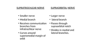
SUPRATROCHLEAR NERVE SUPRAORBITALNERVE
• Smaller nerve
• Medial branch
• Receives communication
branches from
infratrochlear nerve
• Curves around
superomedial margin of
orbit
• Larger nerve
• lateral branch
• Passes through
supraorbital notch
• Divides in medial and
lateral branches.
39.
SUPRATROCHLEAR NERVE SUPRAORBITALNERVE
• supplies: median
conjunctiva, Upper Lid and
lower part of forehead
• Lies between frontalis and
corrugator supercilliary
muscles
• Lies beneath frontalis muscle
• Supplies: conjunctiva,
scalp until vertex , mucous
membrane of frontal sinus
40.
NASOCILLIARY NERVE
• PurelySensory
• Passes through middle part of superior orbital fissure within the
tendonous ring .
• Runs along medial wall of orbit between Superior Oblique and Medial
Rectus
• Divides into Anterior Ethmoidal and External Nasal
• 5 branches in orbit.
41.
1. Short CilliaryNerves: Fibers reaches eyeball and also contains
fibers from Cilliary Ganglion
2. Long Cilliary Nerves : 2 or 3in no. supply to Iris and Cornea.
3. Post Ethmoidal Nerve: passes through posterior ethmoidal foramen
to supply the Ethmoid and Sphenoid PNS.
4. Infratrochlear Nerve: appears on face above med angle the eye.
Supplies to skin of lacrimal sac and caruncle.
44.
5. Anterior EthmoidalNerve:
larger terminal branch
Course: anterior ethmoidal foramen and canal
into anterior cranial fossa on sup surf of cribriform plate
Through slit lat to crista galli into nasal cavity
Med internal nasal branch lat internal nasal branch
Supplies ant nasal septum supplies ant part lat nasal
cavity emerges as
external nasal nerve to
skin of ala,vestibule,and
tip of nose
45.
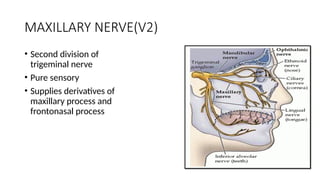
MAXILLARY NERVE(V2)
• Seconddivision of
trigeminal nerve
• Pure sensory
• Supplies derivatives of
maxillary process and
frontonasal process
46.
Course:
Trigeminal ganglion-> Middlecranial fossa
Lateral wall of cavernous sinus
Foramen rotundum
Pterigopalatine fossa
In groove on posterior surface of maxilla
Through inferior orbital fissure into orbit as INFRA ORBITAL N
Through infraorbital foramen on face
49.
Branches
• IN MIDDLECRANIAL FOSSA:
• - Meningeal branch : Travels along the middle meningeal artery and
provides sensory innervation to cranial dura matter.
50.
• IN PTERYGOPALATINEFOSSA:
• 1. Ganglionic branches-
• Arises as 2trunks.Trunks join to form single root within
pterygopalatine ganglion.
• Gives Orbital branches
• Palatine branches,
• Pharyngeal branches,
• Nasal branches
51.
• Gives postganglionicsecretomotor fibers to lacrimal gland via
zygomaticotemporal and lacrimal.
53.
• Orbital branch:Supplies periosteum of orbit
• 3.Nasal branch: Supplies to mucosa of superior and inferior conchae,
posterior ethmiodal sinus and posterior portion of nasal septum. It
also includes Nasopalatine branch.
55.
4. Palatine branch:Arise as greater palatine (anterior) and lesser
palatine (middle and posterior)
• -Greater palatine nerve descends through pterygopalatine canal
from the ganglion and emerges from greater palatine foramen of hard
palate.
• -Middle palatine and posterior palatine emerges from lesser palatine
foramen and supply soft palate and tonsilar region respectively.
58.
5. Pharyngeal branch:It leaves the posterior part of pterygopalatine
ganglion and passes through the pharyngeal canal
• It is distributed to the mucous membrane of the nasal part of
pharynx, posterior to eustachian tube.
59.
Posterior Superior AlveolarNerve
• -It arises from the main trunk of maxillary nerve pterygopalatine
fossa just before the nerve enters the inferior orbital canal
• arises as 2 trunks.
• crosses the pterygoplatine fossa reaching infratemporal surface of
maxilla.
• - 1st trunk continues downwards on posterior surface of maxilla and
provide sensory innervation to buccal gingiva in maxillary molar
region and adjacent facial mucosal surface
60.
• 2nd trunkenters maxila through PSA canal to travel to posterolateral
wall of maxillary sinus providing sensory innervation to sinus mucosa.
62.
Zygomatic nerve
• Itenters orbit through infra orbital fissure
. Zygomaticofacial nerve
-Appears on face through foramen in the zygomatic bone
-Supplies skin on prominence of cheek
63.
Zygomaticotemporal nerve
-Appears ininfratemporal region thru foramen in zygomatic
bone
-Supplies skin of temporal region after peircing temporal fascia 2
cm above zygoma
-Gives communicating branch to lacrimal N suppling parasymp.
Secretomotor fibres to lacrimal gland.
.
64.
IN THE INFRAORBITALCANAL
1.Middle superior alveolar nerve:
runs along lateral wall of maxilla
Participates in superior dental plexus
Supplies premolars.
2. Anterior superior alveolar nerve:
Runs in canal in ant wall of maxilla=canalii sinosus
#Dental branches # nasal branches
Joins sup dental plexus lat wallof inf meatus to
to supply canines opening of max sinus.
65.
• 3. FACIALBRANCHES:
• 1.Palpebral nerves-pierces Orbicularis Occuli and supplies skin of
lower lid.
• 2.Nasal branches-supplies skin of lat wall nose and mobile part of
septum.
• 3. Superior labial nerve- forms infraorbital plexus
• supplies skin and mm of upper lip, cheek and labial glands.
Branches from trunk
•Before dividing into anterior and posterior division it gives 2 branches during its 2-
3mm path
1.Nervous spinosus or Meningeal branch of Mandibular nerve
• It reenters cranial cavity through foramen spinosus along with middle meningial artery
• Supply Dura matter of middle cranial fossa and mastoid air sinus
2.Nerve to medial Pterygoid
• Supplies medial pterygoid
• Through Otic ganglion without interruption to
Tensor tympani
Tensor palatini
73.
Branches from theanterior division
• The anterior division is significantly smaller than posterior.
• After dividing from the main trunk. It runs anteriorly and below the
lateral pterygoid muscle to over its upper border.
74.
• After thisthe nerve is buccal nerve. reach its external surface of
muscle by either passing through two heads or winding
• 1.Nerve to lateral pterygoid: It enters the deep surface of the muscle.
It may arise as independent branch or may arise in common with
buccal nerve.
75.
2.Massetric nerve- Emergesat the upper border of the lateral pterygoid
just in front of TMJ. Passes laterally through mandibular notch along
with massetric vessels, and enters the deep surface of masseter, also
suppliesTMJ
76.
3.Buccal nerve-is theonly sensory branch of ant div. travels betwn 2
heads of lat pterygoid and emerges in cheek at ant border of masseter.
Supplies skin and mucous membrane of cheek.
4.Deep temporal nerve-There are anterior and posterior deep temporal
nerves. Passes between skull, and enters deep surface of the
temporalis. Anterior is often a branch of buccal nerve and the posterior
may arise in common with massetric nerve.
1.Auriculotemporal nerve-
Arises from2 roots which run backwards and encircle the
middle meningeal artery and form single trunk
The trunk passes posterior to lateral pterygoid between neck
of mandible and sphenomandibular ligament superior to 1st
part of maxillary art.
Lies behind the TMJ close to the parotid
Ascends behind superficial temporal vessels and then in
temporal region divides into superficial temporal branches.
79.
Branches Of AuriculotemporalNerve
• Auricular branches- supply tragus, upper part of
aurical,roof of external auditory meatus, anterosuperior part
of tympanic membrane
• Superficial temporal branches-supply skin of temple
• It also supply sensory and secretomotor to parotid.
• Articular branches-supply the TMJ.
80.
2. Inferior alveolarnerve:
• Is mixed nerve
• Runs vertically downwards medial to lateral ptrygoid and
lateroposterior to lingual nerve
• Enters mandible through mandibular foramen
81.
Branches
1.Mylohyoid: Supplies tomylohyoid muscle and anterior belly of
digastric. It is also sensory to skin on inferior and anterior
surface surfaces of mental protuberance. It may provide
sensory innervation to mandibular incisors. There is also
evidence that mylohyoid supply to mesial root of mandibular
frist molar.
82.
2.Branches to lowerteeth and gums.
3.Mental nerve : It exits canal and divides into three branches innervating skin of
chin and skin and mucous membrane of the lower lip.
4.Incisive nerve : It remains within the canal and form plexus that innervates pulpal
tissue of first premolar canine and incisors through dental branches.
84.
3.Lingual nerve:
lies anteriorto inferior alveolar n between lateral
pterygoid and tensor palatini
receives chorda tympani (SVA)
Emerges from inferior border of lateral pterygoid to lie between ramus and
medial pterygoid in peterygomandibular space
moves downwards and forwards deep to pterygomandibular raphe between
origins of supirior constrictor and mylohyoid
Reach to side of base of tongue 1 cm below and behind 3rd
molar just below
mucous membrane of lateral lingual sulcus
85.
-Then proceeds anteriorlyacross the muscles of tongue ,looping
medially and downwards to submandibular duct to deep surface of
submandibular gland where it breaks into terminal branches
-Sensory to anterior 2/3 of tonge along with special sensation also
sensory to floor of mouth and gingiva on lingual side of mandible.
86.
Branches of lingualnerve and its communications:
1.Chorda tympani
2.Communications with submandibular ganglion
3.Hypoglossal nerve
87.
Ganglia Associated WithThe Trigeminal Nerve
1 .Cilliary Ganglion: connected with nasocilliary nerve by ganglionic
branches in orbit,
non synapsing
sensory for orbit
2.Pterygopalatine Ganglion: connected to maxillary nerve in
infratemporal fossa
sensory to orbital septum, orbicularis and nasal cavity, max sinus,
palate, nasopharynx.
88.
3. Otic Ganglion:between trunk of mandibular n and tensor palatini,
nerve to med pterygoid passes thru but does not synapse in the
ganglion.
4.Submandibular Ganglion: related to lingual n, rests on hypoglossus
supplies post gang. Parasym secretomotor fibres to submandibular and
sublingual gland.
89.
CUTANEOUS DISTRIBUTION OFTRIGEMINAL
NERVE
Each half of face is supplied by 13 cutaneous Nerve
1motor and 12 sensory
Of 12 sensory : 11 are from trigeminal Nerve
1 is c2 greater auricular Nerve
Branches of trigeminal N
5 from ophthalmic: lacrimal
supraorbital
supratrocheal
infratrochlear
external nasal
90.
3 from maxillaryN: infra orbital N
zygomaticofacial N
zygomaticotemporal N
3 from mandibular N: buccal N
auriculotemporal N
mental N
92.
Examination of trigeminalnerve
1- Sensation Function
2- Motor Function
3- Corneal reflex
4- Test jaw jerk
93.
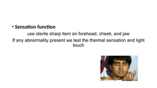
• Sensation function
usesterile sharp item on forehead, cheek, and jaw
If any abnormality present we test the thermal sensation and light
touch
94.
• Corneal reflex
•a clean piece of cotton wool and ask the patient to look away
gently touch the cornea with the cotton wool and the patient will
blink.
95.
• Test jawjerk
• Doctor finger on tip of jaw, grip patellar hammer halfway up
shaft and tap finger lightly usually nothing happens, or just a
slight closure.
96.
Applied Anatomy
. TrigeminalNeuralgia – Tic Douloureux
• Sudden, usually unilateral severe, brief, stabbing lancinating,
recurring pain in the distribution of one or more branches of the
5th Nerve
97.
2. TRIGEMINAL NEUROPATHY
•sensory loss of face or weakness of the jaw muscles
• causes- sjogren syndrome
• herpes zoster, leprosy
• meningioma,schwanomma
98.
4. HERPES ZOSTEROPHTHALMICUS:
• Recurrent neuro-cutaneous infection In opth. division of
trigeminal dermatome, most freq. affecting nasociliary branch
• HHV3 / varicella zoster
• Gasserian ganglion
ophthalmic nerve
Supraorbital N. Infraorbital N.
Supratrochlear N.
Infratrochlear N.
Nasal N.
99.
5. Cavernous sinussyndrome
• Multiple cranial neuropathies
• Exophthalmos, ocular motor defects, sensory loss in V1 and / or V2.
• Pupils may be spared or involved.
causes: bacterial thrombophlebitis
actinomycosis
rhinocerebellar mucormycosis
aspergillosis
tolosa hunt syndrome
neoplasms
vascular lesions
100.
6.Gradenigos syndrome
• GradenigoSyndrome (GS) is classically described as a clinical triad of
otitis media, facial pain and abducens palsy that is most commonly
developed from infection in the petrous temporal bone
• Infection is commonly caused by Streptococcus
pneumoniae, Haemophilus influenzae, Pseudomonas, and
Staphylococcus aureus
Summary
• Since Trigeminalnerve is mixed nerve, suplies mainly head and neck
region.
• Hence as a dentist one should know thoroughly about intracranial and
extracranial course and distribution of Trigeminal nerve, to diagnose
the pathologies associated with Trigeminal nerve and for appropriate
treatment.
Editor's Notes
#19 with motor root lying ventromedially to the sensory root.
#27 , just lateral to posterior part of lateral wall of the cavernous sinus.
#47 After leaving foramen rotundum it moves anteriorly in the uppermost part of pterygopalatine fossa.
As it passes through pterygopalatine fossa it also gives branches to sphenopalatine ganglion, posterior superior alveolar nerve and zygomatic branches.
It then moves laterally and moves in a groove on posterior surface of maxilla.
Then enters orbit through infra orbital fissure and moves through infra orbital groove where it is called as Infraorbital nerve and emerges on face from infra orbital foramen.
#54 It passes across roof of nasal cavity downwards and forwards lying between mucosa and periosteum of nasal septum.
Reaches to floor of nasal cavity n give branch to anterior part of nasal septum and floor of nasal cavity.
Enters Incisive canal and enters oral cavity through insicive foramen
It provides sensation to palatal mucosa of premaxilla region.
#56 -Then moves anteriorly between mucoperiostem and hard palate upto 1st premolar supplying sensory innervation to palatal soft tissue and bone. Then communicates with nasopalatine
#61 Continuing downwards this also provides sensory innervation to alveoli, PDL, pulp of molar tooth.
#81 . Then moves between the sphenomandibular ligament and medial surface of mandibular ramus
#82 Arises just before the nerve enters mandibular foramen. It pierces the sphenomandibular ligament along with mylohyoid muscle and runs in the mylohyoid groove.
#101 The full triad of GS however may not always be present especially in the post-antibiotic era .