This document provides an overview of the trigeminal nerve, which is the fifth cranial nerve. It begins with an introduction to cranial nerves and then discusses the specific nuclei, ganglia, and divisions of the trigeminal nerve. The three divisions - ophthalmic, maxillary, and mandibular nerves - are described in detail regarding their course, branches, and sensory and motor functions. Key structures discussed include the trigeminal ganglion, gasserian ganglion, pterygopalatine ganglion, and submandibular ganglion. The document concludes with a recap of the main branches of the trigeminal nerve.
This presentation contains the detailed description about the courses, branches and supply of the Trigeminal Nerve, contains variations of maxillary nerve & Mandibular Nerve, and the detail about trigeminal Neurolgia and its managements
This presentation contains the detailed description about the courses, branches and supply of the Trigeminal Nerve, contains variations of maxillary nerve & Mandibular Nerve, and the detail about trigeminal Neurolgia and its managements
The facial nerve is the seventh cranial nerve, or simply CN VII. It emerges from the pons of the brainstem, controls the muscles of facial expression, and functions in the conveyance of taste sensations from the anterior two-thirds of the tongue.
The Indian Dental Academy is the Leader in continuing dental education , training dentists in all aspects of dentistry and
offering a wide range of dental certified courses in different formats.for more details please visit
www.indiandentalacademy.com
this presentation consist of introduction to types of nerves, structure of nerve and cranial nerves. there is a detail description about, origin , course of the trigeminal nerve and its branches and the structures supplying the nerve. it also contains applied anatomy of the nerve and its importance of the nerve in oral and maxillofacial surgeries. a detail description about the examination of the trigeminal nerve is also mentioned in the presentation. hoping that it would be useful to the students and people seeking for knowledge about the trigeminal nerve.
micro teaching on communication m.sc nursing.pdfAnurag Sharma
Microteaching is a unique model of practice teaching. It is a viable instrument for the. desired change in the teaching behavior or the behavior potential which, in specified types of real. classroom situations, tends to facilitate the achievement of specified types of objectives.
Ozempic: Preoperative Management of Patients on GLP-1 Receptor Agonists Saeid Safari
Preoperative Management of Patients on GLP-1 Receptor Agonists like Ozempic and Semiglutide
ASA GUIDELINE
NYSORA Guideline
2 Case Reports of Gastric Ultrasound
Prix Galien International 2024 Forum ProgramLevi Shapiro
June 20, 2024, Prix Galien International and Jerusalem Ethics Forum in ROME. Detailed agenda including panels:
- ADVANCES IN CARDIOLOGY: A NEW PARADIGM IS COMING
- WOMEN’S HEALTH: FERTILITY PRESERVATION
- WHAT’S NEW IN THE TREATMENT OF INFECTIOUS,
ONCOLOGICAL AND INFLAMMATORY SKIN DISEASES?
- ARTIFICIAL INTELLIGENCE AND ETHICS
- GENE THERAPY
- BEYOND BORDERS: GLOBAL INITIATIVES FOR DEMOCRATIZING LIFE SCIENCE TECHNOLOGIES AND PROMOTING ACCESS TO HEALTHCARE
- ETHICAL CHALLENGES IN LIFE SCIENCES
- Prix Galien International Awards Ceremony
TEST BANK for Operations Management, 14th Edition by William J. Stevenson, Ve...kevinkariuki227
TEST BANK for Operations Management, 14th Edition by William J. Stevenson, Verified Chapters 1 - 19, Complete Newest Version.pdf
TEST BANK for Operations Management, 14th Edition by William J. Stevenson, Verified Chapters 1 - 19, Complete Newest Version.pdf
Lung Cancer: Artificial Intelligence, Synergetics, Complex System Analysis, S...Oleg Kshivets
RESULTS: Overall life span (LS) was 2252.1±1742.5 days and cumulative 5-year survival (5YS) reached 73.2%, 10 years – 64.8%, 20 years – 42.5%. 513 LCP lived more than 5 years (LS=3124.6±1525.6 days), 148 LCP – more than 10 years (LS=5054.4±1504.1 days).199 LCP died because of LC (LS=562.7±374.5 days). 5YS of LCP after bi/lobectomies was significantly superior in comparison with LCP after pneumonectomies (78.1% vs.63.7%, P=0.00001 by log-rank test). AT significantly improved 5YS (66.3% vs. 34.8%) (P=0.00000 by log-rank test) only for LCP with N1-2. Cox modeling displayed that 5YS of LCP significantly depended on: phase transition (PT) early-invasive LC in terms of synergetics, PT N0—N12, cell ratio factors (ratio between cancer cells- CC and blood cells subpopulations), G1-3, histology, glucose, AT, blood cell circuit, prothrombin index, heparin tolerance, recalcification time (P=0.000-0.038). Neural networks, genetic algorithm selection and bootstrap simulation revealed relationships between 5YS and PT early-invasive LC (rank=1), PT N0—N12 (rank=2), thrombocytes/CC (3), erythrocytes/CC (4), eosinophils/CC (5), healthy cells/CC (6), lymphocytes/CC (7), segmented neutrophils/CC (8), stick neutrophils/CC (9), monocytes/CC (10); leucocytes/CC (11). Correct prediction of 5YS was 100% by neural networks computing (area under ROC curve=1.0; error=0.0).
CONCLUSIONS: 5YS of LCP after radical procedures significantly depended on: 1) PT early-invasive cancer; 2) PT N0--N12; 3) cell ratio factors; 4) blood cell circuit; 5) biochemical factors; 6) hemostasis system; 7) AT; 8) LC characteristics; 9) LC cell dynamics; 10) surgery type: lobectomy/pneumonectomy; 11) anthropometric data. Optimal diagnosis and treatment strategies for LC are: 1) screening and early detection of LC; 2) availability of experienced thoracic surgeons because of complexity of radical procedures; 3) aggressive en block surgery and adequate lymph node dissection for completeness; 4) precise prediction; 5) adjuvant chemoimmunoradiotherapy for LCP with unfavorable prognosis.
New Directions in Targeted Therapeutic Approaches for Older Adults With Mantl...i3 Health
i3 Health is pleased to make the speaker slides from this activity available for use as a non-accredited self-study or teaching resource.
This slide deck presented by Dr. Kami Maddocks, Professor-Clinical in the Division of Hematology and
Associate Division Director for Ambulatory Operations
The Ohio State University Comprehensive Cancer Center, will provide insight into new directions in targeted therapeutic approaches for older adults with mantle cell lymphoma.
STATEMENT OF NEED
Mantle cell lymphoma (MCL) is a rare, aggressive B-cell non-Hodgkin lymphoma (NHL) accounting for 5% to 7% of all lymphomas. Its prognosis ranges from indolent disease that does not require treatment for years to very aggressive disease, which is associated with poor survival (Silkenstedt et al, 2021). Typically, MCL is diagnosed at advanced stage and in older patients who cannot tolerate intensive therapy (NCCN, 2022). Although recent advances have slightly increased remission rates, recurrence and relapse remain very common, leading to a median overall survival between 3 and 6 years (LLS, 2021). Though there are several effective options, progress is still needed towards establishing an accepted frontline approach for MCL (Castellino et al, 2022). Treatment selection and management of MCL are complicated by the heterogeneity of prognosis, advanced age and comorbidities of patients, and lack of an established standard approach for treatment, making it vital that clinicians be familiar with the latest research and advances in this area. In this activity chaired by Michael Wang, MD, Professor in the Department of Lymphoma & Myeloma at MD Anderson Cancer Center, expert faculty will discuss prognostic factors informing treatment, the promising results of recent trials in new therapeutic approaches, and the implications of treatment resistance in therapeutic selection for MCL.
Target Audience
Hematology/oncology fellows, attending faculty, and other health care professionals involved in the treatment of patients with mantle cell lymphoma (MCL).
Learning Objectives
1.) Identify clinical and biological prognostic factors that can guide treatment decision making for older adults with MCL
2.) Evaluate emerging data on targeted therapeutic approaches for treatment-naive and relapsed/refractory MCL and their applicability to older adults
3.) Assess mechanisms of resistance to targeted therapies for MCL and their implications for treatment selection
ARTIFICIAL INTELLIGENCE IN HEALTHCARE.pdfAnujkumaranit
Artificial intelligence (AI) refers to the simulation of human intelligence processes by machines, especially computer systems. It encompasses tasks such as learning, reasoning, problem-solving, perception, and language understanding. AI technologies are revolutionizing various fields, from healthcare to finance, by enabling machines to perform tasks that typically require human intelligence.
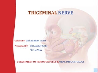
1. TRIGEMINAL NERVE
Guided By- DR.SHUBHRA VAISH
Presented BY – DR.Lakshay Sethi
DEPARTMENT OF PERIODONTOLGY & ORAL IMPLANTOLOGY
PG 1st Year
2. oCRANIAL NERVES AND ITS PHYSIOLOGY
oINTRODUCTION
oTRIGEMINAL NUCLEI
oCOURSE AND DISTRIBUTION
oTRIGEMINAL GANGLION
oDIVISIONS OF TRIGEMINAL NERVE
oAPPLIED ANATOMY
oEXAMINATION OF TRIGEMINAL NERVE
oCONCLUSION
oBIBLIOGRAPHY
CONTENTS
3.
4. CRANIAL NERVES
The 12 pairs of nerves are:
I Olfactory
II Optic
III Oculomotor
IV Trochlear
V Trigeminal
VI Abducent
VII Facial
VIII Vestibulo-cochlear
IX Glossopharyngeal
X Vagus
XI Accessory
XII Hypoglossal
• There are 12 pairs of the cranial
Nerves.
• They are numbered 1 to 12 in the
craniocaudal sequence of their
attachment on the brain.
5. It is Fifth (5th) of twelve pairs of cranial nerves.
Also Called Trigeminus or Trifacial Nerve
Largest Cranial Nerve
first described by Gabriele Fallopius
Trigeminal nerve was proposed by Jacob Benignus
Winslow
Nerve OF 1st Branchial Arch.
INTRODUCTION
7. • A cranial nerve consists of motor fibres (motor nerve) or
sensory fibres (sensory nerve) or both the motor and sensory
fibres (mixed nerve).
MOTOR FIBRES SENSORY FIBRES
Somatic efferent (SE) or
general somatic efferent
(GSE)
Special visceral efferent
(SVE)
General somatic afferent
(GSA)
General visceral efferent
(GVE)
General visceral afferent
(GVA)
Special visceral afferent
(SVA)
Special Somatic Afferent
(SSA)
8. • Mixed Nerve – I.e Both
Sensory
Motor
• Sensory To -
Skin of the anterior part of
the head Oral and Nasal Cavities Teeth and the Meninges
• MOTOR TO -
Muscles of
Mastication
Anterior Belly of
Digastric
Tensor Tympani,
Tensor Veli Palatini
9.
10. Relations :
a) Lateral – middle meningeal artery .
b) Medial – internal carotid artery, cavernous sinus .
c) Inferior – foramen lacerum, greater petrosal nerve,
motor root of trigeminal nerve .
d) Superior – parahippocampal gyrus .
12. (Semilunar Ganglion; ganglion semilunare; Gasseri; Gasserian
ganglion) occupies a cavity (cavum Meckelii) in the dura
mater covering the trigeminal impression near the apex of the
petrous part of the temporal bone.
It is somewhat crescentic in shape, with its convexity directed
forward: medially, it is in relation with the internal carotid
artery and the posterior part of the cavernous sinus.
The motor root runs in front of and medial to the sensory root,
and passes beneath the ganglion; it leaves the skull through
the foramen ovale, and, immediately below this foramen,
joins the mandibular nerve. The greater superficial petrosal
nerve lies also underneath the ganglion.
13. Relations
medial: motor root of the trigeminal nerve and sphenoid bone
lateral: posterior part of the cavernous sinus, petrous apex and petrous
segment of the internal carotid artery
anterior: cavernous sinus and cavernous segment of the ICA
posterior: prepontine cistern
inferior: greater petrosal nerve and middle cranial fossa .
Blood supply
small ganglionic branches of the cavernous portion of the ICA
accessory meningeal artery (from the maxillary artery, via the foramen
ovale) .
Innervation
The ganglionic epineurium is innervated by the nervus spinosus from
the mandibular division of the trigeminal nerve which re-enters the
skull via the foramen spinosum.
14.
15. • The trigeminal nerve originates from three sensory nuclei (mesencephalic,
principal sensory, spinal nuclei of trigeminal nerve) and one motor
nucleus (motor nucleus of the trigeminal nerve) extending from the
midbrain to the medulla.
• At the level of the pons, the sensory nuclei merge to form a sensory root.
• The motor nucleus continues to form a motor root. These roots are
analogous to the dorsal and ventral roots of the spinal cord.
• In middle cranial fossa, the sensory root expands into the trigeminal
ganglion.
• The peripheral aspect of the trigeminal ganglion gives rise to 3
divisions:
• Ophthalmic (V1),
• Maxillary (V2)
• Mandibular (V3).
16. OPTHALMIC NERVE (V1)
• First division of the trigeminal nerve
• ORIGIN –TRIGEMINAL GANGLION
• TYPE - It is a purely sensory nerve
• FUNCTION - Carries afferent stimuli of pain, light touch,
and temperature from the upper eyelids and supraorbital region of the
face, up to the vertex of the head .
17. • The ophthalmic division (V1) travels forward through the cavernous sinus
where it receives some fibers from the sympathetic plexus traveling with the
internal carotid artery
• In the sinus, the nerve is located inferior to
the trochlear nerve and lateral to the abducent and oculomotor nerves
COURSE
• Trigeminal ganglion -> cavernous sinus -> superior orbital fissure ->
lacrimal, frontal, nasociliary nerves (terminal branches) -> respective
anatomical structures
18. • It subdivides into three terminal branches
1. The Lacrimal,
2. The Frontal
3. Nasociliary Nerve
20. LACRIMAL NERVE
• Most lateral and thinnest branch of the ophthalmic
nerve .
• Passes into orbit through lateral compartment of the Superior
orbital fissure outside the tendinous ring .
Connected with zygomaticotemporal branch of maxillary nerve .
secretomotor fibres to lacrimal gland
Supplies : lacrimal gland and skin of upper eyelid and conjunctiva .
21.
22. FRONTAL NERVE
• Is the middle and thickest branch of the ophthalmic nerve .
• It runs directly beneath the roof of the orbit and superiorly to the superior
palpebral levator muscle .
• DIVIDES Into –
1. SUPRAORBITAL – lateral branch of the frontal nerve.
-- the nerve gives off several palpebral filaments that
supply the conjuctiva and the skin of the upper eyelid.
2. SUPRATROCHLEAR -- The supratrochlear nerve is placed medial to the
supraorbital nerve.
-- Innervate the skin of the dorsum of the nose and
adjacent skin of the upper eyelid.
23. SUPRATROCHLEAR NERVE
Smaller nerve
Medial branch
Receives communication
branches from
infratrochlear nerve
Curves around
superomedial margin of
Orbit
supplies: median
conjunctiva, Upper Lid and
lower part of forehead
Lies between frontalis and
corrugator supercilliary
muscles
SUPRAORBITAL NERVE
Larger nerve
lateral branch
Passes through
supraorbital notch
Divides in medial and
lateral branches
Lies beneath frontalis
Muscle.
Supplies: conjunctiva,
scalp upto vertex , mucous
membrane of frontal sinus
24.
25. NASOCILIARY NERVE
• Nerve is the medial terminal branch of the
ophthalmic nerve .
• It begins in the lateral wall of the anterior part of
the cavernous sinus .
• Crossing over the superior side of the optic nerve it
reaches the anterior ethmoid foramen, where it
divides to its own two terminal branches.
26. • Nasociliary nerve extends to the lateral branches in the following order going
from proximal to distal to the root:
1. Long Ciliary Nerve
2. Short Ciliary Nerve
3. Posterior ethmoid nerve
In the area of the anterior ethmoid foramen, the nasociliary nerve extends to its
two terminal branches:
1. Anterior Ethmoid Nerve
2. Infratrochlear nerve
27.
28. Ciliary ganglion
• This ganglion belongs to the autonomic nervous system and
is functionally added to the ophthalmic nerve.
• Lies in posterior part of orbital cavity .
• The ciliary ganglion has preganglionic and postganglionic
fibers .
29. • Preganglionic fibers are –
1. Sensory
2. Sympathetic
3. Parasympathetic
• Postganglionic fibers :-
are the short ciliary nerves that extend forward while
grouped around the optic nerve .
30. MAXILLARY NERVE V(2)
• SECOND division of the trigeminal nerve
• ORIGIN -- Trigeminal Ganglion .
• TYPE -- It is a purely sensory nerve .
• Function – SUPPLIES
• Skin of the face over maxilla .
• Teeth of the upper jaw .
• Mucous membrane of the nose .
• The maxillary sinus and palate .
31. COURSE
• Courses forward through the lateral dural wall of the cavernous
sinus, inferiorly and laterally to the ophthalmic nerve.
• The nerve leaves the middle cranial fossa after it passes through
the foramen rotundum and enters the upper part of
the pterygopalatine fossa.
• The fibers of the maxillary nerve leave the fossa by coursing
forward through the pterygomaxillary fissure and then enter
the infratemporal fossa .
32.
33. BRANCHES
1. Branches in middle cranial fossa
i. Meningeal branch
2. Branches arising in pterygopalatine fossa
i. Ganglionic branches
ii. Zygomatic nerve
iii. Posterior superior alveolar nerve
3. Branches arising in infraorbital groove and canal
i. Middle superior alveolar nerve
ii. Anterior superior alveolar nerve
4. Branches of infraorbital nerve in the face
i. Inferior palpebral
ii. Lateral nasal
iii. Superior labial
34.
35.
36. IN MIDDLE CRANIAL FOSSA:
• Meningeal branch: Travels along the middle meningeal
artery and provides sensory innervation to cranial dura
matter.
37. IN PTERIGOPALATINE FOSSA:
1. Ganglionic Branches –
Arises as 2trunks.
• Trunks join to form single root within pterygopalatine ganglion.
• Gives Orital branches,Palatine branches,Pharyngeal
branches,Nasal branches
• Gives postganglionic secretomotor fibers to lacrimal gland
via zygomaticotemporal and lacrimal.
2. Orbital branch: Supplies periosteum of orbit
38. • Nasal branch --
• Supplies to mucosa of superior and inferior conchae,
posterior ethmiodal sinus and posterior portion of nasal
septum. It also includes Nasopalatine branch .
39. B. . ZYGOMATIC NERVE
• It enters the orbit through inferior orbital fissure and runs forward
along its lateral wall .
• Divides into 2 branches :
i. Zygomaticotemporal nerve :
emerges from the temporal surface of the bone
supplies the skin over temple region.
ii. Zygomaticofacial nerve :
emerges from the bone through the zygomaticofacial foramen
present on the lateral surface of the bone
Supplies skin of cheek
40. BRANCHES OF INFRAORBITAL NERVE ON FACE
INFRAORBITAL NERVE
INFERIOR
PALPEBRAL
Supply lower
eyelid
LATERAL NASAL
Supply the skin
on lateral side
of nose
SUPERIOR LABIAL
Supply skin of
upper lip and part
of the cheek
41.
42. Pterygopalatine Ganglion
Largest peripheral ganglion of parasympathetic system
Topographically = maxillary nerve
Functionally = greater petrosal branch of facial nerve
Situation :
i. Lateral to sphenopalatine foramen
ii. Below the
maxillary nerve .
iii. Front of
pterygoid canal .
43. Pterygopalatine Fossa
• The body of the ganglion rests in
the pterygopalatine fossa. The
pterygopalatine fossa is a
depression that lies within the
pterygomaxillary fissure, inferior to
the sphenopalatine foramen.
• This fissure is a natural furrow
that is formed between the
posterosuperior border of
the maxilla and the anterosuperior
border of the pterygoid plates.
44. MANDIBULAR NERVE V3
• THIRD DIVISION OF TRIGEMINAL NERVE
• TYPE -- both sensory and motor.
• SUPPLY --
• Buccal skin
• anterior two-thirds of the tongue,
• temporal region;
• mastication muscles, mylohyoid muscle
• anterior belly of the digastric muscle
45. • Formed by union of two roots .
• Sensory root arises from lateral part of trigeminal
ganglion – leaves the skull through foramen ovale .
• Motor root passes through foramen ovale and unites
with the sensory root just below the foramen .
• Nerve then enters the infratemporal fossa
• After a short downward course, it then divides into a
smaller anterior division and a large posterior division
52. SUBMANDIBULAR GANGLION
The submandibular ganglion is
one of four parasympathetic
ganglia of the head and neck. It
receives parasympathetic fibers
from the facial nerve .
Gross anatomy
• Small ganglion suspended from
the undersurface of the lingual
nerve.
• Inferior to submandibular
duct sitting on the hyoglossus
muscle .
• Supplies secretomotor fibers to
the sublingual and submandibu
lar salivary glands .
53. • Preganglionic parasympathetic fibers derived from the chorda tympani travel in
the lingual nerve and synapse in the submandibular ganglion.
• Some of the postganglionic secretory fibers enter the submandibular gland;
others, by entering the lingual nerve, reach the sublingual gland.
• Postganglionic sympathetic fibers (from the superior cervical ganglion) pass
through the submandibular ganglion and are distributed with the
parasympathetic fibers.
Roots
Branches
Lingual Nerve
56. To be able to distinguish and to cure with some degree of certainty, a disease
that
during the time it lasts is extremely excruciating is an addition, however small,
to John Fothergill (1712–80)
57. 1. Referred pain
2. Mandibular neuralgia .
3. Lesion at foramen ovale .
4. Lingual nerve damage .
5. Referred pain in the ear
CONTENTS
58. EXAMINATION OF TRIGEMINAL NERVE
1. SENSORY
FUNCTION
2. MOTOR
FUNCTION
3. CORNEAL
REFLEX
4. JAW JERK TEST
59. SENSORY
FUNCTION
•Initially test the sensory branches by lightly touching the face with a piece of
cotton wool followed by a blunt pin in three places on each side of the face:
1.Around the jawline
2. on the cheek
3. on the forehead
1 2
3
60. MOTOR FUNCTION
• Inspect for wasting of the temporal and masseter muscles.
• Ask patient to clench their teeth and palpate for contraction of the
temporal and masseter muscles.
• Ask the patient then to open their mouth against resistance
61. CORNEAL REFLEX
Ask the patient to look up and away, touch the cornea .
Reflex blinking of both eyes is a normal response.
62. JAWJERK TEST
• Ask the patient to open the mouth fully, and close halfway, place
index finger on the chin and tap with a patella hammer.
• When it is normal, tapping the mandible produces a brisk
contraction.
63. Mechanisms OF Injury to
TRIGEMINAL NERVE
1. Local anaesthetic injection
2. Third molar surgery
3. Maxillofacial trauma
4. Orthognathic surgery
5. Maxillofacial pathology
6. Endodontic and chemical injury
64. LOCAL ANESTHETIC SOLUTION
Incidence – between 1:26,762 and 1:160,571
Harn and Durham – 1990 – transient sensory disturbance – 3.62%
Causes :
i. Direct penetration of the nerve by needle
ii. Hematoma formation from vessel laceration
iii. Direct laceration of the nerve from a barb on the tip of needle
after repeated injections.
iv. Needle contact with cortical bone.
v. Chemical injury from intraneural injection.
65. Maxillofacial Trauma
• Neurosensory impairment due to trauma – 70.9%
• Neurosensory alterations caused by – laceration,
traction or compression of the IAN from bony
segment displacement
• Reduction of fractures with alignment of segments
and removal of loose bony segments that impinge on
nerve will assist in spontaneous neurosensory
recovery .
66. Orthognathic surgery
• During SSO, nerves could be injured at various locations
• Incidence of neurosensory alterations during mandibular
orthognathic surgery has been found to increase with
intraoperative complications .
• During maxillary or mid face procedures, ION is at risk
for injury because of soft tissue flap retraction .
70. Trigeminal Neuralgia (Fothergills Disease)
or
Tic Douloreux)
• Trigeminal neuralgia is defined as sudden, usually unilateral,
severe, brief, stabbing, lancinating, paroxysmal, recurring pain
in the distribution of one or more branches of 5th cranial nerve
.
• John Locke – 1677 – first full description with its treatment.
• Nicholas Andre – 1756 – Tic Douloureux (painful jerking)
• John Fothergill – 1773 – published detailed description of TN,
thus, Fothergill’s disease .
72. GENERAL CHARACTERISTICS
Incidence : rare
Age : 5th or 6th decade
Sex predilection : female predisposition (58%)
Affliction for side : right side (60%)
Trigeminal nerve involvement : V3>V2>V1
73. Clinical Features :
Refractory period can be as short as a couple of seconds
With each attack, pain seems to become more intense and
unbearable
Pain rarely crosses midline.
Pain is of short duration & lasts for a few seconds
extreme cases – frozen or mask like face .
different stimuli can elicit pain :
i. Touching or applying heat or cold to the cheek or gum
ii. Chewing, yawning or talking
iii. Wind blowing on face
iv. Gustatory stimuli and vibration
v. Smiling / brushing / shaving / washing face
74. Diagnosis – Imaging
1. CT scan – poor resolution in posterior
fossa.
2. MRI – imaging modality of choice;
reveal MS plaques and pontine
gliomas
.
1. Conventional angiogram – useful
only to detect vascular malformation
.
75. SWEET DIAGNOSTIC CRITERIA
1. Pain is PAROXYSMAL
2. Pain is provoked by light touch to the face
3. Pain is confined to trigeminal distribution
4. Pain is unilateral
5. Clinical sensory examination is normal
76. MEDICAL TREATMENT
• First line of approach
• TN does not respond to analgesics
1. Carbamazepine is used as a standard drug. Adult dose 100mg,
thrice daily initially.
• Started as small dose & gradually increased to prevent side
effects.
ADVERSE EFFECTS -- include dizziness, ataxia, vertigo, skin rashes etc
When carbamazepine is contraindicated, clonazepam 1.5mg/day
can be used
2. Phenytoin Sodium
usually used in combination of carbamazepine
100mg thrice a day
side effects: gum hyperplasia, swelling of lymph glands
3. Gabapentin
300mg/day
used with caution in patients with renal & hepatic disease
4. Gaba agonist
these drugs reduce the central projection of painful impulses
eg. Baclofen , adult dose being 5-10 mg TDS.
77. 1. Peripheral nerve injections
a) Long acting anesthetic agents without
adrenaline (bupivacaine)
b) Alcohol block - 0.5 -2ml of 95% absolute
alcohol.
2. Peripheral neurectomy
oldest and most effective
Acts by interrupting the flow of afferent
impulses to central trigeminal apparatus
3. Cryotherapy or Cryoneurolysis
Direct applications of cryotherapy probe
at temperatures colder than -60 degrees
Celsius .
In this the nerve is not sectioned but
destroyed
4. Peripheral radiofrequency neurolysis
(thermocoagulation)
Gregg and Small (1986)
Radiofrequency electrode has the
capacity to destroy the pain fibres
78. • 5 Gasserian ganglion procedures
• Glycerol injection
• thermocoagulation
• Balloon compression
• 6.
• Open procedures (intracranial procedures)
microvascular decompression of sensory root .
1967-1976 by Jannetta
most commonly performed intracranial open
procedure .
open craniotomy approach is used to gain
access to the trigeminal root entry zone and
adjacent brain stem
Compressing branch of superior cerebellar
artery is carefully separated from the nerve
.
79. • Ophthalmic zoster is a disease characterised by
reactivation of varicella zoster virus that is inactive in
dorsal root or cranial root ganglion, after primary infection
Oral and facial lesions result from HZ of 2nd and 3rd divisions
of trigeminal nerve, but involvement of 1st division is
considerably more common esp. nasociliary nerve
HZ has been associated with dental anomalies and severe
scarring of facial skin when trigeminal HZ occurs during tooth
formation
HERPES ZOSTER OPHTHALMICUS
80. • Diagnosis :
history of pain.
unilateral nature .
Segmental distribution of
lesions.
vesicles are present.
81. Treatment :
• Acyclovir 800mg, 5 times/day for a week,
within 4 days of onset of rash
• Valacyclovir 1,000mg, 3times/day for a week
• Analgesics
• Antibiotic ointments
• Systemic steroids 60mg/day
• Corneal grafting
82. TRIGEMINAL NEUROPATHY
• Characterized by numbness in the skin or
mucosal membranes in the distribution of the
trigeminal nerve Neuropathic weakness in the
muscles of mastication.
• TNO should not be confused with trigeminal
neuralgia (TNA)
• Brief attacks of lancinating pain but without
sensory impairment or motor weakness.
83. • In TNO, pain may dominate the clinical picture
• As disorder progresses and neurons are destroyed,
numbness and weakness usually appear.
• In untreated idiopathic TNA, neurons are preserved,
although their myelin sheaths may be destroyed and there
is ultimately gain of function.
• In TNO as the condition advances, loss of function in the
affected nerve branches becomes evident .
84. Wallenberg syndrome
Neurological disorder causing a range of
symptoms due to ischemia in the lateral
part of the medulla oblongata in
the brainstem.
The ischemia is a result of a blockage in
the posterior inferior cerebellar artery or
one of its branches.
Wallenberg syndrome is also called
Lateral medullary syndrome, posterior
inferior cerebellar artery syndrome and
Vertebral artery syndrome
85. CAUSE
Occlusion of the posterior inferior cerebellar artery or one of
its branches or of the vertebral artery, in which the lateral part
of the medulla oblongata infarcts, resulting in a typical
pattern.
DIAGNOSIS
Diagnosis is usually done by assessing vestibular-related
symptoms in order to determine where in the medulla that
the infarction has occurred.
Head Impulsive Nystagmus Test of Skew (HINTS)
examination of oculomotor function
Computed tomography (CT)
Magnetic resonance imaging (MRI) to assist in stroke
detection
86. • Treatment involves focusing on relief of symptoms and
active rehabilitation
Speech Therapy - common form of rehabilitation
In more severe cases, a feeding tube maybe inserted through
the mouth or a gastrostomy may be necessary if swallowing is
impaired.
Medication may be used to reduce or eliminate residual pain -
anti-epileptics such as gabapentin. Antiplatelets like aspirin or
clopidogrel and statin regimen. Warfarin is used if atrial fibrillation is
present.
• One of the most unusual and difficult to treat - violent hiccups.
struggle to eat
sleep
carry on conversations
Unfortunately there are very few successful medications
available to mediate the inconvenience of constant hiccups.
Treatment for this disorder can be disconcerting because some
individuals will always have residual symptoms due to the severity of
the blockage as well as the location of the infarction.
87.
88. • Nerve blocks .
• Flap retraction during
periodontal surgery .
• Implant surgery .
• Maxillary sinus surgery .
89.
90. Photograph showing the osteology involved in an inferior
alveolar nerve
injection and an inset showing an actual injection .
93. A = the orthopantomograph, B and C = cone beam
computed tomography shows full dental implant
intrusion into mandibular canal in 35 jaw dental
segment region. There is direct mechanical
trauma - IAN transection.
94. In a recent study, Kim et al classified the buccolingual location of IAN
into 3 types :
1. Type 1 (70%) : IAN canal follows the lingual cortical plate of the
mandibular ramus and body
2. Type 2 (15%) : IAN canal is located in the middle of the mandibular
ramus posterior to the second molar. It then runs lingually to follow
the lingual plate
3. Type 3 (15%) : IAN canal is located near the middle of the ramus
and body
Bifid canal
i. Nortje et al – 0.9%
ii. Grover et al – 0.08% of radiographs suggestive of bifurcation
of IAN
iii. Langlais et al – 0.95% cases had bifid canals
96. Treatment Approaches
FACTORS DECIDING THE MOST
APPROPRIATE SURGICAL
APPROACH
1. PATIENT’S ANATOMY
2. SURGEON’S EXPERIENCE
3. MECHANISM OF INJURY
4. LOCATION OF INJURY
97. APPROACHES FOR DIFFERENT NERVES
LINGUAL NERVE
Approached intraorally via a paralingual or lingual gingival
sulcus incision
INFERIOR ALVEOLAR NERVE
vestibular incision with identification of MN and lateral
decortication to expose a portion of IAN
in cases of limited mouth opening, extraoral approach
applied
IAN is approached via lateral decortication techniques
INFRAORBITAL NERVE
transorally via a maxillary vestibular incision at the time of
maxillary or zygomaticomaxillary complex fracture repair
98. Outcomes of Trigeminal Nerve
injuries and Surgical Intervention .
• Injury to trigeminal nerve maybe associated with impairment of
speech, taste, mastication and impact on quality of life.
• Based on recent studies, success of microneurosurgical
reconstruction of the TN injuries could be estimated to be between
30% and 50% for all degrees of injury and types of reconstruction.
• Gap repair with an autogenous sural nerve graft may provide a
better outcome because the graft itself provides a rich source of
Schwann cells and neurotropic and neurotrophic factors that assist
in the degeneration and regeneration process of neural recovery.
99. CONCLUSION
Since Trigeminal nerve is a mixed nerve and supplies mainly head and
neck region, one should know thoroughly about its cranial and
extracranial course and distribution, to diagnose the pathologies
associated with nerve and for appropriate treatment .
100. BIBLIOGRAPHY
• Anatomy for dental students – Inderbir Singh
• Grant’s Atlas of Anatomy – 10th edition
• Atlas of Human Anatomy 5th edition – Frank H.
Netter
• The Clinical Anatomy Of Cranial Nerves – Joel A.
Vilenski
• Anand’s Human Anatomy for dental students 2nd
edition
• Textbook of oral and maxillofacial surgery – Balaji
2007 edition
• Essentials of Human Anatomy – A.K. Datta
• Monheim’s Local Anaesthesia and Pain Control in
Dental Practice - 7th edition
• Oral and Maxillofacial Trauma – Fonseca – 4th
Edition
• Handbook of Local Anesthesia - Malamed
101. • Inderbir Singh’s Human Embryology, 11th edition
• BD Charausia , 6th edition
• Textbook of anatomy, Vishram Singh, vol III, 2nd
edition
• Textbook of oral medicine, oral diagnosis and oral
radiology – Ravikiran Ongole, 2nd edition
• Burket’s oral medicine and diagnosis – 9th edition
•
• Textbook of oral and maxillofacial surgery, Neelima
Malik, 3rd edition
Editor's Notes
INFERIOR VIEW OF HUMAN SKULL DEPICTING ALL FOSSAS AND FORAMEN PRSNT .
Coronal cross-sectional view of the cavernous sinus highlighting the location of
V1 and V2 vis-à-vis the other cranial nerves and blood vessels located within the sinus.