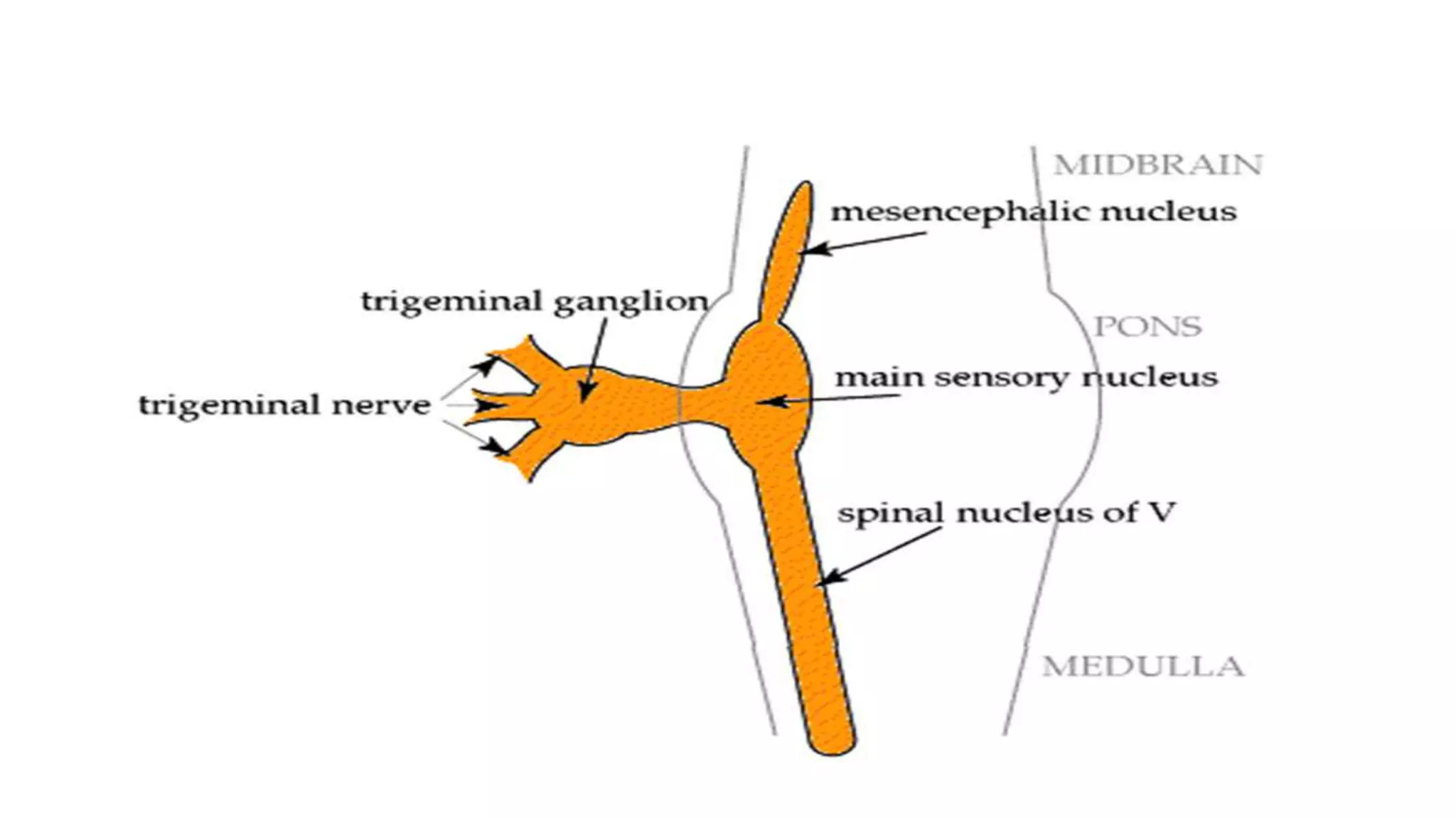
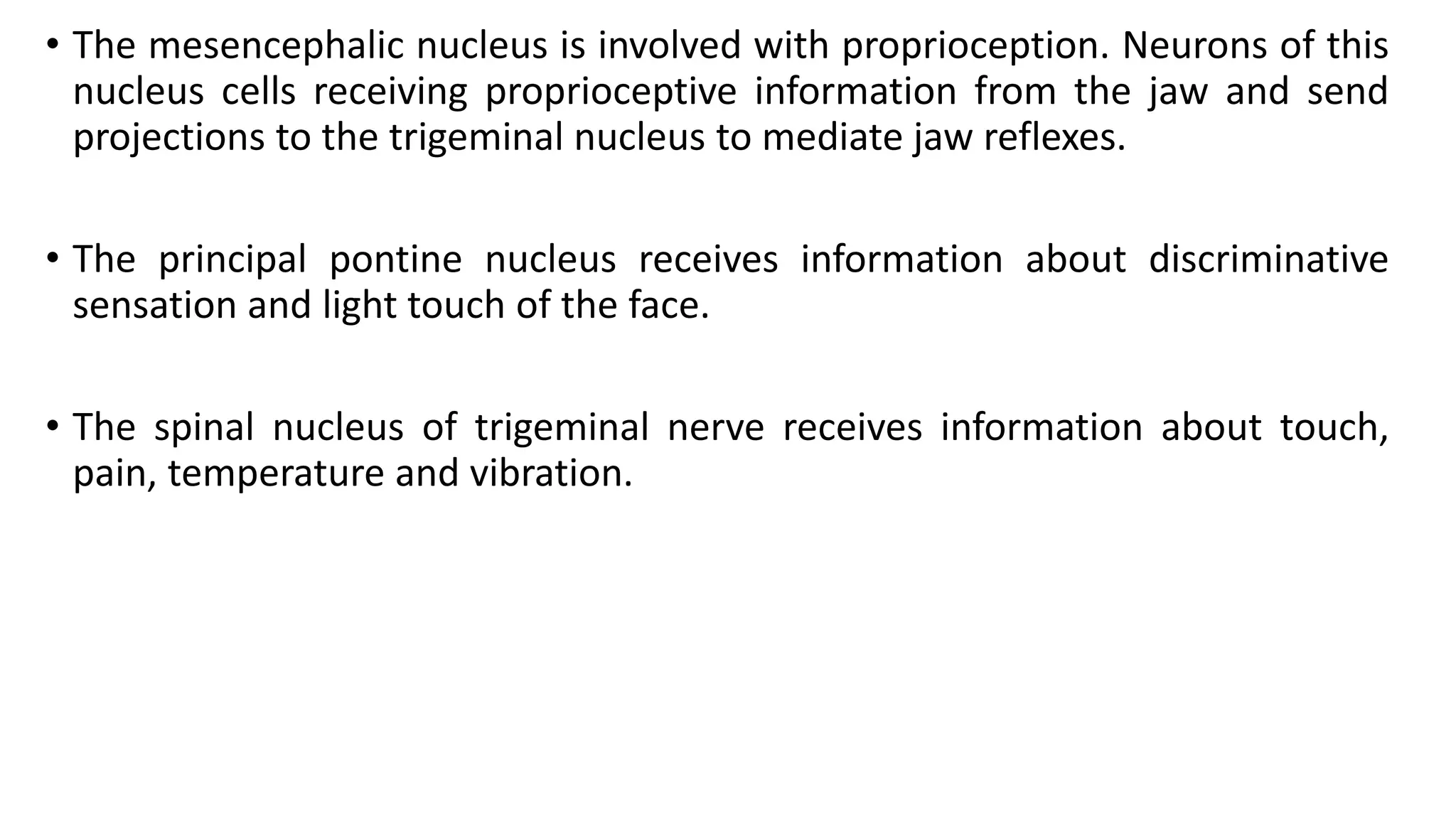
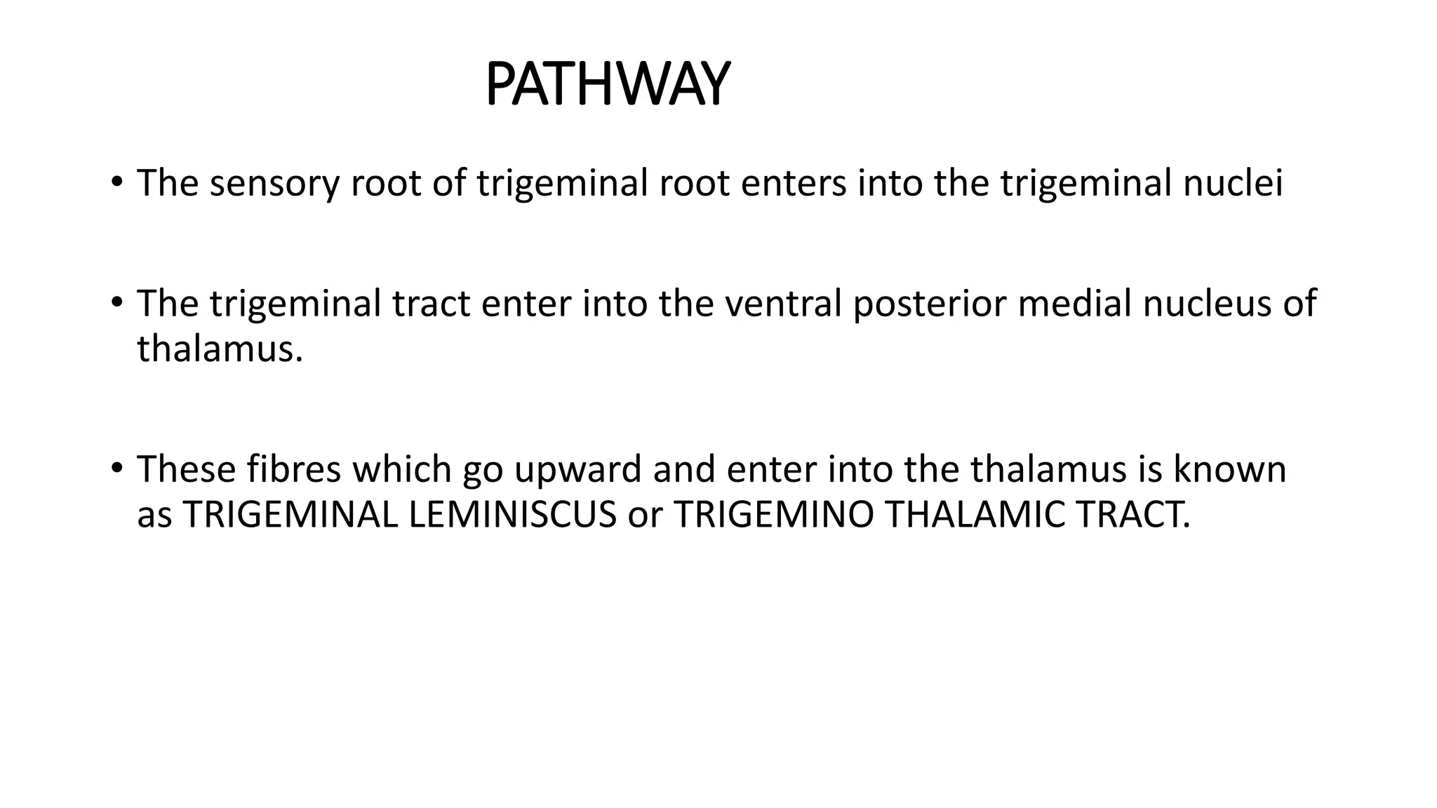
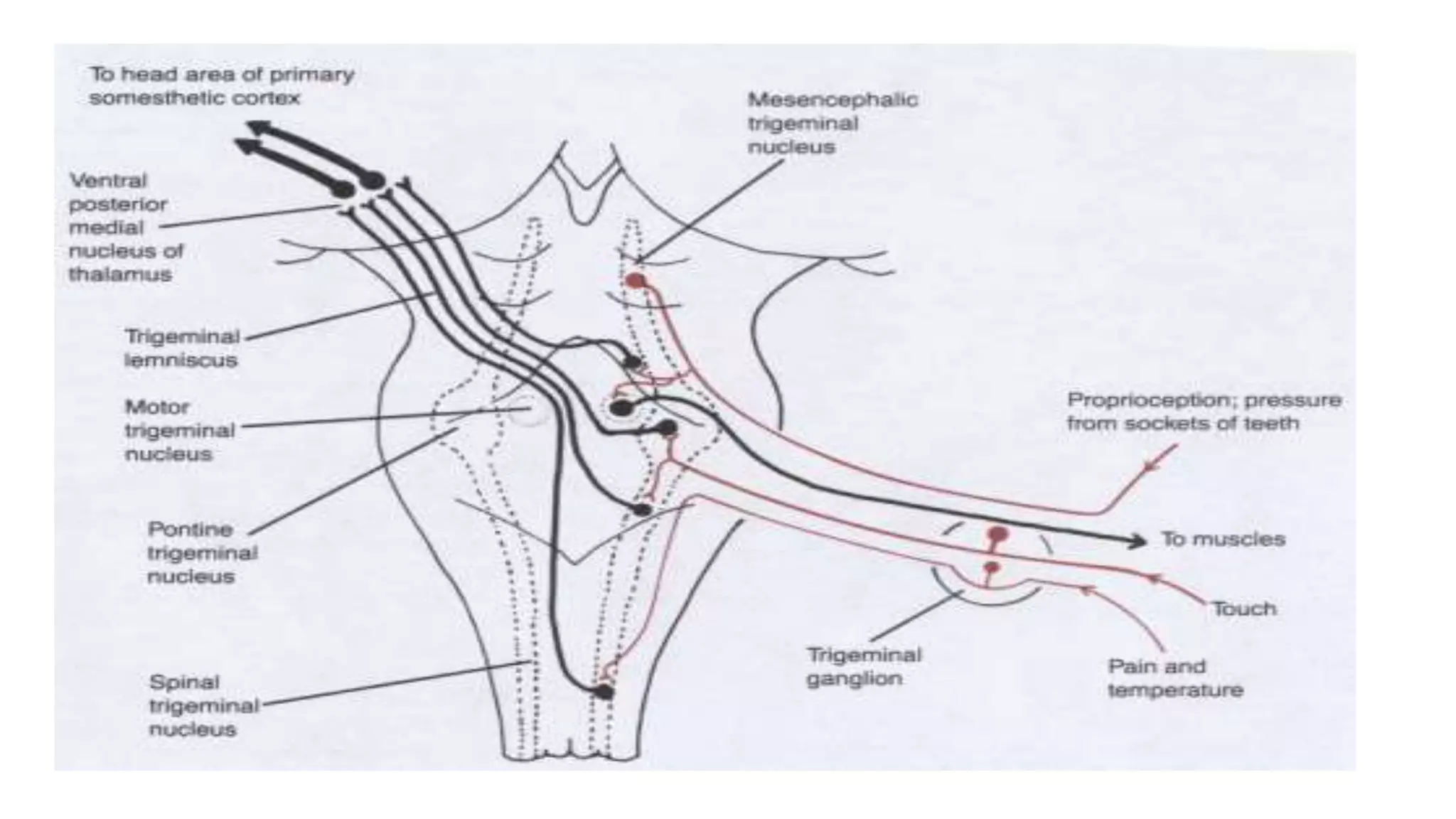
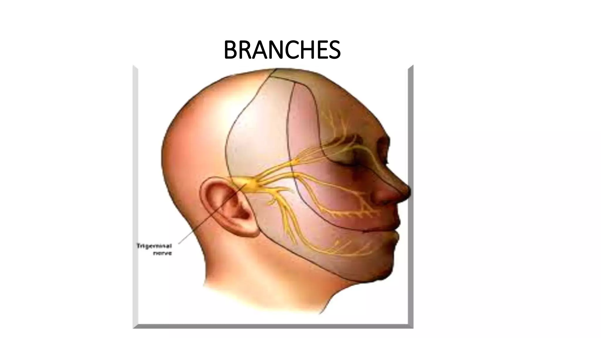
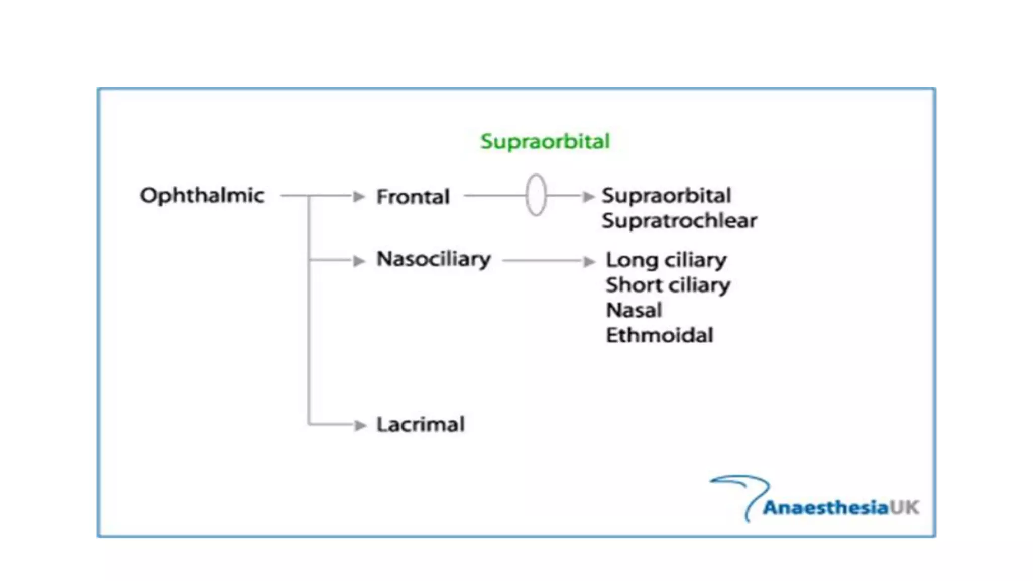
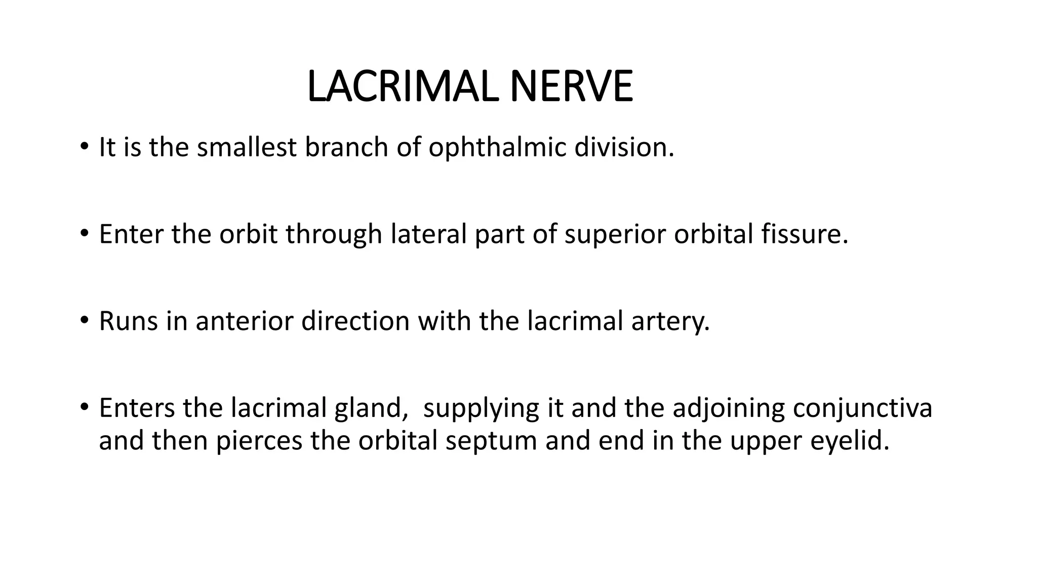
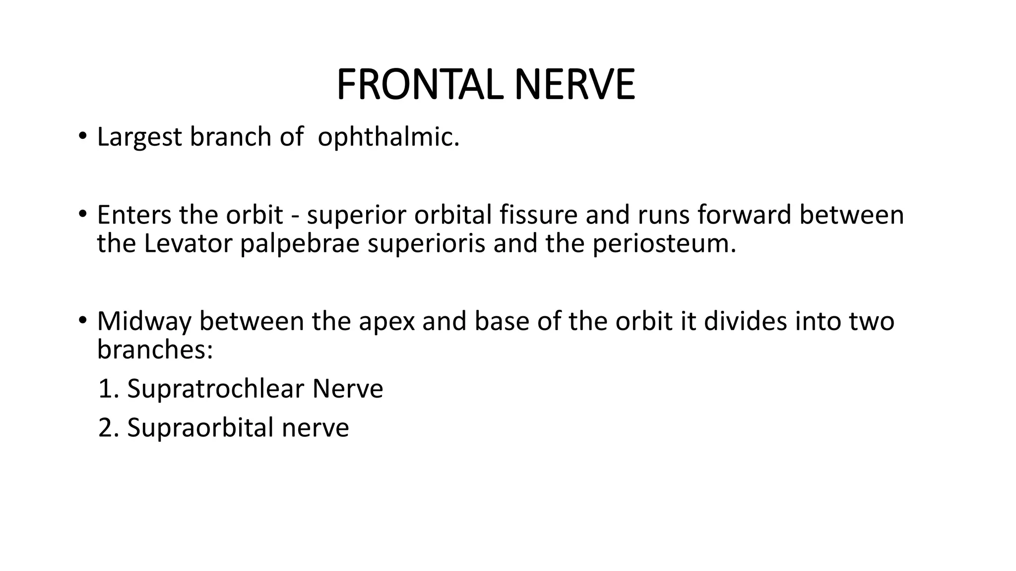
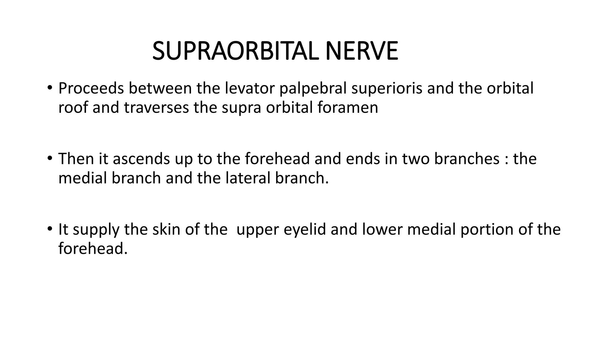
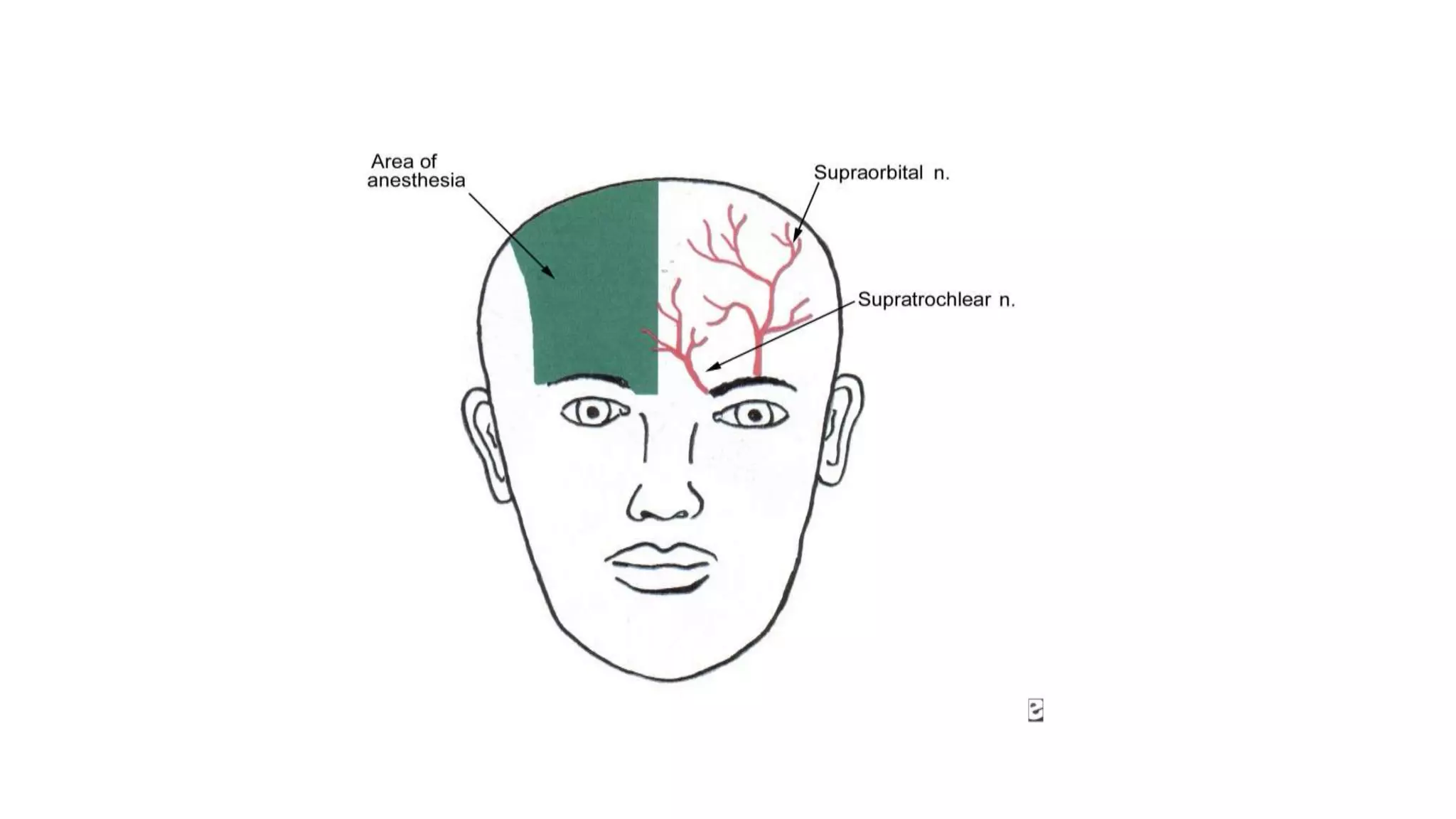
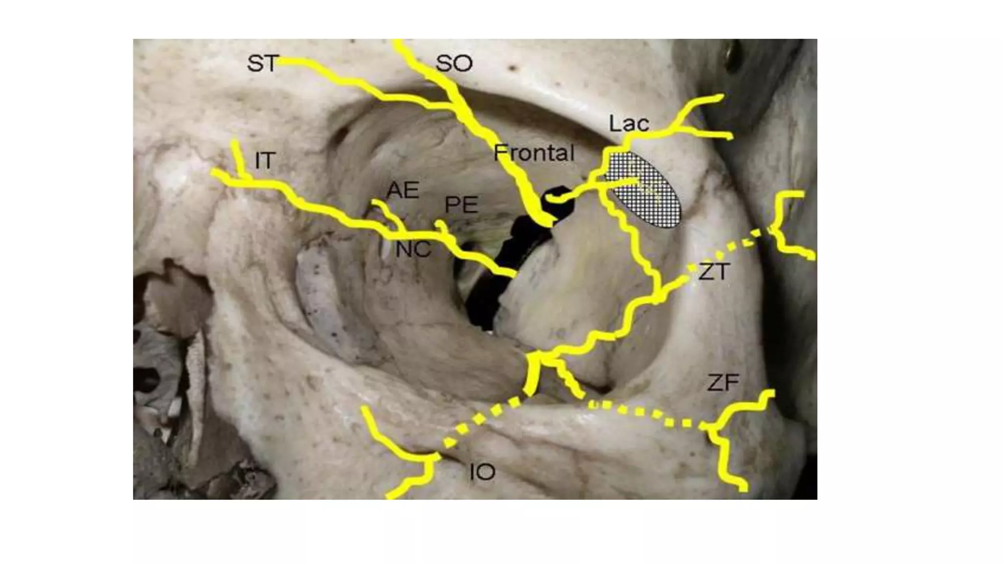
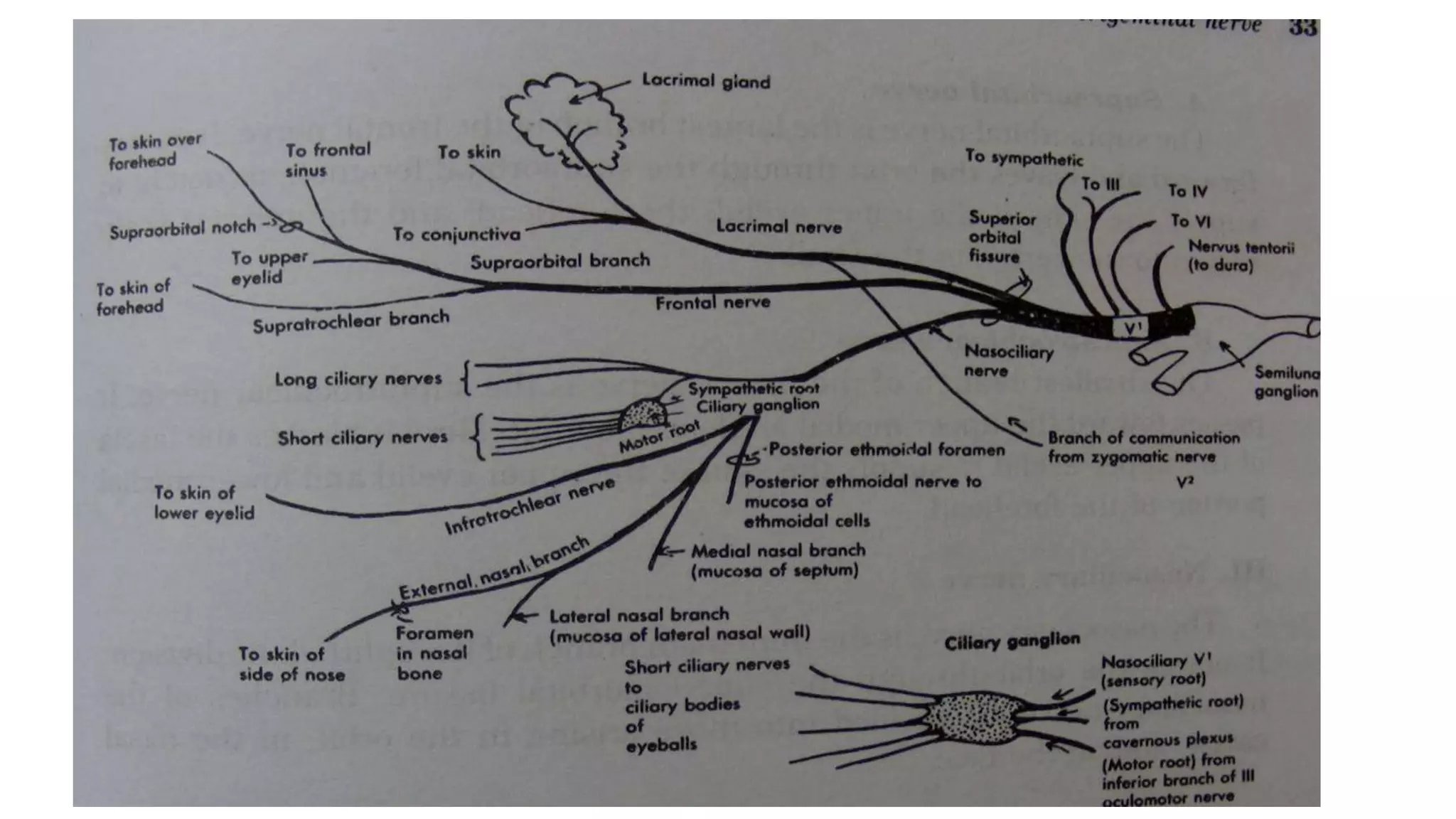
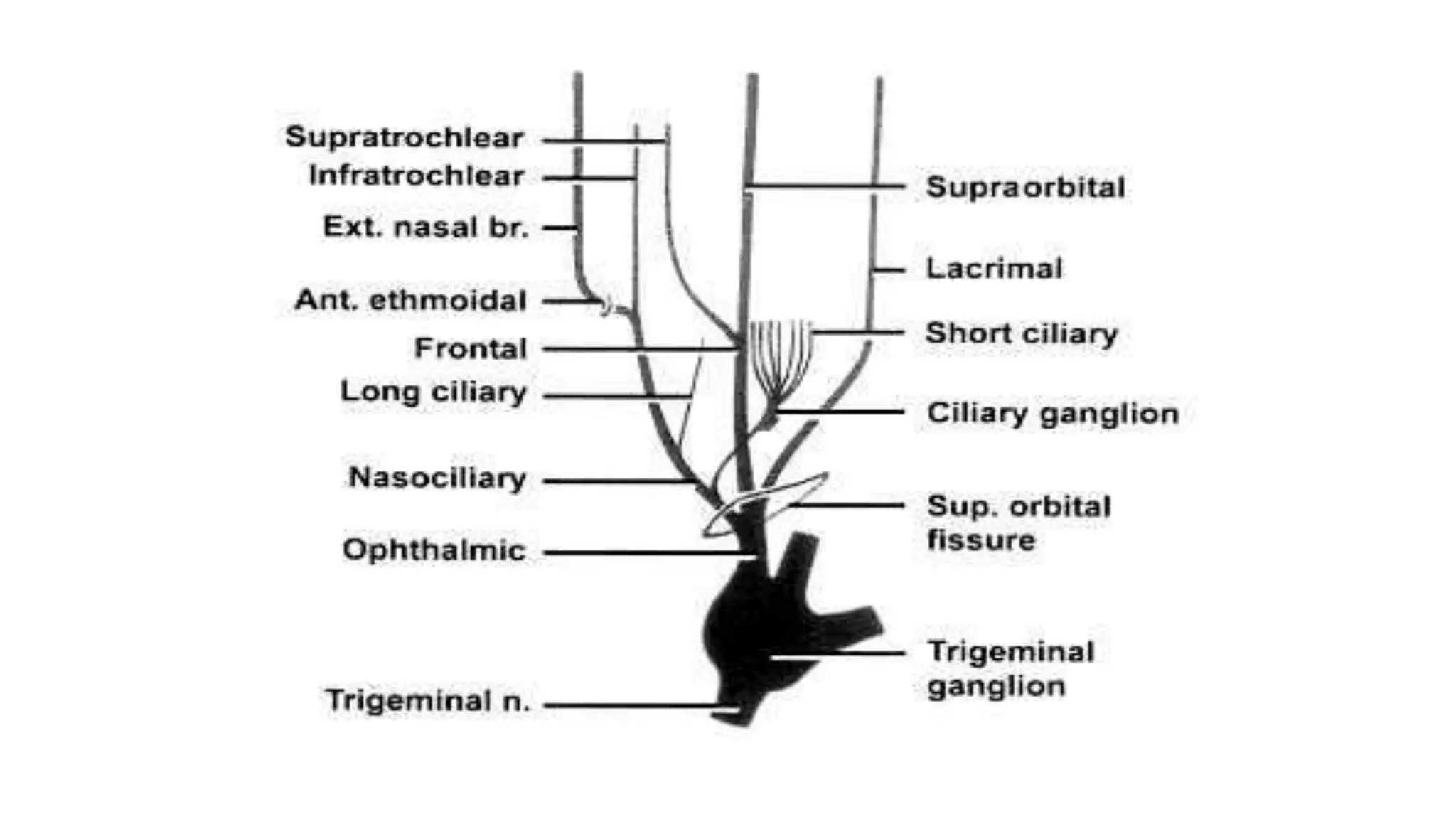
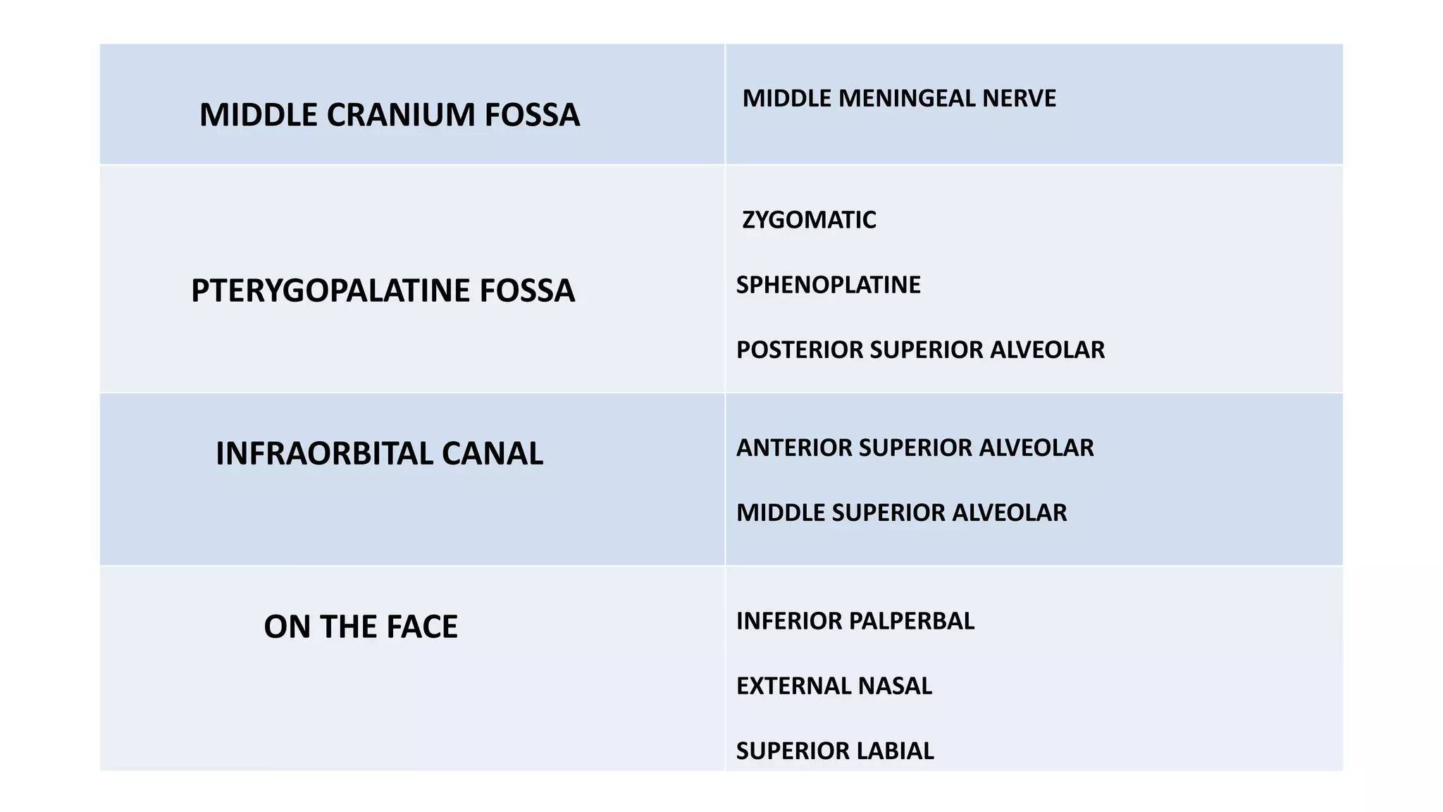
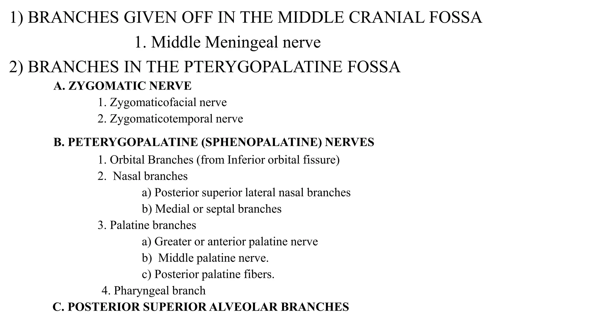
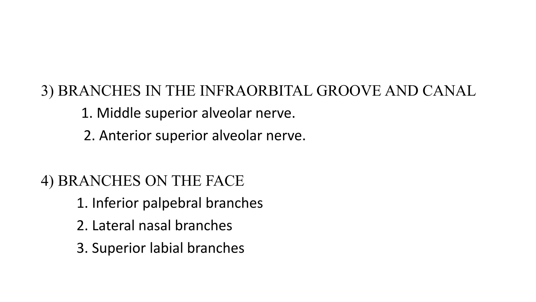
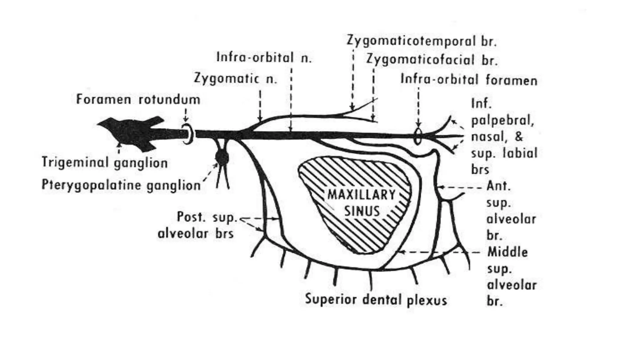
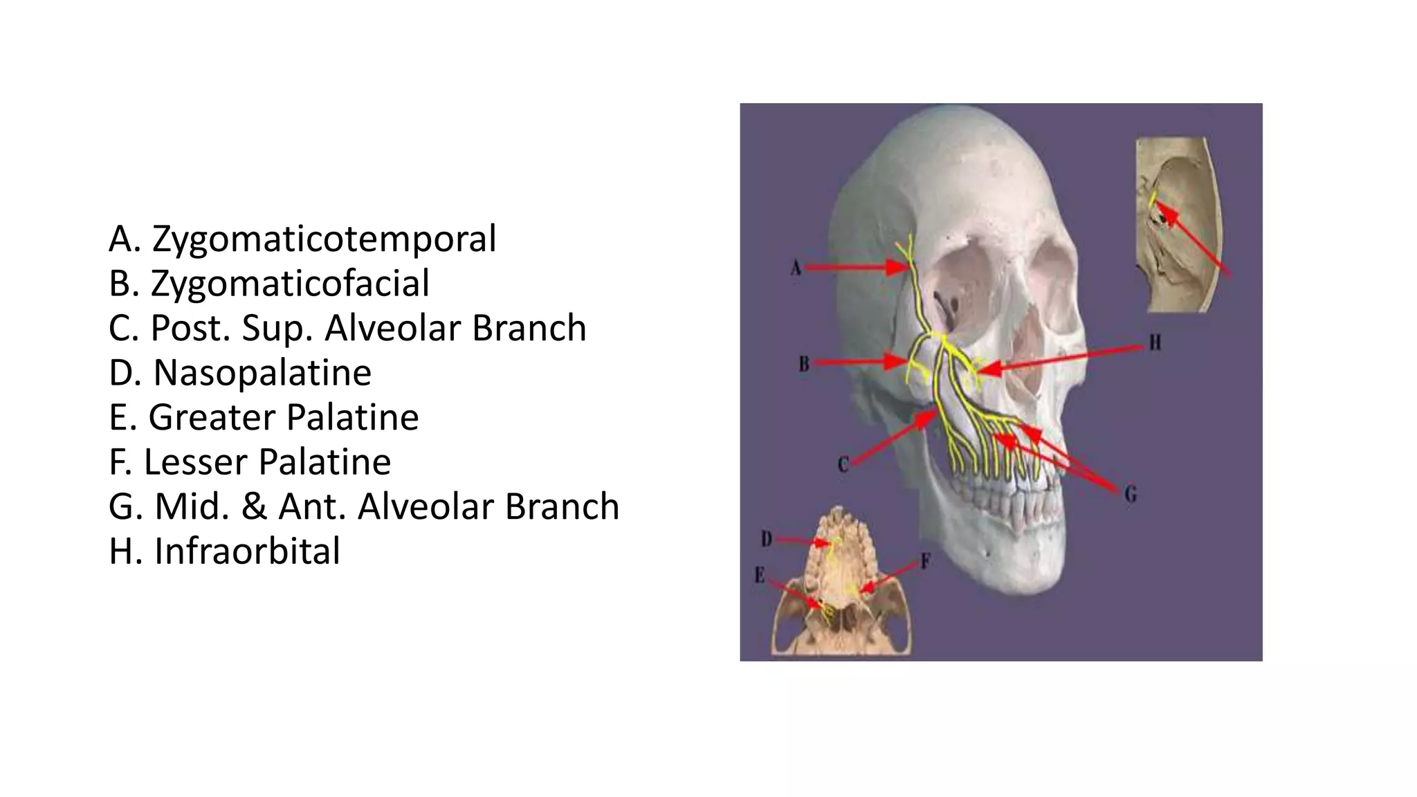
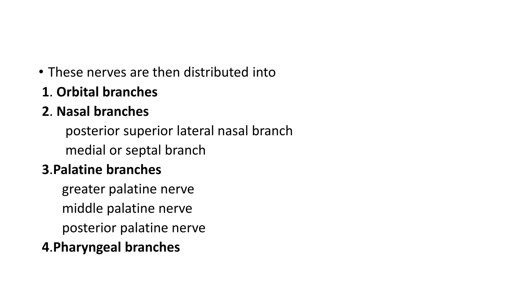
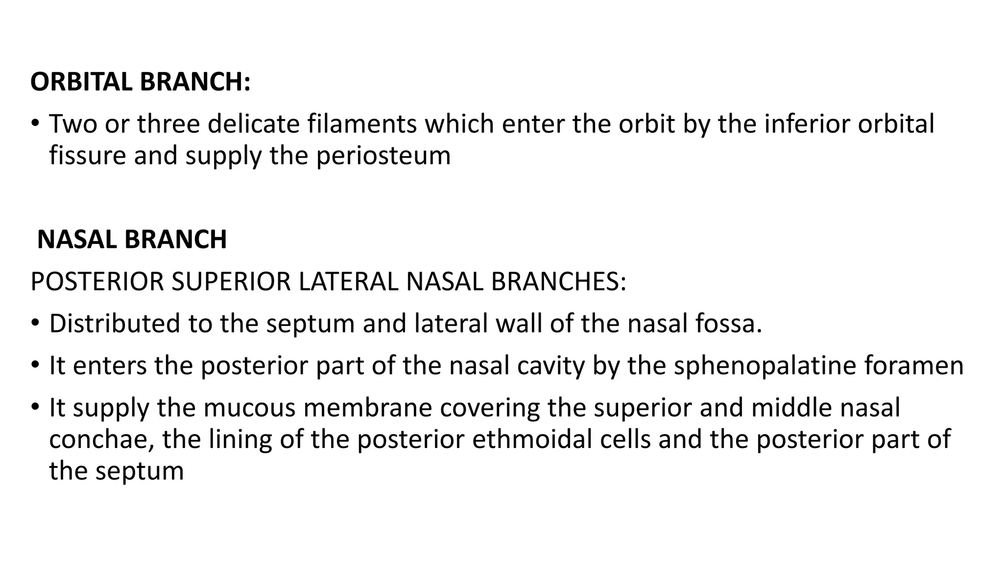
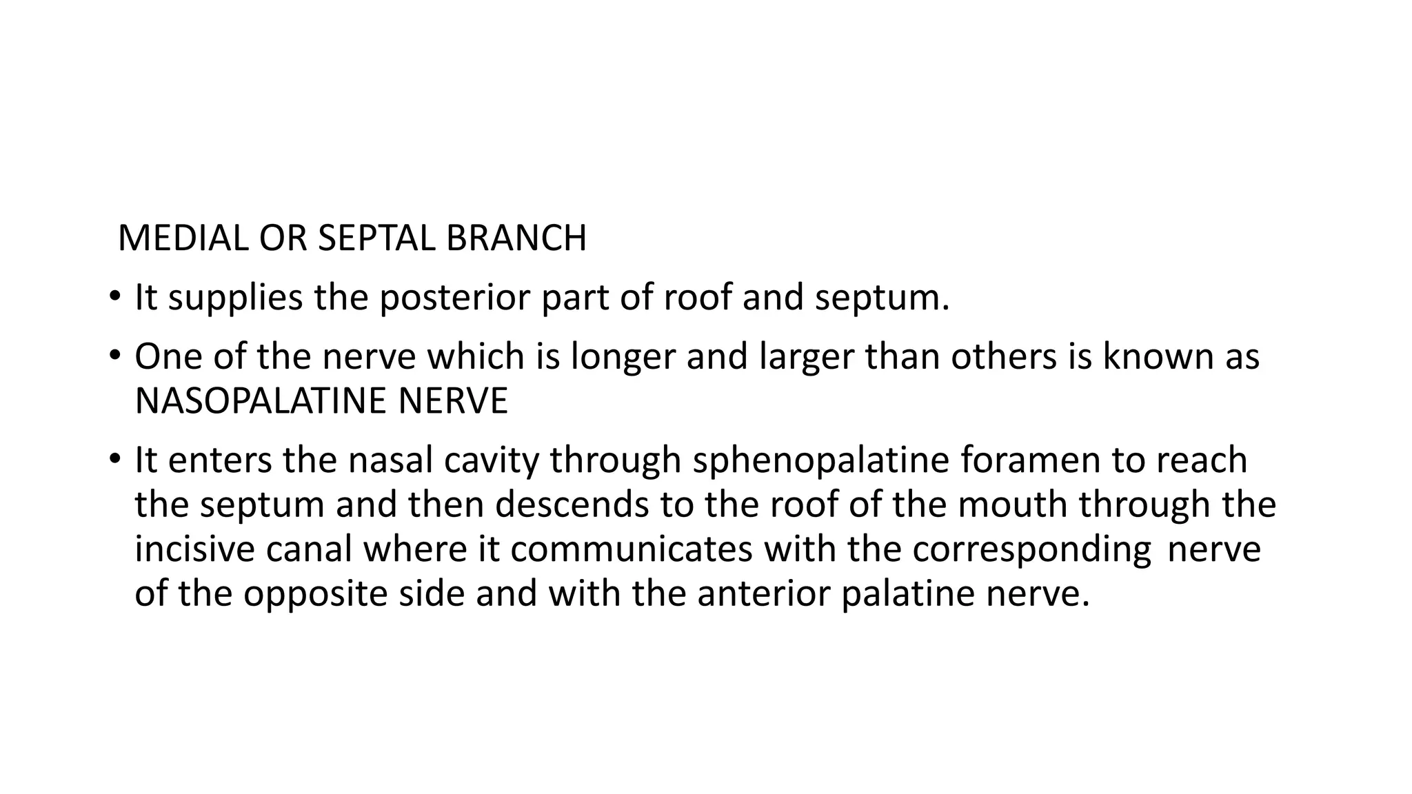
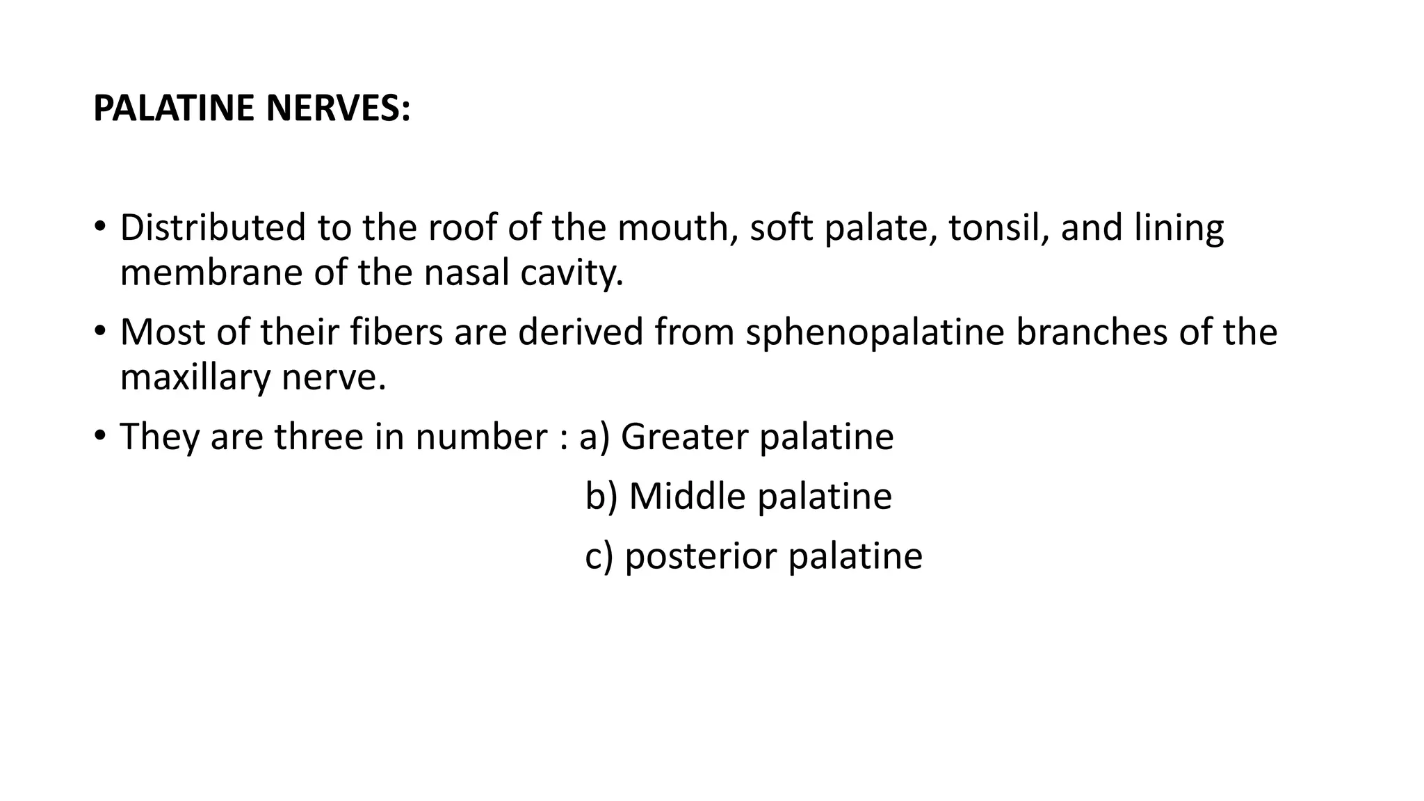
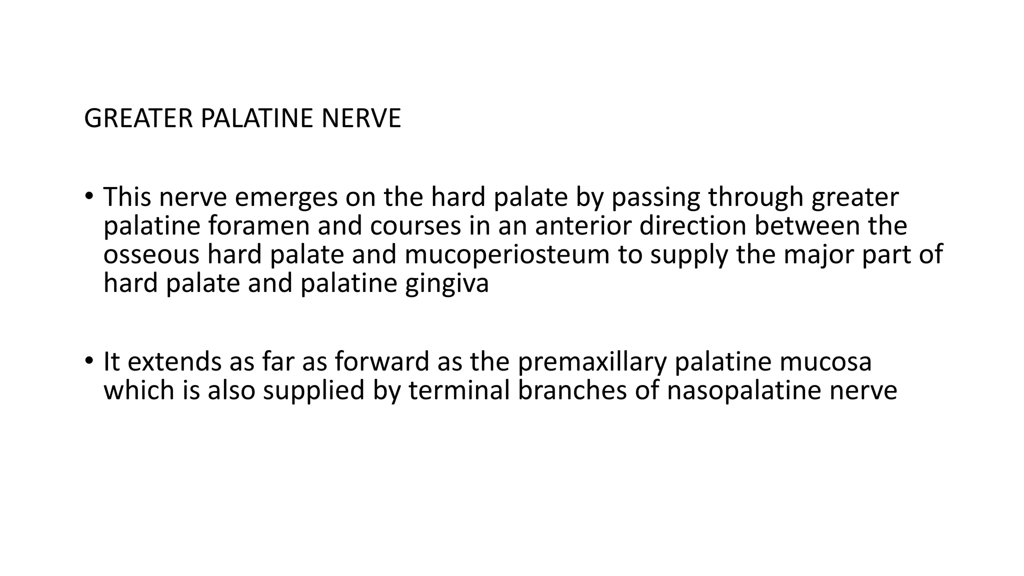
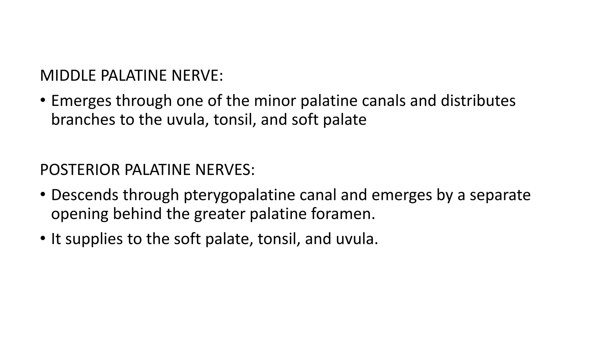
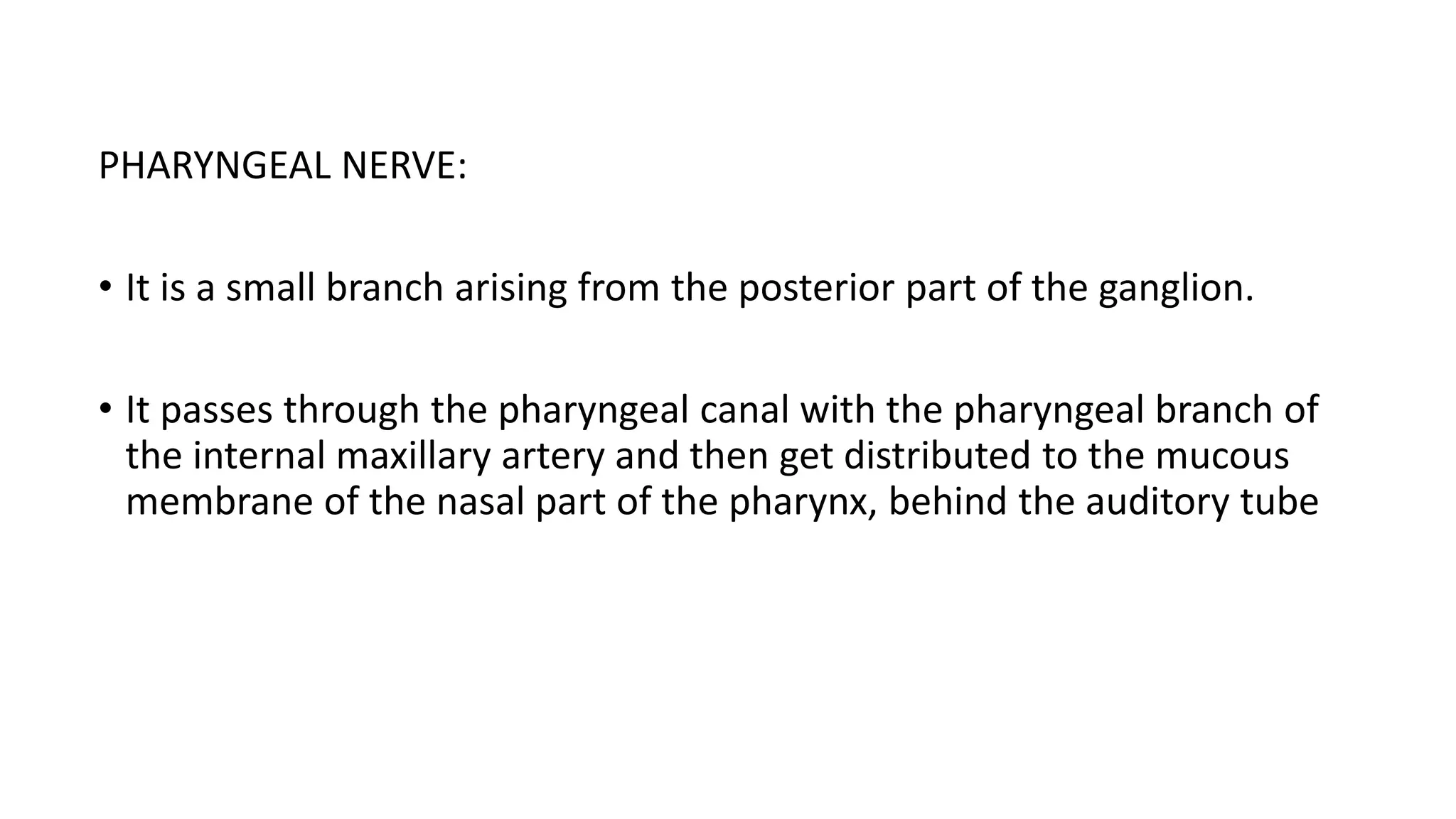
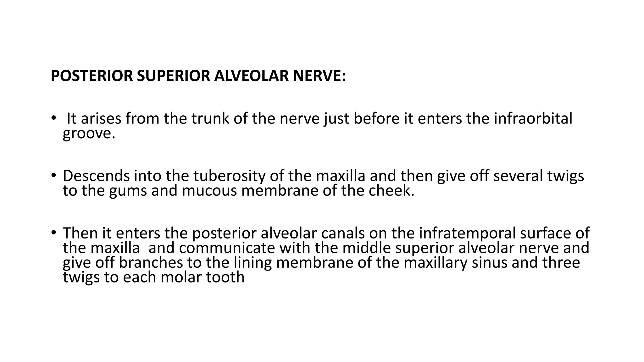
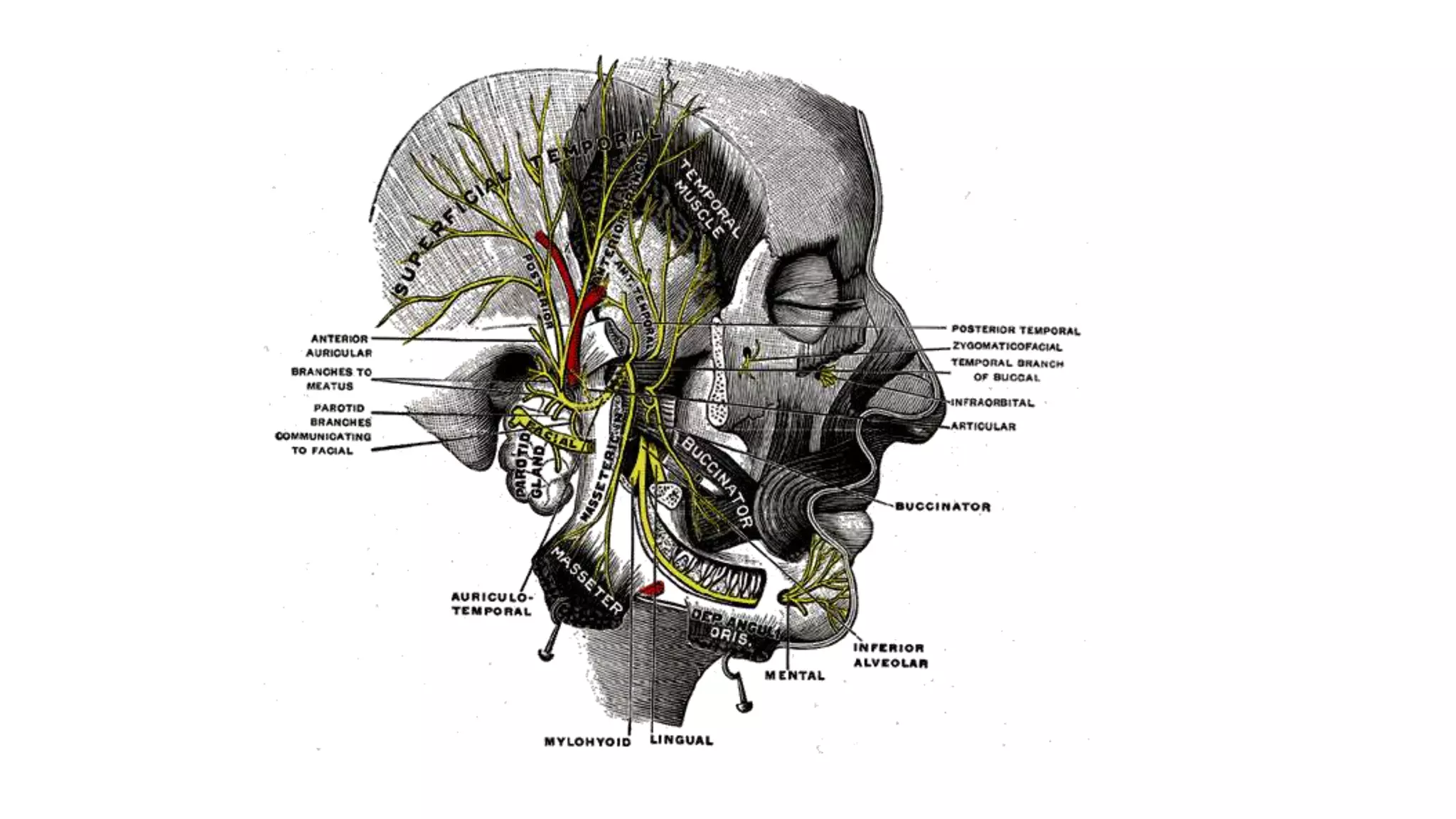
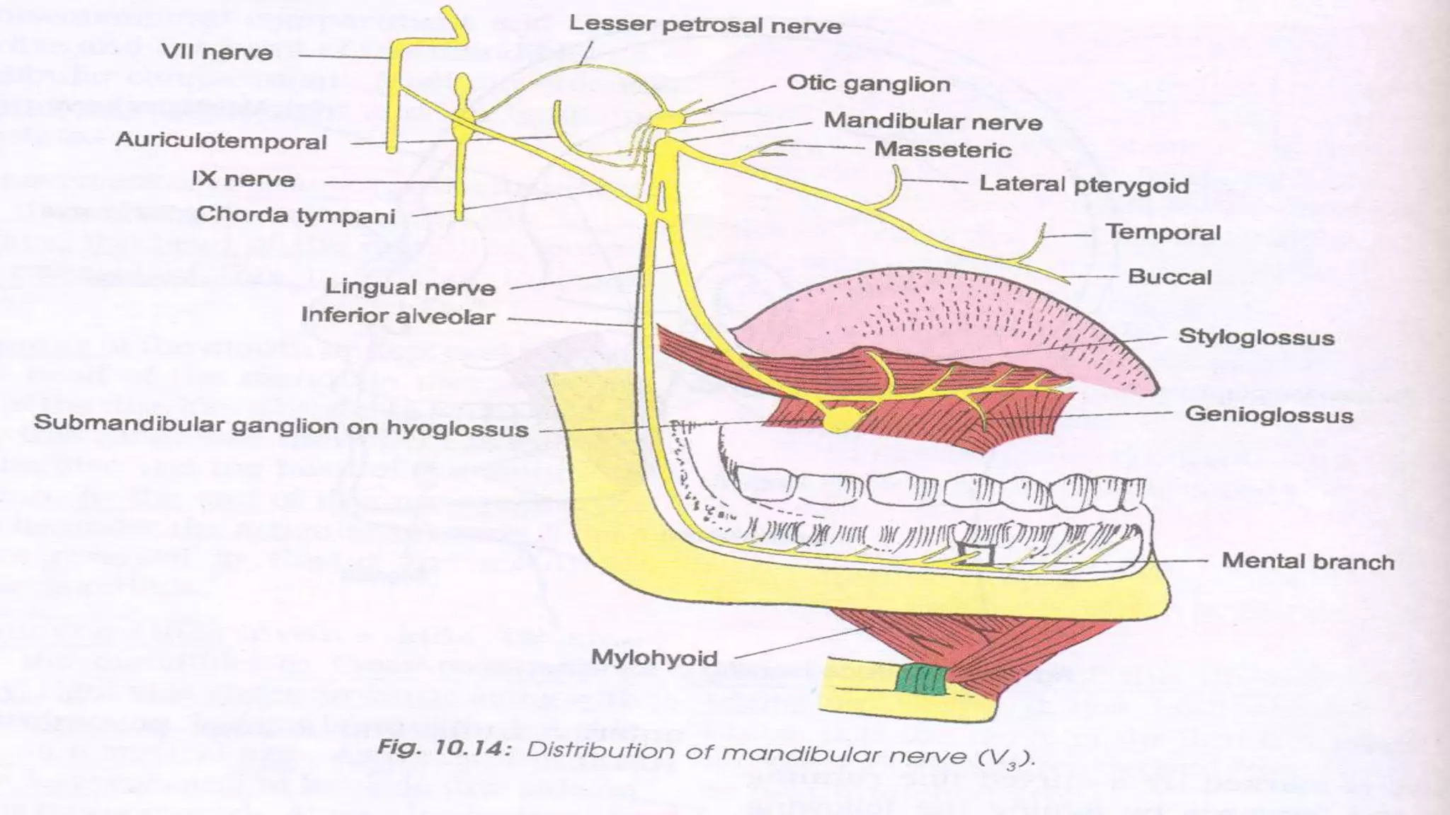
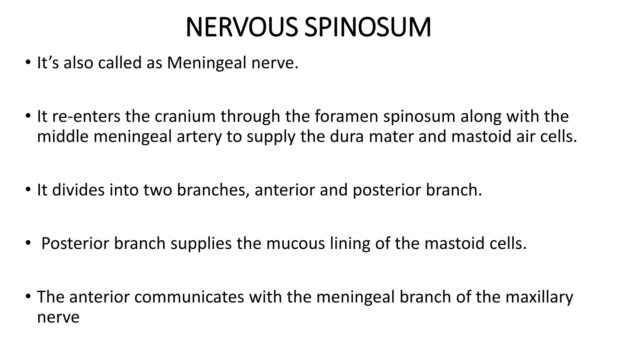
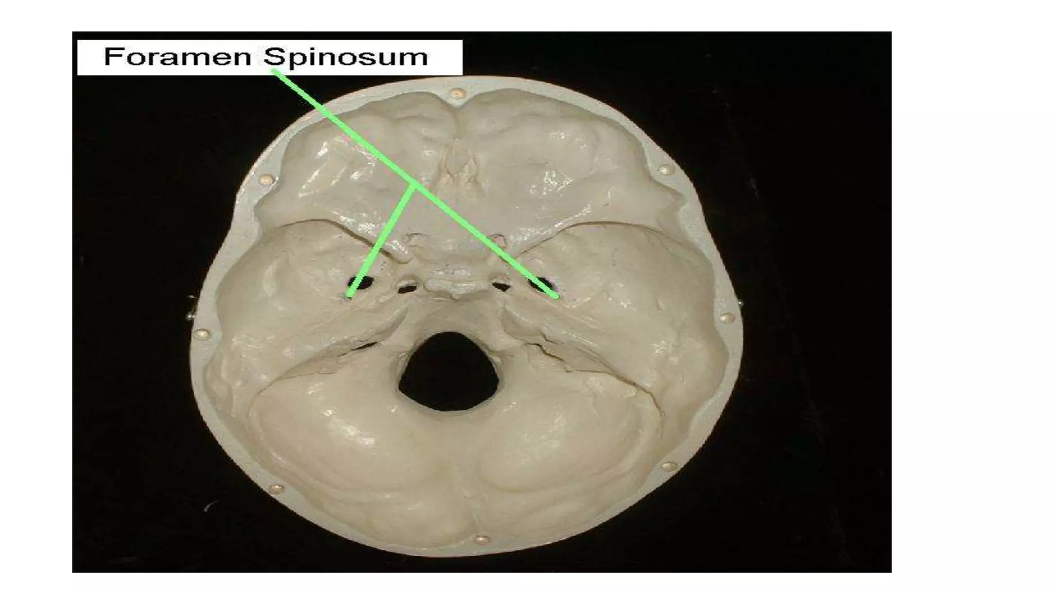
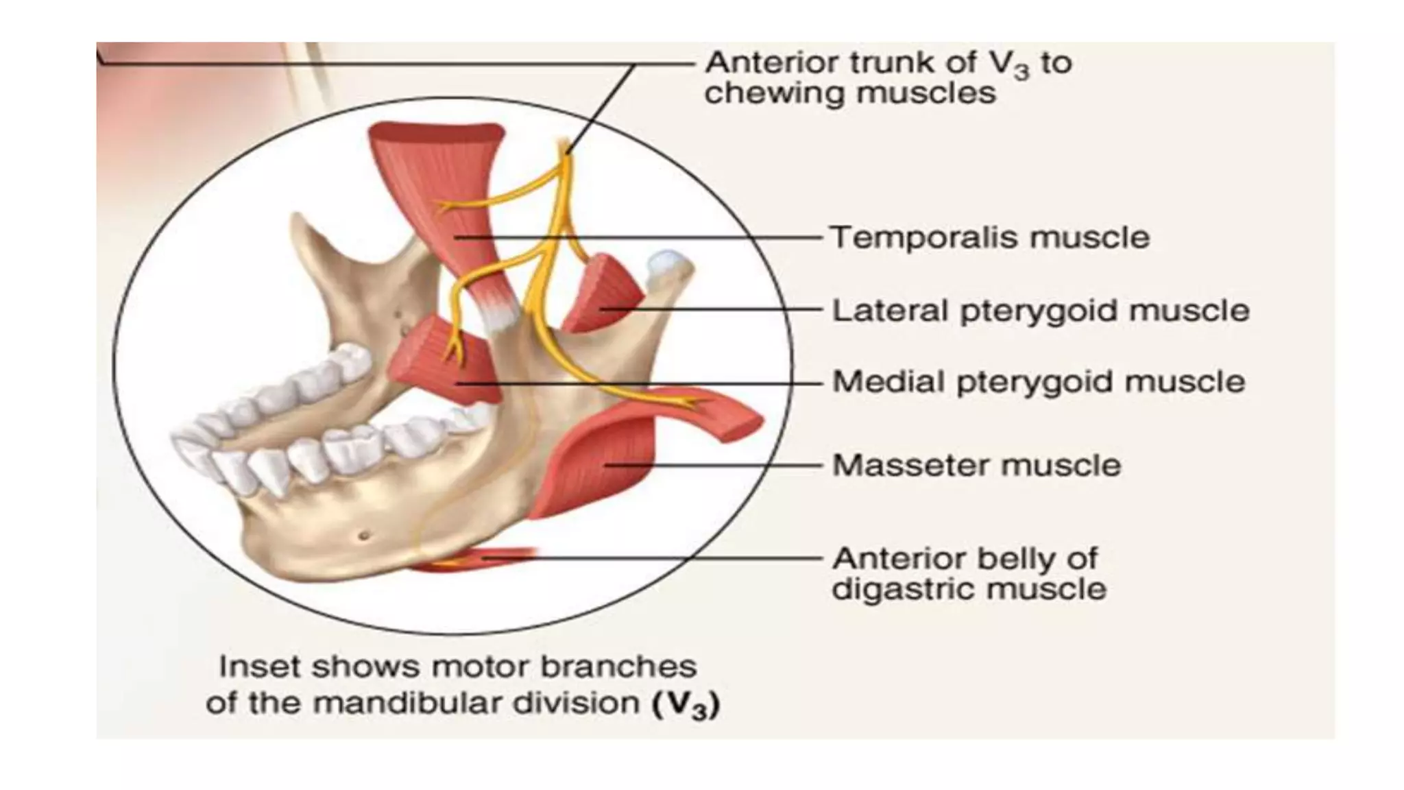
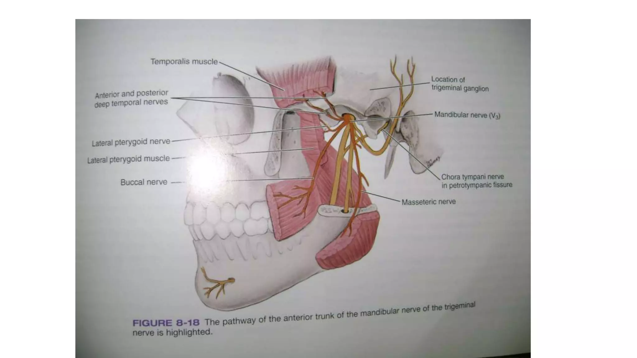
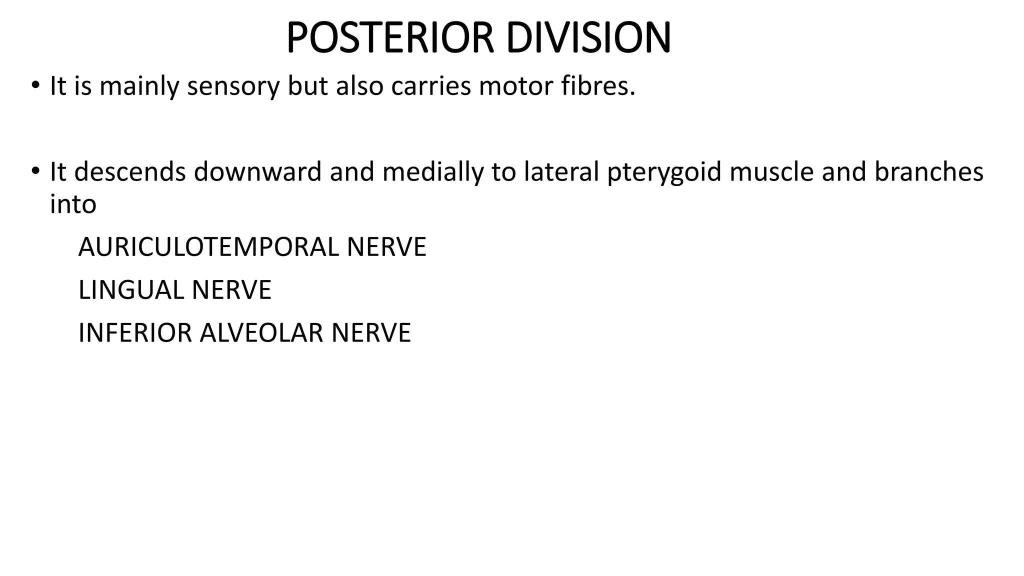
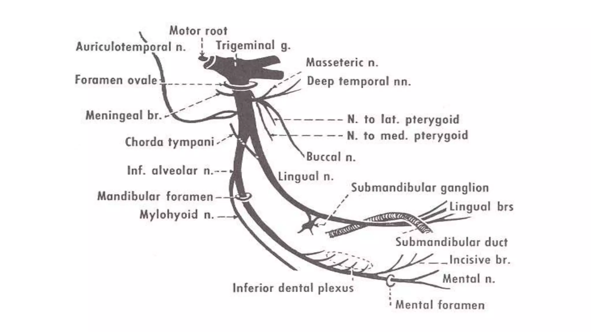
SlideShare a Scribd company logo
1 of 105
TRIGEMINAL NERVE
MODERATOR : Dr Gaurav
PRESENTED BY: Dr Md Mahbubul Hoda
CONTENTS
• INTRODUCTION
• TRIGEMINAL NERVE
• EMBRYOLOGY
• TRIGEMINAL GANGLION
• DIVISIONS
• BRANCHES
• GANGLIONS ASSOCIATED
• APPLIED ANATOMY
• REFERENCES
TRIGEMINAL NERVE
• The name TRIGEMINAL was given by “WINSLOW”
• It is the fifth cranial nerve.
• It is the largest and one of the most complex cranial nerves.
• It consists of two roots : a) sensory b) motor
• The sensory component supplies the skin of the entire face and the
mucous membrane of the cranial viscera, except for the pharynx and
the base of the tongue
• The motor component supplies the muscles of mastication (masseter,
temporalis, lateral pterygoid and medial pterygoid), muscle of ear
(tensor tympani), muscle of soft palate (tensor veli palatini), mylohoid
muscle and anterior belly of digastric.
EMBRYOLOGY
• The Trigeminal nerve arises from nuclei located in the brain stem.
• The sensory nucleus arises from metencephalon and myelencephalon
but are later displaced partially in mesencephalon.
• The motor nucleus arise from metencephalon and stays there.
• The Trigeminal nerve is related to four nucleus:
1.Mesencephalic nucleus
2.Main Sensory nucleus
3.Spinal nucleus
4.Motor nucleus
• The sensory trigeminal nerve nuclei are the largest of the cranial
nerve nuclei and extend through whole of the brainstem.
• The nucleus is divided in three parts:
a) Mesencephalic trigeminal nucleus
b) Principal pontine nucleus
c) Spinal nucleus of trigeminal nerve
• The mesencephalic nucleus is involved with proprioception. Neurons of this
nucleus cells receiving proprioceptive information from the jaw and send
projections to the trigeminal nucleus to mediate jaw reflexes.
• The principal pontine nucleus receives information about discriminative
sensation and light touch of the face.
• The spinal nucleus of trigeminal nerve receives information about touch,
pain, temperature and vibration.
PATHWAY
• The sensory root of trigeminal root enters into the trigeminal nuclei
• The trigeminal tract enter into the ventral posterior medial nucleus of
thalamus.
• These fibres which go upward and enter into the thalamus is known
as TRIGEMINAL LEMINISCUS or TRIGEMINO THALAMIC TRACT.
TRIGEMINAL GANGLION
• Trigeminal ganglion is also known as semilunar ganglion or gasserian
ganglion and is cresentric in shape.
• It is convex anterolaterally and concave posteromedially.
• It lies in posterior most part of middle cranial fossa (Meckel’s cave or
cavum trigeminale).
• It lies in a bony fossa at the apex of petrous part of temporal bone on
the floor of middle cranial fossa just lateral to posterior part of lateral
wall of cavernous sinus
DIVISION
• It has three division :-
a) V1 - Ophthalmic division
b) V2 - Maxillary division
c) V3 - Mandibular division
• The ophthalmic division enters orbital cavity through superior orbital
fissure.
• The maxillary division exits from cranial cavity through foramen rotundum.
• The mandibular division passes through foramen ovale.
• The trigeminal ganglion arises from sensory root which is connected
to lateral part of mid pons of brain
• The motor root also comes from lateral part of mid pons of brain and
leaves cranial cavity with mandibular division through foramen ovale.
BRANCHES
OPHTHALMIC NERVE
• The ophthalmic nerve or first division of the trigeminal is a sensory
nerve.
• It is the smallest of the 3 divisions of trigeminal nerve.
• It arises from the anteromedial part of trigeminal ganglion.
• Its about 2.5cm in length and passes forward along the lateral wall of
cavernous sinus, below the occulomotor and trochlear nerve
• Finally it enters the orbit through superior orbital fissure and divides
into three branches
1. Lacrimal
2. Frontal
3. Nasociliary
I) LACRIMAL NERVE
II) FRONTAL NERVE
A. Supraorbital nerve.
B. Supratrochlear nerve.
III) NASOCILIARY NERVE
A. Branches in the orbit
1. Short ciliary nerves.
2. Long ciliary nerves.
3. Posterior ethmoid nerve.
4. Anterior ethmoid nerve.
a) Internal nasal branches
b) External nasal branches
B. Branches arising in the nasal cavity
C. Terminal branches of the ophthalmic division on the face
LACRIMAL NERVE
• It is the smallest branch of ophthalmic division.
• Enter the orbit through lateral part of superior orbital fissure.
• Runs in anterior direction with the lacrimal artery.
• Enters the lacrimal gland, supplying it and the adjoining conjunctiva
and then pierces the orbital septum and end in the upper eyelid.
FRONTAL NERVE
• Largest branch of ophthalmic.
• Enters the orbit - superior orbital fissure and runs forward between
the Levator palpebrae superioris and the periosteum.
• Midway between the apex and base of the orbit it divides into two
branches:
1. Supratrochlear Nerve
2. Supraorbital nerve
SUPRATROCHLEAR NERVE
• Smaller of the two - passes above the superior Oblique muscle and
gives off a descending filament to join the infratrochlear branch of the
nasociliary nerve.
• Escapes from the orbit between the superior oblique and the
supraorbital foramen.
• Supplies - skin of the lower part of the forehead close to the middle
line and sends filament to the conjunctiva and skin of the upper
eyelid.
SUPRAORBITAL NERVE
• Proceeds between the levator palpebral superioris and the orbital
roof and traverses the supra orbital foramen
• Then it ascends up to the forehead and ends in two branches : the
medial branch and the lateral branch.
• It supply the skin of the upper eyelid and lower medial portion of the
forehead.
NASOCILIARY NERVE
• It originates from ophthalmic nerve in the anterior part of cavernous
sinus.
• It enters the orbit through superior orbital fissure.
• Branches of the nasociliary nerve are divided into those:
1.Branches in the orbit.
2.Branches in the nasal cavity
3.Branches on the face
BRANCHES IN ORBIT
1.Short ciliary nerve.
- pass through the ganglion without synapsing.
- continue to the eyeball.
2.Long ciliary nerve.
-branches are 2-3 in Nos.
-iris and cornea.
3.Posterior ethmoid nerve.
- This nerve enters the posterior ethmoidal canal.
- Supply to ethmoidal & sphenoidal sinuses.
4.Anterior ethmoid nerve.
- supply the frontal sinus.
- divides into internal and external nasal branches.
- Internal nasal:-septal branch and lateral branch.
- External nasal:-ala of nose and tip of nose.
BRANCHES IN NASAL CAVITY
• Supplies the mucous membrane lining the cavity.
BRANCHES ON FACE
• Skin of medial parts of both eyelids, the lacrimal sac and lacrimal
caruncle and side of bridge of nose
APPLIED ANATOMY
• Superior orbital fissure syndrome
1. Ophthalmoplegia
2. Pupillary constriction
3. Enopthalmos
4. Ptosis of eyelid
MAXILLARY NERVE
• It is the second division of trigeminal nerve.
• It is a purely sensory nerve.
• It is intermediate both in position and size between the ophthalmic and
mandibular nerve.
• It originates from the middle of the semilunar ganglion and continues forward
in the lateral part of cavernous sinus, it then passes through the foramen
rotundum.
• Through which it takes the cranial fossa and enters the
pterygopalatine fossa.
• It enters the inferior orbital fissure to pass into the orbital cavity, here
it turns laterally in a groove on the orbital surface of the maxilla called
the infraorbital groove, continues forwards, the second division
emerges on the anterior surface of the maxilla through the
infraorbital where it divides.
• It then divides into branches which spread out upon the side of the
nose, lower eyelid and the upper lip.
BRANCHES OF MAXILLARY NERVE
• It is divided into four groups, as they are given off:
1. Middle Cranial Fossa
2. Pterygopalatine Fossa
3. Infraorbital Canal and Groove
4. On the Face
MIDDLE CRANIUM FOSSA
MIDDLE MENINGEAL NERVE
PTERYGOPALATINE FOSSA
ZYGOMATIC
SPHENOPLATINE
POSTERIOR SUPERIOR ALVEOLAR
INFRAORBITAL CANAL ANTERIOR SUPERIOR ALVEOLAR
MIDDLE SUPERIOR ALVEOLAR
ON THE FACE INFERIOR PALPERBAL
EXTERNAL NASAL
SUPERIOR LABIAL
1) BRANCHES GIVEN OFF IN THE MIDDLE CRANIAL FOSSA
1. Middle Meningeal nerve
2) BRANCHES IN THE PTERYGOPALATINE FOSSA
A. ZYGOMATIC NERVE
1. Zygomaticofacial nerve
2. Zygomaticotemporal nerve
B. PETERYGOPALATINE (SPHENOPALATINE) NERVES
1. Orbital Branches (from Inferior orbital fissure)
2. Nasal branches
a) Posterior superior lateral nasal branches
b) Medial or septal branches
3. Palatine branches
a) Greater or anterior palatine nerve
b) Middle palatine nerve.
c) Posterior palatine fibers.
4. Pharyngeal branch
C. POSTERIOR SUPERIOR ALVEOLAR BRANCHES
3) BRANCHES IN THE INFRAORBITAL GROOVE AND CANAL
1. Middle superior alveolar nerve.
2. Anterior superior alveolar nerve.
4) BRANCHES ON THE FACE
1. Inferior palpebral branches
2. Lateral nasal branches
3. Superior labial branches
A. Zygomaticotemporal
B. Zygomaticofacial
C. Post. Sup. Alveolar Branch
D. Nasopalatine
E. Greater Palatine
F. Lesser Palatine
G. Mid. & Ant. Alveolar Branch
H. Infraorbital
MIDDLE MENINGEAL NERVE:
• It is given off from the maxillary nerve directly after its origin from the
semilunar ganglion.
• It accompanies the middle meningeal artery and supplies the dura
mater.
ZYGOMATIC NERVE:
• It arises from pterygopalatine fossa.
• Enters the orbit through inferior orbital fissure and then divides into
two branches
1.Zygomaticotemporal
2.Zygomaticofacial
ZYGOMATICO-TEMPORAL BRANCH:
• It runs along the lateral wall of the orbit in a groove in the zygomatic
bone.
• It receives communication from the lacrimal and passing through a
foramen in the Zygomatic bone it enters the temporal fossa.
• Ascends between the bone and substance of the Temporalis muscle
and pierces the temporal fascia about 2.5 cm above the Zygomatic
arch and then get distributed to the skin of the side of the forehead
and communicates with the facial nerve
ZYGOMATICO-FACIAL BRANCH:
• Passes along the inferio-lateral angle of the orbit.
• Emerges from the face through a foramen in the Zygomatic bone.
• Perforating the Orbicularis oculi it supplies to the skin on the
prominence of the cheek.
SPHENOPALATINE BRANCHES:
• Two in number.
• Descend to the sphenopalatine ganglion.
• These nerves are then distributed into
1. Orbital branches
2. Nasal branches
posterior superior lateral nasal branch
medial or septal branch
3.Palatine branches
greater palatine nerve
middle palatine nerve
posterior palatine nerve
4.Pharyngeal branches
ORBITAL BRANCH:
• Two or three delicate filaments which enter the orbit by the inferior orbital
fissure and supply the periosteum
NASAL BRANCH
POSTERIOR SUPERIOR LATERAL NASAL BRANCHES:
• Distributed to the septum and lateral wall of the nasal fossa.
• It enters the posterior part of the nasal cavity by the sphenopalatine foramen
• It supply the mucous membrane covering the superior and middle nasal
conchae, the lining of the posterior ethmoidal cells and the posterior part of
the septum
MEDIAL OR SEPTAL BRANCH
• It supplies the posterior part of roof and septum.
• One of the nerve which is longer and larger than others is known as
NASOPALATINE NERVE
• It enters the nasal cavity through sphenopalatine foramen to reach
the septum and then descends to the roof of the mouth through the
incisive canal where it communicates with the corresponding nerve
of the opposite side and with the anterior palatine nerve.
PALATINE NERVES:
• Distributed to the roof of the mouth, soft palate, tonsil, and lining
membrane of the nasal cavity.
• Most of their fibers are derived from sphenopalatine branches of the
maxillary nerve.
• They are three in number : a) Greater palatine
b) Middle palatine
c) posterior palatine
GREATER PALATINE NERVE
• This nerve emerges on the hard palate by passing through greater
palatine foramen and courses in an anterior direction between the
osseous hard palate and mucoperiosteum to supply the major part of
hard palate and palatine gingiva
• It extends as far as forward as the premaxillary palatine mucosa
which is also supplied by terminal branches of nasopalatine nerve
MIDDLE PALATINE NERVE:
• Emerges through one of the minor palatine canals and distributes
branches to the uvula, tonsil, and soft palate
POSTERIOR PALATINE NERVES:
• Descends through pterygopalatine canal and emerges by a separate
opening behind the greater palatine foramen.
• It supplies to the soft palate, tonsil, and uvula.
PHARYNGEAL NERVE:
• It is a small branch arising from the posterior part of the ganglion.
• It passes through the pharyngeal canal with the pharyngeal branch of
the internal maxillary artery and then get distributed to the mucous
membrane of the nasal part of the pharynx, behind the auditory tube
POSTERIOR SUPERIOR ALVEOLAR NERVE:
• It arises from the trunk of the nerve just before it enters the infraorbital
groove.
• Descends into the tuberosity of the maxilla and then give off several twigs
to the gums and mucous membrane of the cheek.
• Then it enters the posterior alveolar canals on the infratemporal surface of
the maxilla and communicate with the middle superior alveolar nerve and
give off branches to the lining membrane of the maxillary sinus and three
twigs to each molar tooth
MIDDLE SUPERIOR ALVEOLAR NERVE:
• It is given off from the nerve in the posterior part of the infraorbital canal.
• It runs and forward in a canal and enter the lateral wall of the maxillary
sinus to supply the two premolar teeth.
• It forms the superior dental plexus with the anterior and posterior superior
alveolar branches.
ANTERIOR SUPERIOR ALVEOLAR NERVE:
• It is given off from the nerve just before its exits from the infraorbital
foramen.
• It descends in a canal in the anterior wall of the maxillary sinus and divides
into branches which supply the incisor and canine teeth.
• It then communicates with middle superior alveolar branch and gives off a
nasal branch which passes through a minute canal in the lateral wall of the
inferior meatus.
• It supplies the Mucous membrane of the anterior part of the inferior meatus
and floor of the nasal cavity.
INFERIOR PALPEBRAL BRANCH:
• It ascends behind the Orbicularis oculi.
• It supplies the skin and conjunctiva of the lower eyelid.
• Join at the lateral angle of the orbit with the facial and zygomaticofacial
nerves.
EXTERNAL NASAL BRANCHES:
• It supplies the skin of the side of the nose and join with the terminal twigs
of the nasociliary nerve.
SUPERIOR LABIAL BRANCHES:
• The largest and most numerous.
• It descends behind the Quadratus labii superioris.
• Distributed over the skin of the upper lip, the mucous membrane of the
mouth and labial glands.
• They are joined immediately beneath the orbit, by filaments from the facial
nerve forming with them the infraorbital plexus.
MANDIBULAR NERVE
• It is largest of all three divisions of trigeminal nerve.
• It is derived from 1st brachial arch.
• Mixed nerve
• Made of two roots : sensory and motor
• A large sensory root proceeding from the semilunar ganglion.
• A small motor root which passes beneath the ganglion and unites
with the sensory root just after its exit from foramen ovale.
• Immediately beneath the base of the skull it gives off a recurrent
branch (nervus spinosus) and the nerve to the Pterygoideus internus
and then divides into two trunks - an anterior and a posterior.
• Trunk remains undivided for 2-3mm.
• Then it divides into small anterior and large posterior division.
• Branches given off are:-
1. Undivided Nerve
2. Anterior division
3. Posterior division
BRANCHES
1.UNDIVIDED NERVE/TRUNK
Nervous spinosis
Nerve to medial Pterygoid
2. DIVIDED NERVE
a) Anterior Division
Masseteric Nerve
Deep temporal Nerve
Nerve to lateral Pterygoid
Buccal Nerve/Buccinator
b) Post. Division
Auriculotemporal
Lingual Nerve
Inferior Alveolar Nerve
NERVOUS SPINOSUM
• It’s also called as Meningeal nerve.
• It re-enters the cranium through the foramen spinosum along with the
middle meningeal artery to supply the dura mater and mastoid air cells.
• It divides into two branches, anterior and posterior branch.
• Posterior branch supplies the mucous lining of the mastoid cells.
• The anterior communicates with the meningeal branch of the maxillary
nerve
NERVE TO MEDIAL PTERYGIOD
• It supplies to medial pterygoid muscle.
• It also gives of small branches to tensor veli palatini and tensor
tympani through otic ganglion without interruption.
ANTERIOR DIVISION
• The anterior division is smaller than the posterior division.
• It runs forward under lateral pterygoid muscle and give branches :-
Masseteric Nerve
Deep temporal Nerve
Nerve to lateral Pterygoid
Buccal Nerve/Buccinator
MASSETERIC NERVE
• It emerges at the upper border of the lateral pterygoid just infront of the
TMJ, passes laterally through the mandibular notch and enters the deep
surface of the masseter muscle.it also supplies the TMJ.
DEEP TEMPORAL NERVE
• It ascend deep to lateral pterygoid and supply temporalis muscle.
NERVE TO LATERAL PTERYGOID
• It enters the medial side of the lateral pterygoid muscle to provide its motor
nerve supply
BUCCAL NERVE
• It passes downward anteriorly & laterally between the two heads of
the external pterygoid muscle.
• At about the level of the occlusal plane of the mandibular 2nd & 3rd
molars, it divides into several branches that divide on the buccinator
muscle.
• It does not supply the buccinator muscle.
• It supplies the skin of cheek and buccal gingiva of mandibular molars
and mucobuccal fold
POSTERIOR DIVISION
• It is mainly sensory but also carries motor fibres.
• It descends downward and medially to lateral pterygoid muscle and branches
into
AURICULOTEMPORAL NERVE
LINGUAL NERVE
INFERIOR ALVEOLAR NERVE
AURICULOTEMPORAL NERVE
• It arises from 2 roots which encircle the middle meningeal artery
• The trunk passes posterior to lateral pterygoid between the neck of mandible
and sphenomandibular ligament superior to 1st part of maxillary artery
• It lies behind the TMJ close to the parotid
• Ascends behind superficial temporal vessels and then in temporal region
divides into superficial temporal branches
COMMUNICATION
• 2 roots of the nerve:- both these roots receive communications from
the OTIC ganglion. And receive postganglionic secretomotor fibres for
the parotid gland they control the secretions from the gland.
• Communicating branches to the facial nerve: These are purely
sensory from the Auriculotemporal nerve.
BRANCHES
1. Parotid branches - sensory, secretomotor, vasomotor.
2. Articular branches - to the TMJ.
3. Auricular branches - to the skin of the auricle & tragus.
4. Meatal branches - Meatus of the tympanic membrane
5. Terminal branches - Scalp over the temporal region
LINGUAL NERVE
• It originates from the posterior trunk of mandibular nerve in the infratemporal
fossa.
• It is the smallest of the two terminal branches.
• It supplies mucous membrane of the anterior two-thirds of the tongue, floor of
the mouth, tonsil and gum.
• It lies anterior to inferior alveolar and between lateral pterygoid and tensor
palatini which receives through chorda tympani
• It then emerges from inferior border of lateral pterygoid to lie between
ramus and medial pterygoid
• Between origins of superior constrictor and mylohyoid it reaches the side of
the tongue below and behind 3rd molar
• It lies just below mucous membrane in lateral lingual sulcus, proceeds
anteriorly looping downward and medial to submandibular duct to deep
surface of submandibular gland
COMMUNICATION
• As the lingual nerve passes medially to the lateral pterygoid, it is
joined from behind by the chorda tympani. This nerve conveys
secretory fibres from the facial nerve.
• The parasympathetic secretory fibres control the submandibular &
sublingual salivary glands.
INFERIOR ALVEOLAR NERVE
• It is the largest branch of the posterior division.
• It passes downward on the medial side of the external pterygoid muscle &
passes between sphenomandibular ligament and ramus of mandible upto
mandibular foramen
• It then enters the mandibular canal and runs below the teeth as for as mental
foramen and terminates by dividing into branches.
• Branches of the inferior alveolar nerve are the
1. Mylohyoid
2. Dental
3. Incisive
4. Mental.
MYLOHYOID NERVE:
• Derived - inferior alveolar just before it enters the mandibular foramen.
• It descends - groove on the deep surface of the ramus of the mandible -
reaching the under surface of the Mylohyoid - supplies this muscle and the
anterior belly of the Digastric.
DENTAL BRANCHES:
• Supply - molar and premolar teeth
• Above the alveolar nerve they form an inferior dental plexus.
INCISIVE BRANCH:
• Continued onward within the bone and supplies the canine and incisor
teeth.
MENTAL NERVE:
• Emerges at the mental foramen and divides beneath the Triangularis
muscle into three branches.
1.One descends to the skin of the chin.
2.Two ascend to the skin and mucous membrane of the lower lip.
GANGLIONS
CILIARY GANGLION:
• Parasympathetic Ganglion placed in a course of the occulomotor nerve.
• It lies in the apex of orbit between the optic nerve and tendon of the lateral
rectus muscle.
• It has sensory, motor, and sympathetic roots.
sensory - from nasociliary
motor - from occulomotor, preganglionic fibres from Edinger westphal nucleus
to supply sphincter pupillae and ciliaris muscle.
sympathetic - from post ganglionic fibres of superior cervical ganglion to
supply the blood vessels of eye ball and dilator pupillae
PTERYGOPALATINE GANGLION:
• Largest parasympathetic ganglion
• Connected to maxillary nerve in infratemporal fossa
• Sensory to periosteum of the orbit, orbicularis and nasal
cavity,maxillary sinus , palate , nasopharynx.
• Topographically related to maxillary nerve, but functionally related
facial nerve
• The preganglionic fibres are from superior salivatory nucleus
OTIC GANGLION:
• Topographically related to mandibular nerve, but functionally is a part
of glossopharyngeal nerve
• Situated in infratemporal fossa below the foramen ovale and
surrounds the origin of nerve to medial pterygoid.
• Preganglionic parasympathetic fibres from inferior salivatory nucleus
to reach the ganglion
SUBMANDIBULAR GANGLION:
• Topographically related to lingual nerve, functionally it is connected
to the facial nerve
• It is a relay station for secretomotor fibres to the submandibular and
sublingual glands.
• The preganglionic fibres are from superior salivatory nucleus
APPLIED ANATOMY
• Trigeminal neuralgia or tic douloreux is described as the most excruciating
pain.
• This intense, stabbing, electric shock-like pain is caused by irritation of the
trigeminal nerve
• Anticonvulsive medications are normally the first treatment choice.
• Initially short, mild attacks, but trigeminal neuralgia can progress,causing
longer, more frequent bouts of searing pain.
• They may also be provoked by even mild stimulation of face
PRECIPITATING FACTORS:
• Shaving
• Stroking your face
• Eating
• Drinking
• Brushing your teeth
• Talking
• Putting on makeup
• Encountering a breeze
• Smiling
Treatment-
Medical: carbamazepine(Tegretol), phenytoin,
valproate.
SURGERY:
• Alcohol injection
• Glycerol injection OR Percutaneous Glycerol Rhizotomy (PGR)
• Balloon compression. Also called as percutaneous balloon compression of
the trigeminal nerve (PBCTN)
• Electric current. Also called as percutaneous stereotactic radiofrequency
thermal rhizotomy (PSRTR)
• Microvascular Decompression (MVD )
• Severing the nerve. A procedure called Partial Sensory Rhizotomy (PSR)
• Radiation. GAMMA-KNIFE RADIOSURGERY (GKR)
SPHENOPALATINE NEURALGIA
• ETIOLOGY vasodilatation of the internal maxillary artery near the region of
Sphenopalatine region.
• Unilateral paroxysms of intersperse pain in the region of eyes,maxilla,
ear,mastoid base of the nose and beneath the zygoma
• There are no TRIGGER ZONES
• Seen in persons below 40 years
• Treatment
- Alcohol injection of the ganglion
- Resection of the ganglion
AURICULOTEMPORAL SYNDROME
ETIOLOGY
• It results from damage to the Auriculotemporal nerve. The usually
syndrome follows some surgical operation
CLINICAL FEATURES
• Flushing and sweating of the involved side of the face
TREATMENT
• Intracranial division of Auriculotemporal nerve
CROCODILE TEARS
• Exhibits profuse lacrimation during eating especially hot or spicy
food..It follows facial paralysis
• Or as a result of herpes zoster ,head injury or intra cranial operative
trauma
• TREATMENT- Intracranial division of Auriculotemporal nerve
COMPLEX REGION PAIN SYNDROME
• Is a burning pain and paresthesia associated with deformation of
nerves Seen after surgical tooth extraction
TREATMENT
• Injection of procaine ,alcohol
• Surgical curettage of bone or Resection of nerves
REFERENCES
• GRAY’S ANATOMY BY HENRY GRAY
• HUMAN ANATOMY BY BD CHAURASIA
• HUMAN EMBRYOLOGY BY I.B SINGH
• LOCAL ANESTHESIA BY STANLEY F. MALAMED
• LOCAL ANESTHESIA AND PAIN CONTROL IN DENTAL PRACTICE BY
MONHEIM’S
• CUNNIGHAM’S MANUAL OF PRACTICAL ANATOMY
THANK YOU

More Related Content

What's hot (20)

Facial nerve
Facial nerveFacial nerve
Facial nerve
 
Trigeminal nerve 1
Trigeminal nerve 1Trigeminal nerve 1
Trigeminal nerve 1
 
Facial artery
Facial arteryFacial artery
Facial artery
 
Maxillary artery
Maxillary arteryMaxillary artery
Maxillary artery
 
Anatomy of mandible
Anatomy of mandibleAnatomy of mandible
Anatomy of mandible
 
Facial nerve
Facial nerveFacial nerve
Facial nerve
 
Trigeminal nerve
Trigeminal nerveTrigeminal nerve
Trigeminal nerve
 
blood supply nerve supply of face
blood supply nerve supply of faceblood supply nerve supply of face
blood supply nerve supply of face
 
Development of face, palate and jaw
Development of face, palate and jawDevelopment of face, palate and jaw
Development of face, palate and jaw
 
Trigeminal Nerve Anatomy
Trigeminal Nerve AnatomyTrigeminal Nerve Anatomy
Trigeminal Nerve Anatomy
 
Facial nerve
Facial nerveFacial nerve
Facial nerve
 
Infra temporal fossa
Infra temporal fossaInfra temporal fossa
Infra temporal fossa
 
Mandibular nerve
Mandibular nerveMandibular nerve
Mandibular nerve
 
Tongue development, applied anatomy and prosthetic implications
Tongue development, applied anatomy and prosthetic implicationsTongue development, applied anatomy and prosthetic implications
Tongue development, applied anatomy and prosthetic implications
 
External carotid artery
External carotid arteryExternal carotid artery
External carotid artery
 
Trigeminal nerve maxillary nerve and clinical implication
Trigeminal nerve maxillary nerve and clinical implicationTrigeminal nerve maxillary nerve and clinical implication
Trigeminal nerve maxillary nerve and clinical implication
 
PTERYGOPALATINE FOSSA.pptx
PTERYGOPALATINE FOSSA.pptxPTERYGOPALATINE FOSSA.pptx
PTERYGOPALATINE FOSSA.pptx
 
Muscles of mastication
Muscles of masticationMuscles of mastication
Muscles of mastication
 
venous supply of head & neck
venous supply of head & neckvenous supply of head & neck
venous supply of head & neck
 
Trigeminal nerve
Trigeminal nerveTrigeminal nerve
Trigeminal nerve
 

Similar to Trigeminal nerve

Trigeminal Nerve Dr.AD.pptx
Trigeminal Nerve Dr.AD.pptxTrigeminal Nerve Dr.AD.pptx
Trigeminal Nerve Dr.AD.pptxArunCreations
 
trigeminal-neuralgia.pptx
trigeminal-neuralgia.pptxtrigeminal-neuralgia.pptx
trigeminal-neuralgia.pptxssuser20cec1
 
trigeminal-neuralgia (1).pptx
trigeminal-neuralgia (1).pptxtrigeminal-neuralgia (1).pptx
trigeminal-neuralgia (1).pptxAkshaySingh503850
 
Trigeminal nerve and its course
Trigeminal nerve and its courseTrigeminal nerve and its course
Trigeminal nerve and its courseShruti MISHRA
 
Trigeminal Nerve and Applied Anatomy
Trigeminal Nerve and Applied AnatomyTrigeminal Nerve and Applied Anatomy
Trigeminal Nerve and Applied AnatomyAbhijith P Ajith
 
MICROSURGICAL ANATOMY OF CRANIAL NERVES
MICROSURGICAL ANATOMY OF CRANIAL NERVESMICROSURGICAL ANATOMY OF CRANIAL NERVES
MICROSURGICAL ANATOMY OF CRANIAL NERVESpankaj patel
 
Cranial Nerve V Trigemial
Cranial Nerve V Trigemial Cranial Nerve V Trigemial
Cranial Nerve V Trigemial Anam_ Khan
 
Trigeminal and facial nerve.pptx
Trigeminal and facial nerve.pptxTrigeminal and facial nerve.pptx
Trigeminal and facial nerve.pptxAnkur Rathaur
 
Trigeminal and facial nerve.pptx
Trigeminal and facial nerve.pptxTrigeminal and facial nerve.pptx
Trigeminal and facial nerve.pptxAnkur Rathaur
 
Trigeminal nerve- anatomy
Trigeminal nerve- anatomy Trigeminal nerve- anatomy
Trigeminal nerve- anatomy spsangeetaporiya
 
cranial nerve 5.pptx
cranial nerve 5.pptxcranial nerve 5.pptx
cranial nerve 5.pptxDeepthinker14
 
Branches of Trigeminal Nerve
Branches of Trigeminal NerveBranches of Trigeminal Nerve
Branches of Trigeminal NerveDr. Julius Kwedhi
 
Trigeminal nerve and its applied anatomy
Trigeminal nerve and its applied  anatomyTrigeminal nerve and its applied  anatomy
Trigeminal nerve and its applied anatomyKhadeeja Kulood
 

Similar to Trigeminal nerve (20)

Trigeminal Nerve Dr.AD.pptx
Trigeminal Nerve Dr.AD.pptxTrigeminal Nerve Dr.AD.pptx
Trigeminal Nerve Dr.AD.pptx
 
Trigeminal nerve final
Trigeminal nerve finalTrigeminal nerve final
Trigeminal nerve final
 
trigeminal-neuralgia.pptx
trigeminal-neuralgia.pptxtrigeminal-neuralgia.pptx
trigeminal-neuralgia.pptx
 
trigeminal-neuralgia (1).pptx
trigeminal-neuralgia (1).pptxtrigeminal-neuralgia (1).pptx
trigeminal-neuralgia (1).pptx
 
Trigeminal nerve and its course
Trigeminal nerve and its courseTrigeminal nerve and its course
Trigeminal nerve and its course
 
Trigeminal Nerve and Applied Anatomy
Trigeminal Nerve and Applied AnatomyTrigeminal Nerve and Applied Anatomy
Trigeminal Nerve and Applied Anatomy
 
Trigeminal nerve ppt
Trigeminal nerve ppt  Trigeminal nerve ppt
Trigeminal nerve ppt
 
MICROSURGICAL ANATOMY OF CRANIAL NERVES
MICROSURGICAL ANATOMY OF CRANIAL NERVESMICROSURGICAL ANATOMY OF CRANIAL NERVES
MICROSURGICAL ANATOMY OF CRANIAL NERVES
 
Trigeminal n.
Trigeminal n.Trigeminal n.
Trigeminal n.
 
Cranial Nerve V Trigemial
Cranial Nerve V Trigemial Cranial Nerve V Trigemial
Cranial Nerve V Trigemial
 
Trigeminal and facial nerve.pptx
Trigeminal and facial nerve.pptxTrigeminal and facial nerve.pptx
Trigeminal and facial nerve.pptx
 
Trigeminal and facial nerve.pptx
Trigeminal and facial nerve.pptxTrigeminal and facial nerve.pptx
Trigeminal and facial nerve.pptx
 
Mandibular nerve.pptx
Mandibular nerve.pptxMandibular nerve.pptx
Mandibular nerve.pptx
 
Trigeminal nerve- anatomy
Trigeminal nerve- anatomy Trigeminal nerve- anatomy
Trigeminal nerve- anatomy
 
15.pptx
15.pptx15.pptx
15.pptx
 
cranial nerve 5.pptx
cranial nerve 5.pptxcranial nerve 5.pptx
cranial nerve 5.pptx
 
Branches of Trigeminal Nerve
Branches of Trigeminal NerveBranches of Trigeminal Nerve
Branches of Trigeminal Nerve
 
Trigeminal nerve
Trigeminal nerveTrigeminal nerve
Trigeminal nerve
 
Trigeminal nerve and its applied anatomy
Trigeminal nerve and its applied  anatomyTrigeminal nerve and its applied  anatomy
Trigeminal nerve and its applied anatomy
 
Trigeminal nerve
Trigeminal nerveTrigeminal nerve
Trigeminal nerve
 

More from Dr. Md Mahbubul Hoda (6)

Odontogenic infections
Odontogenic infectionsOdontogenic infections
Odontogenic infections
 
Tmj final)
Tmj final)Tmj final)
Tmj final)
 
ECA
ECAECA
ECA
 
Sterilization and disinfection
Sterilization and disinfectionSterilization and disinfection
Sterilization and disinfection
 
Complication of la
Complication of laComplication of la
Complication of la
 
Nerve blocks
Nerve blocksNerve blocks
Nerve blocks
 

Recently uploaded

Call Girls Cuttack Just Call 9907093804 Top Class Call Girl Service Available
Call Girls Cuttack Just Call 9907093804 Top Class Call Girl Service AvailableCall Girls Cuttack Just Call 9907093804 Top Class Call Girl Service Available
Call Girls Cuttack Just Call 9907093804 Top Class Call Girl Service AvailableDipal Arora
 
Call Girls Ludhiana Just Call 9907093804 Top Class Call Girl Service Available
Call Girls Ludhiana Just Call 9907093804 Top Class Call Girl Service AvailableCall Girls Ludhiana Just Call 9907093804 Top Class Call Girl Service Available
Call Girls Ludhiana Just Call 9907093804 Top Class Call Girl Service AvailableDipal Arora
 
VIP Service Call Girls Sindhi Colony 📳 7877925207 For 18+ VIP Call Girl At Th...
VIP Service Call Girls Sindhi Colony 📳 7877925207 For 18+ VIP Call Girl At Th...VIP Service Call Girls Sindhi Colony 📳 7877925207 For 18+ VIP Call Girl At Th...
VIP Service Call Girls Sindhi Colony 📳 7877925207 For 18+ VIP Call Girl At Th...jageshsingh5554
 
♛VVIP Hyderabad Call Girls Chintalkunta🖕7001035870🖕Riya Kappor Top Call Girl ...
♛VVIP Hyderabad Call Girls Chintalkunta🖕7001035870🖕Riya Kappor Top Call Girl ...♛VVIP Hyderabad Call Girls Chintalkunta🖕7001035870🖕Riya Kappor Top Call Girl ...
♛VVIP Hyderabad Call Girls Chintalkunta🖕7001035870🖕Riya Kappor Top Call Girl ...astropune
 
Best Rate (Hyderabad) Call Girls Jahanuma ⟟ 8250192130 ⟟ High Class Call Girl...
Best Rate (Hyderabad) Call Girls Jahanuma ⟟ 8250192130 ⟟ High Class Call Girl...Best Rate (Hyderabad) Call Girls Jahanuma ⟟ 8250192130 ⟟ High Class Call Girl...
Best Rate (Hyderabad) Call Girls Jahanuma ⟟ 8250192130 ⟟ High Class Call Girl...astropune
 
Bangalore Call Girls Majestic 📞 9907093804 High Profile Service 100% Safe
Bangalore Call Girls Majestic 📞 9907093804 High Profile Service 100% SafeBangalore Call Girls Majestic 📞 9907093804 High Profile Service 100% Safe
Bangalore Call Girls Majestic 📞 9907093804 High Profile Service 100% Safenarwatsonia7
 
Lucknow Call girls - 8800925952 - 24x7 service with hotel room
Lucknow Call girls - 8800925952 - 24x7 service with hotel roomLucknow Call girls - 8800925952 - 24x7 service with hotel room
Lucknow Call girls - 8800925952 - 24x7 service with hotel roomdiscovermytutordmt
 
Call Girls Horamavu WhatsApp Number 7001035870 Meeting With Bangalore Escorts
Call Girls Horamavu WhatsApp Number 7001035870 Meeting With Bangalore EscortsCall Girls Horamavu WhatsApp Number 7001035870 Meeting With Bangalore Escorts
Call Girls Horamavu WhatsApp Number 7001035870 Meeting With Bangalore Escortsvidya singh
 
Premium Call Girls Cottonpet Whatsapp 7001035870 Independent Escort Service
Premium Call Girls Cottonpet Whatsapp 7001035870 Independent Escort ServicePremium Call Girls Cottonpet Whatsapp 7001035870 Independent Escort Service
Premium Call Girls Cottonpet Whatsapp 7001035870 Independent Escort Servicevidya singh
 
CALL ON ➥9907093804 🔝 Call Girls Baramati ( Pune) Girls Service
CALL ON ➥9907093804 🔝 Call Girls Baramati ( Pune)  Girls ServiceCALL ON ➥9907093804 🔝 Call Girls Baramati ( Pune)  Girls Service
CALL ON ➥9907093804 🔝 Call Girls Baramati ( Pune) Girls ServiceMiss joya
 
Bangalore Call Girl Whatsapp Number 100% Complete Your Sexual Needs
Bangalore Call Girl Whatsapp Number 100% Complete Your Sexual NeedsBangalore Call Girl Whatsapp Number 100% Complete Your Sexual Needs
Bangalore Call Girl Whatsapp Number 100% Complete Your Sexual NeedsGfnyt
 
Call Girls Service Navi Mumbai Samaira 8617697112 Independent Escort Service ...
Call Girls Service Navi Mumbai Samaira 8617697112 Independent Escort Service ...Call Girls Service Navi Mumbai Samaira 8617697112 Independent Escort Service ...
Call Girls Service Navi Mumbai Samaira 8617697112 Independent Escort Service ...Call girls in Ahmedabad High profile
 
Call Girls Darjeeling Just Call 9907093804 Top Class Call Girl Service Available
Call Girls Darjeeling Just Call 9907093804 Top Class Call Girl Service AvailableCall Girls Darjeeling Just Call 9907093804 Top Class Call Girl Service Available
Call Girls Darjeeling Just Call 9907093804 Top Class Call Girl Service AvailableDipal Arora
 
Low Rate Call Girls Patna Anika 8250192130 Independent Escort Service Patna
Low Rate Call Girls Patna Anika 8250192130 Independent Escort Service PatnaLow Rate Call Girls Patna Anika 8250192130 Independent Escort Service Patna
Low Rate Call Girls Patna Anika 8250192130 Independent Escort Service Patnamakika9823
 
Call Girl Number in Vashi Mumbai📲 9833363713 💞 Full Night Enjoy
Call Girl Number in Vashi Mumbai📲 9833363713 💞 Full Night EnjoyCall Girl Number in Vashi Mumbai📲 9833363713 💞 Full Night Enjoy
Call Girl Number in Vashi Mumbai📲 9833363713 💞 Full Night Enjoybabeytanya
 
Aspirin presentation slides by Dr. Rewas Ali
Aspirin presentation slides by Dr. Rewas AliAspirin presentation slides by Dr. Rewas Ali
Aspirin presentation slides by Dr. Rewas AliRewAs ALI
 
Call Girls Service Jaipur Grishma WhatsApp ❤8445551418 VIP Call Girls Jaipur
Call Girls Service Jaipur Grishma WhatsApp ❤8445551418 VIP Call Girls JaipurCall Girls Service Jaipur Grishma WhatsApp ❤8445551418 VIP Call Girls Jaipur
Call Girls Service Jaipur Grishma WhatsApp ❤8445551418 VIP Call Girls Jaipurparulsinha
 

Recently uploaded (20)

Escort Service Call Girls In Sarita Vihar,, 99530°56974 Delhi NCR
Escort Service Call Girls In Sarita Vihar,, 99530°56974 Delhi NCREscort Service Call Girls In Sarita Vihar,, 99530°56974 Delhi NCR
Escort Service Call Girls In Sarita Vihar,, 99530°56974 Delhi NCR
 
Call Girls Cuttack Just Call 9907093804 Top Class Call Girl Service Available
Call Girls Cuttack Just Call 9907093804 Top Class Call Girl Service AvailableCall Girls Cuttack Just Call 9907093804 Top Class Call Girl Service Available
Call Girls Cuttack Just Call 9907093804 Top Class Call Girl Service Available
 
Call Girls Ludhiana Just Call 9907093804 Top Class Call Girl Service Available
Call Girls Ludhiana Just Call 9907093804 Top Class Call Girl Service AvailableCall Girls Ludhiana Just Call 9907093804 Top Class Call Girl Service Available
Call Girls Ludhiana Just Call 9907093804 Top Class Call Girl Service Available
 
VIP Service Call Girls Sindhi Colony 📳 7877925207 For 18+ VIP Call Girl At Th...
VIP Service Call Girls Sindhi Colony 📳 7877925207 For 18+ VIP Call Girl At Th...VIP Service Call Girls Sindhi Colony 📳 7877925207 For 18+ VIP Call Girl At Th...
VIP Service Call Girls Sindhi Colony 📳 7877925207 For 18+ VIP Call Girl At Th...
 
♛VVIP Hyderabad Call Girls Chintalkunta🖕7001035870🖕Riya Kappor Top Call Girl ...
♛VVIP Hyderabad Call Girls Chintalkunta🖕7001035870🖕Riya Kappor Top Call Girl ...♛VVIP Hyderabad Call Girls Chintalkunta🖕7001035870🖕Riya Kappor Top Call Girl ...
♛VVIP Hyderabad Call Girls Chintalkunta🖕7001035870🖕Riya Kappor Top Call Girl ...
 
Best Rate (Hyderabad) Call Girls Jahanuma ⟟ 8250192130 ⟟ High Class Call Girl...
Best Rate (Hyderabad) Call Girls Jahanuma ⟟ 8250192130 ⟟ High Class Call Girl...Best Rate (Hyderabad) Call Girls Jahanuma ⟟ 8250192130 ⟟ High Class Call Girl...
Best Rate (Hyderabad) Call Girls Jahanuma ⟟ 8250192130 ⟟ High Class Call Girl...
 
Bangalore Call Girls Majestic 📞 9907093804 High Profile Service 100% Safe
Bangalore Call Girls Majestic 📞 9907093804 High Profile Service 100% SafeBangalore Call Girls Majestic 📞 9907093804 High Profile Service 100% Safe
Bangalore Call Girls Majestic 📞 9907093804 High Profile Service 100% Safe
 
Lucknow Call girls - 8800925952 - 24x7 service with hotel room
Lucknow Call girls - 8800925952 - 24x7 service with hotel roomLucknow Call girls - 8800925952 - 24x7 service with hotel room
Lucknow Call girls - 8800925952 - 24x7 service with hotel room
 
Call Girls Horamavu WhatsApp Number 7001035870 Meeting With Bangalore Escorts
Call Girls Horamavu WhatsApp Number 7001035870 Meeting With Bangalore EscortsCall Girls Horamavu WhatsApp Number 7001035870 Meeting With Bangalore Escorts
Call Girls Horamavu WhatsApp Number 7001035870 Meeting With Bangalore Escorts
 
Premium Call Girls Cottonpet Whatsapp 7001035870 Independent Escort Service
Premium Call Girls Cottonpet Whatsapp 7001035870 Independent Escort ServicePremium Call Girls Cottonpet Whatsapp 7001035870 Independent Escort Service
Premium Call Girls Cottonpet Whatsapp 7001035870 Independent Escort Service
 
CALL ON ➥9907093804 🔝 Call Girls Baramati ( Pune) Girls Service
CALL ON ➥9907093804 🔝 Call Girls Baramati ( Pune)  Girls ServiceCALL ON ➥9907093804 🔝 Call Girls Baramati ( Pune)  Girls Service
CALL ON ➥9907093804 🔝 Call Girls Baramati ( Pune) Girls Service
 
Bangalore Call Girl Whatsapp Number 100% Complete Your Sexual Needs
Bangalore Call Girl Whatsapp Number 100% Complete Your Sexual NeedsBangalore Call Girl Whatsapp Number 100% Complete Your Sexual Needs
Bangalore Call Girl Whatsapp Number 100% Complete Your Sexual Needs
 
sauth delhi call girls in Bhajanpura 🔝 9953056974 🔝 escort Service
sauth delhi call girls in Bhajanpura 🔝 9953056974 🔝 escort Servicesauth delhi call girls in Bhajanpura 🔝 9953056974 🔝 escort Service
sauth delhi call girls in Bhajanpura 🔝 9953056974 🔝 escort Service
 
Call Girls Service Navi Mumbai Samaira 8617697112 Independent Escort Service ...
Call Girls Service Navi Mumbai Samaira 8617697112 Independent Escort Service ...Call Girls Service Navi Mumbai Samaira 8617697112 Independent Escort Service ...
Call Girls Service Navi Mumbai Samaira 8617697112 Independent Escort Service ...
 
Call Girls Darjeeling Just Call 9907093804 Top Class Call Girl Service Available
Call Girls Darjeeling Just Call 9907093804 Top Class Call Girl Service AvailableCall Girls Darjeeling Just Call 9907093804 Top Class Call Girl Service Available
Call Girls Darjeeling Just Call 9907093804 Top Class Call Girl Service Available
 
Russian Call Girls in Delhi Tanvi ➡️ 9711199012 💋📞 Independent Escort Service...
Russian Call Girls in Delhi Tanvi ➡️ 9711199012 💋📞 Independent Escort Service...Russian Call Girls in Delhi Tanvi ➡️ 9711199012 💋📞 Independent Escort Service...
Russian Call Girls in Delhi Tanvi ➡️ 9711199012 💋📞 Independent Escort Service...
 
Low Rate Call Girls Patna Anika 8250192130 Independent Escort Service Patna
Low Rate Call Girls Patna Anika 8250192130 Independent Escort Service PatnaLow Rate Call Girls Patna Anika 8250192130 Independent Escort Service Patna
Low Rate Call Girls Patna Anika 8250192130 Independent Escort Service Patna
 
Call Girl Number in Vashi Mumbai📲 9833363713 💞 Full Night Enjoy
Call Girl Number in Vashi Mumbai📲 9833363713 💞 Full Night EnjoyCall Girl Number in Vashi Mumbai📲 9833363713 💞 Full Night Enjoy
Call Girl Number in Vashi Mumbai📲 9833363713 💞 Full Night Enjoy
 
Aspirin presentation slides by Dr. Rewas Ali
Aspirin presentation slides by Dr. Rewas AliAspirin presentation slides by Dr. Rewas Ali
Aspirin presentation slides by Dr. Rewas Ali
 
Call Girls Service Jaipur Grishma WhatsApp ❤8445551418 VIP Call Girls Jaipur
Call Girls Service Jaipur Grishma WhatsApp ❤8445551418 VIP Call Girls JaipurCall Girls Service Jaipur Grishma WhatsApp ❤8445551418 VIP Call Girls Jaipur
Call Girls Service Jaipur Grishma WhatsApp ❤8445551418 VIP Call Girls Jaipur
 

Trigeminal nerve

  • 1. TRIGEMINAL NERVE MODERATOR : Dr Gaurav PRESENTED BY: Dr Md Mahbubul Hoda
  • 2. CONTENTS • INTRODUCTION • TRIGEMINAL NERVE • EMBRYOLOGY • TRIGEMINAL GANGLION • DIVISIONS • BRANCHES • GANGLIONS ASSOCIATED • APPLIED ANATOMY • REFERENCES
  • 3. TRIGEMINAL NERVE • The name TRIGEMINAL was given by “WINSLOW” • It is the fifth cranial nerve. • It is the largest and one of the most complex cranial nerves. • It consists of two roots : a) sensory b) motor
  • 4. • The sensory component supplies the skin of the entire face and the mucous membrane of the cranial viscera, except for the pharynx and the base of the tongue • The motor component supplies the muscles of mastication (masseter, temporalis, lateral pterygoid and medial pterygoid), muscle of ear (tensor tympani), muscle of soft palate (tensor veli palatini), mylohoid muscle and anterior belly of digastric.
  • 5. EMBRYOLOGY • The Trigeminal nerve arises from nuclei located in the brain stem. • The sensory nucleus arises from metencephalon and myelencephalon but are later displaced partially in mesencephalon. • The motor nucleus arise from metencephalon and stays there.
  • 6.
  • 7. • The Trigeminal nerve is related to four nucleus: 1.Mesencephalic nucleus 2.Main Sensory nucleus 3.Spinal nucleus 4.Motor nucleus
  • 8. • The sensory trigeminal nerve nuclei are the largest of the cranial nerve nuclei and extend through whole of the brainstem. • The nucleus is divided in three parts: a) Mesencephalic trigeminal nucleus b) Principal pontine nucleus c) Spinal nucleus of trigeminal nerve
  • 9.
  • 10. • The mesencephalic nucleus is involved with proprioception. Neurons of this nucleus cells receiving proprioceptive information from the jaw and send projections to the trigeminal nucleus to mediate jaw reflexes. • The principal pontine nucleus receives information about discriminative sensation and light touch of the face. • The spinal nucleus of trigeminal nerve receives information about touch, pain, temperature and vibration.
  • 11. PATHWAY • The sensory root of trigeminal root enters into the trigeminal nuclei • The trigeminal tract enter into the ventral posterior medial nucleus of thalamus. • These fibres which go upward and enter into the thalamus is known as TRIGEMINAL LEMINISCUS or TRIGEMINO THALAMIC TRACT.
  • 12.
  • 13. TRIGEMINAL GANGLION • Trigeminal ganglion is also known as semilunar ganglion or gasserian ganglion and is cresentric in shape. • It is convex anterolaterally and concave posteromedially. • It lies in posterior most part of middle cranial fossa (Meckel’s cave or cavum trigeminale). • It lies in a bony fossa at the apex of petrous part of temporal bone on the floor of middle cranial fossa just lateral to posterior part of lateral wall of cavernous sinus
  • 14.
  • 15. DIVISION • It has three division :- a) V1 - Ophthalmic division b) V2 - Maxillary division c) V3 - Mandibular division • The ophthalmic division enters orbital cavity through superior orbital fissure. • The maxillary division exits from cranial cavity through foramen rotundum. • The mandibular division passes through foramen ovale.
  • 16. • The trigeminal ganglion arises from sensory root which is connected to lateral part of mid pons of brain • The motor root also comes from lateral part of mid pons of brain and leaves cranial cavity with mandibular division through foramen ovale.
  • 17.
  • 19. OPHTHALMIC NERVE • The ophthalmic nerve or first division of the trigeminal is a sensory nerve. • It is the smallest of the 3 divisions of trigeminal nerve. • It arises from the anteromedial part of trigeminal ganglion. • Its about 2.5cm in length and passes forward along the lateral wall of cavernous sinus, below the occulomotor and trochlear nerve
  • 20. • Finally it enters the orbit through superior orbital fissure and divides into three branches 1. Lacrimal 2. Frontal 3. Nasociliary
  • 21. I) LACRIMAL NERVE II) FRONTAL NERVE A. Supraorbital nerve. B. Supratrochlear nerve. III) NASOCILIARY NERVE A. Branches in the orbit 1. Short ciliary nerves. 2. Long ciliary nerves. 3. Posterior ethmoid nerve. 4. Anterior ethmoid nerve. a) Internal nasal branches b) External nasal branches B. Branches arising in the nasal cavity C. Terminal branches of the ophthalmic division on the face
  • 22.
  • 23. LACRIMAL NERVE • It is the smallest branch of ophthalmic division. • Enter the orbit through lateral part of superior orbital fissure. • Runs in anterior direction with the lacrimal artery. • Enters the lacrimal gland, supplying it and the adjoining conjunctiva and then pierces the orbital septum and end in the upper eyelid.
  • 24. FRONTAL NERVE • Largest branch of ophthalmic. • Enters the orbit - superior orbital fissure and runs forward between the Levator palpebrae superioris and the periosteum. • Midway between the apex and base of the orbit it divides into two branches: 1. Supratrochlear Nerve 2. Supraorbital nerve
  • 25. SUPRATROCHLEAR NERVE • Smaller of the two - passes above the superior Oblique muscle and gives off a descending filament to join the infratrochlear branch of the nasociliary nerve. • Escapes from the orbit between the superior oblique and the supraorbital foramen. • Supplies - skin of the lower part of the forehead close to the middle line and sends filament to the conjunctiva and skin of the upper eyelid.
  • 26. SUPRAORBITAL NERVE • Proceeds between the levator palpebral superioris and the orbital roof and traverses the supra orbital foramen • Then it ascends up to the forehead and ends in two branches : the medial branch and the lateral branch. • It supply the skin of the upper eyelid and lower medial portion of the forehead.
  • 27.
  • 28.
  • 29. NASOCILIARY NERVE • It originates from ophthalmic nerve in the anterior part of cavernous sinus. • It enters the orbit through superior orbital fissure. • Branches of the nasociliary nerve are divided into those: 1.Branches in the orbit. 2.Branches in the nasal cavity 3.Branches on the face
  • 30. BRANCHES IN ORBIT 1.Short ciliary nerve. - pass through the ganglion without synapsing. - continue to the eyeball. 2.Long ciliary nerve. -branches are 2-3 in Nos. -iris and cornea. 3.Posterior ethmoid nerve. - This nerve enters the posterior ethmoidal canal. - Supply to ethmoidal & sphenoidal sinuses. 4.Anterior ethmoid nerve. - supply the frontal sinus. - divides into internal and external nasal branches. - Internal nasal:-septal branch and lateral branch. - External nasal:-ala of nose and tip of nose.
  • 31. BRANCHES IN NASAL CAVITY • Supplies the mucous membrane lining the cavity. BRANCHES ON FACE • Skin of medial parts of both eyelids, the lacrimal sac and lacrimal caruncle and side of bridge of nose
  • 32.
  • 33.
  • 34. APPLIED ANATOMY • Superior orbital fissure syndrome 1. Ophthalmoplegia 2. Pupillary constriction 3. Enopthalmos 4. Ptosis of eyelid
  • 35.
  • 36. MAXILLARY NERVE • It is the second division of trigeminal nerve. • It is a purely sensory nerve. • It is intermediate both in position and size between the ophthalmic and mandibular nerve. • It originates from the middle of the semilunar ganglion and continues forward in the lateral part of cavernous sinus, it then passes through the foramen rotundum.
  • 37. • Through which it takes the cranial fossa and enters the pterygopalatine fossa. • It enters the inferior orbital fissure to pass into the orbital cavity, here it turns laterally in a groove on the orbital surface of the maxilla called the infraorbital groove, continues forwards, the second division emerges on the anterior surface of the maxilla through the infraorbital where it divides. • It then divides into branches which spread out upon the side of the nose, lower eyelid and the upper lip.
  • 38.
  • 39.
  • 40. BRANCHES OF MAXILLARY NERVE • It is divided into four groups, as they are given off: 1. Middle Cranial Fossa 2. Pterygopalatine Fossa 3. Infraorbital Canal and Groove 4. On the Face
  • 41. MIDDLE CRANIUM FOSSA MIDDLE MENINGEAL NERVE PTERYGOPALATINE FOSSA ZYGOMATIC SPHENOPLATINE POSTERIOR SUPERIOR ALVEOLAR INFRAORBITAL CANAL ANTERIOR SUPERIOR ALVEOLAR MIDDLE SUPERIOR ALVEOLAR ON THE FACE INFERIOR PALPERBAL EXTERNAL NASAL SUPERIOR LABIAL
  • 42. 1) BRANCHES GIVEN OFF IN THE MIDDLE CRANIAL FOSSA 1. Middle Meningeal nerve 2) BRANCHES IN THE PTERYGOPALATINE FOSSA A. ZYGOMATIC NERVE 1. Zygomaticofacial nerve 2. Zygomaticotemporal nerve B. PETERYGOPALATINE (SPHENOPALATINE) NERVES 1. Orbital Branches (from Inferior orbital fissure) 2. Nasal branches a) Posterior superior lateral nasal branches b) Medial or septal branches 3. Palatine branches a) Greater or anterior palatine nerve b) Middle palatine nerve. c) Posterior palatine fibers. 4. Pharyngeal branch C. POSTERIOR SUPERIOR ALVEOLAR BRANCHES
  • 43. 3) BRANCHES IN THE INFRAORBITAL GROOVE AND CANAL 1. Middle superior alveolar nerve. 2. Anterior superior alveolar nerve. 4) BRANCHES ON THE FACE 1. Inferior palpebral branches 2. Lateral nasal branches 3. Superior labial branches
  • 44.
  • 45. A. Zygomaticotemporal B. Zygomaticofacial C. Post. Sup. Alveolar Branch D. Nasopalatine E. Greater Palatine F. Lesser Palatine G. Mid. & Ant. Alveolar Branch H. Infraorbital
  • 46. MIDDLE MENINGEAL NERVE: • It is given off from the maxillary nerve directly after its origin from the semilunar ganglion. • It accompanies the middle meningeal artery and supplies the dura mater. ZYGOMATIC NERVE: • It arises from pterygopalatine fossa. • Enters the orbit through inferior orbital fissure and then divides into two branches 1.Zygomaticotemporal 2.Zygomaticofacial
  • 47. ZYGOMATICO-TEMPORAL BRANCH: • It runs along the lateral wall of the orbit in a groove in the zygomatic bone. • It receives communication from the lacrimal and passing through a foramen in the Zygomatic bone it enters the temporal fossa. • Ascends between the bone and substance of the Temporalis muscle and pierces the temporal fascia about 2.5 cm above the Zygomatic arch and then get distributed to the skin of the side of the forehead and communicates with the facial nerve
  • 48. ZYGOMATICO-FACIAL BRANCH: • Passes along the inferio-lateral angle of the orbit. • Emerges from the face through a foramen in the Zygomatic bone. • Perforating the Orbicularis oculi it supplies to the skin on the prominence of the cheek. SPHENOPALATINE BRANCHES: • Two in number. • Descend to the sphenopalatine ganglion.
  • 49. • These nerves are then distributed into 1. Orbital branches 2. Nasal branches posterior superior lateral nasal branch medial or septal branch 3.Palatine branches greater palatine nerve middle palatine nerve posterior palatine nerve 4.Pharyngeal branches
  • 50. ORBITAL BRANCH: • Two or three delicate filaments which enter the orbit by the inferior orbital fissure and supply the periosteum NASAL BRANCH POSTERIOR SUPERIOR LATERAL NASAL BRANCHES: • Distributed to the septum and lateral wall of the nasal fossa. • It enters the posterior part of the nasal cavity by the sphenopalatine foramen • It supply the mucous membrane covering the superior and middle nasal conchae, the lining of the posterior ethmoidal cells and the posterior part of the septum
  • 51. MEDIAL OR SEPTAL BRANCH • It supplies the posterior part of roof and septum. • One of the nerve which is longer and larger than others is known as NASOPALATINE NERVE • It enters the nasal cavity through sphenopalatine foramen to reach the septum and then descends to the roof of the mouth through the incisive canal where it communicates with the corresponding nerve of the opposite side and with the anterior palatine nerve.
  • 52. PALATINE NERVES: • Distributed to the roof of the mouth, soft palate, tonsil, and lining membrane of the nasal cavity. • Most of their fibers are derived from sphenopalatine branches of the maxillary nerve. • They are three in number : a) Greater palatine b) Middle palatine c) posterior palatine
  • 53. GREATER PALATINE NERVE • This nerve emerges on the hard palate by passing through greater palatine foramen and courses in an anterior direction between the osseous hard palate and mucoperiosteum to supply the major part of hard palate and palatine gingiva • It extends as far as forward as the premaxillary palatine mucosa which is also supplied by terminal branches of nasopalatine nerve
  • 54. MIDDLE PALATINE NERVE: • Emerges through one of the minor palatine canals and distributes branches to the uvula, tonsil, and soft palate POSTERIOR PALATINE NERVES: • Descends through pterygopalatine canal and emerges by a separate opening behind the greater palatine foramen. • It supplies to the soft palate, tonsil, and uvula.
  • 55. PHARYNGEAL NERVE: • It is a small branch arising from the posterior part of the ganglion. • It passes through the pharyngeal canal with the pharyngeal branch of the internal maxillary artery and then get distributed to the mucous membrane of the nasal part of the pharynx, behind the auditory tube
  • 56. POSTERIOR SUPERIOR ALVEOLAR NERVE: • It arises from the trunk of the nerve just before it enters the infraorbital groove. • Descends into the tuberosity of the maxilla and then give off several twigs to the gums and mucous membrane of the cheek. • Then it enters the posterior alveolar canals on the infratemporal surface of the maxilla and communicate with the middle superior alveolar nerve and give off branches to the lining membrane of the maxillary sinus and three twigs to each molar tooth
  • 57. MIDDLE SUPERIOR ALVEOLAR NERVE: • It is given off from the nerve in the posterior part of the infraorbital canal. • It runs and forward in a canal and enter the lateral wall of the maxillary sinus to supply the two premolar teeth. • It forms the superior dental plexus with the anterior and posterior superior alveolar branches. ANTERIOR SUPERIOR ALVEOLAR NERVE: • It is given off from the nerve just before its exits from the infraorbital foramen. • It descends in a canal in the anterior wall of the maxillary sinus and divides into branches which supply the incisor and canine teeth.
  • 58. • It then communicates with middle superior alveolar branch and gives off a nasal branch which passes through a minute canal in the lateral wall of the inferior meatus. • It supplies the Mucous membrane of the anterior part of the inferior meatus and floor of the nasal cavity. INFERIOR PALPEBRAL BRANCH: • It ascends behind the Orbicularis oculi. • It supplies the skin and conjunctiva of the lower eyelid. • Join at the lateral angle of the orbit with the facial and zygomaticofacial nerves.
  • 59. EXTERNAL NASAL BRANCHES: • It supplies the skin of the side of the nose and join with the terminal twigs of the nasociliary nerve. SUPERIOR LABIAL BRANCHES: • The largest and most numerous. • It descends behind the Quadratus labii superioris. • Distributed over the skin of the upper lip, the mucous membrane of the mouth and labial glands. • They are joined immediately beneath the orbit, by filaments from the facial nerve forming with them the infraorbital plexus.
  • 60.
  • 61.
  • 62. MANDIBULAR NERVE • It is largest of all three divisions of trigeminal nerve. • It is derived from 1st brachial arch. • Mixed nerve • Made of two roots : sensory and motor
  • 63. • A large sensory root proceeding from the semilunar ganglion. • A small motor root which passes beneath the ganglion and unites with the sensory root just after its exit from foramen ovale. • Immediately beneath the base of the skull it gives off a recurrent branch (nervus spinosus) and the nerve to the Pterygoideus internus and then divides into two trunks - an anterior and a posterior.
  • 64. • Trunk remains undivided for 2-3mm. • Then it divides into small anterior and large posterior division. • Branches given off are:- 1. Undivided Nerve 2. Anterior division 3. Posterior division
  • 65. BRANCHES 1.UNDIVIDED NERVE/TRUNK Nervous spinosis Nerve to medial Pterygoid 2. DIVIDED NERVE a) Anterior Division Masseteric Nerve Deep temporal Nerve Nerve to lateral Pterygoid Buccal Nerve/Buccinator b) Post. Division Auriculotemporal Lingual Nerve Inferior Alveolar Nerve
  • 66.
  • 67.
  • 68.
  • 69. NERVOUS SPINOSUM • It’s also called as Meningeal nerve. • It re-enters the cranium through the foramen spinosum along with the middle meningeal artery to supply the dura mater and mastoid air cells. • It divides into two branches, anterior and posterior branch. • Posterior branch supplies the mucous lining of the mastoid cells. • The anterior communicates with the meningeal branch of the maxillary nerve
  • 70.
  • 71. NERVE TO MEDIAL PTERYGIOD • It supplies to medial pterygoid muscle. • It also gives of small branches to tensor veli palatini and tensor tympani through otic ganglion without interruption.
  • 72. ANTERIOR DIVISION • The anterior division is smaller than the posterior division. • It runs forward under lateral pterygoid muscle and give branches :- Masseteric Nerve Deep temporal Nerve Nerve to lateral Pterygoid Buccal Nerve/Buccinator
  • 73. MASSETERIC NERVE • It emerges at the upper border of the lateral pterygoid just infront of the TMJ, passes laterally through the mandibular notch and enters the deep surface of the masseter muscle.it also supplies the TMJ. DEEP TEMPORAL NERVE • It ascend deep to lateral pterygoid and supply temporalis muscle. NERVE TO LATERAL PTERYGOID • It enters the medial side of the lateral pterygoid muscle to provide its motor nerve supply
  • 74. BUCCAL NERVE • It passes downward anteriorly & laterally between the two heads of the external pterygoid muscle. • At about the level of the occlusal plane of the mandibular 2nd & 3rd molars, it divides into several branches that divide on the buccinator muscle. • It does not supply the buccinator muscle. • It supplies the skin of cheek and buccal gingiva of mandibular molars and mucobuccal fold
  • 75.
  • 76.
  • 77. POSTERIOR DIVISION • It is mainly sensory but also carries motor fibres. • It descends downward and medially to lateral pterygoid muscle and branches into AURICULOTEMPORAL NERVE LINGUAL NERVE INFERIOR ALVEOLAR NERVE
  • 78.
  • 79. AURICULOTEMPORAL NERVE • It arises from 2 roots which encircle the middle meningeal artery • The trunk passes posterior to lateral pterygoid between the neck of mandible and sphenomandibular ligament superior to 1st part of maxillary artery • It lies behind the TMJ close to the parotid • Ascends behind superficial temporal vessels and then in temporal region divides into superficial temporal branches
  • 80. COMMUNICATION • 2 roots of the nerve:- both these roots receive communications from the OTIC ganglion. And receive postganglionic secretomotor fibres for the parotid gland they control the secretions from the gland. • Communicating branches to the facial nerve: These are purely sensory from the Auriculotemporal nerve.
  • 81.
  • 82. BRANCHES 1. Parotid branches - sensory, secretomotor, vasomotor. 2. Articular branches - to the TMJ. 3. Auricular branches - to the skin of the auricle & tragus. 4. Meatal branches - Meatus of the tympanic membrane 5. Terminal branches - Scalp over the temporal region
  • 83. LINGUAL NERVE • It originates from the posterior trunk of mandibular nerve in the infratemporal fossa. • It is the smallest of the two terminal branches. • It supplies mucous membrane of the anterior two-thirds of the tongue, floor of the mouth, tonsil and gum.
  • 84. • It lies anterior to inferior alveolar and between lateral pterygoid and tensor palatini which receives through chorda tympani • It then emerges from inferior border of lateral pterygoid to lie between ramus and medial pterygoid • Between origins of superior constrictor and mylohyoid it reaches the side of the tongue below and behind 3rd molar • It lies just below mucous membrane in lateral lingual sulcus, proceeds anteriorly looping downward and medial to submandibular duct to deep surface of submandibular gland
  • 85. COMMUNICATION • As the lingual nerve passes medially to the lateral pterygoid, it is joined from behind by the chorda tympani. This nerve conveys secretory fibres from the facial nerve. • The parasympathetic secretory fibres control the submandibular & sublingual salivary glands.
  • 86. INFERIOR ALVEOLAR NERVE • It is the largest branch of the posterior division. • It passes downward on the medial side of the external pterygoid muscle & passes between sphenomandibular ligament and ramus of mandible upto mandibular foramen • It then enters the mandibular canal and runs below the teeth as for as mental foramen and terminates by dividing into branches.
  • 87. • Branches of the inferior alveolar nerve are the 1. Mylohyoid 2. Dental 3. Incisive 4. Mental.
  • 88. MYLOHYOID NERVE: • Derived - inferior alveolar just before it enters the mandibular foramen. • It descends - groove on the deep surface of the ramus of the mandible - reaching the under surface of the Mylohyoid - supplies this muscle and the anterior belly of the Digastric. DENTAL BRANCHES: • Supply - molar and premolar teeth • Above the alveolar nerve they form an inferior dental plexus.
  • 89. INCISIVE BRANCH: • Continued onward within the bone and supplies the canine and incisor teeth. MENTAL NERVE: • Emerges at the mental foramen and divides beneath the Triangularis muscle into three branches. 1.One descends to the skin of the chin. 2.Two ascend to the skin and mucous membrane of the lower lip.
  • 90.
  • 91.
  • 92. GANGLIONS CILIARY GANGLION: • Parasympathetic Ganglion placed in a course of the occulomotor nerve. • It lies in the apex of orbit between the optic nerve and tendon of the lateral rectus muscle. • It has sensory, motor, and sympathetic roots. sensory - from nasociliary motor - from occulomotor, preganglionic fibres from Edinger westphal nucleus to supply sphincter pupillae and ciliaris muscle. sympathetic - from post ganglionic fibres of superior cervical ganglion to supply the blood vessels of eye ball and dilator pupillae
  • 93. PTERYGOPALATINE GANGLION: • Largest parasympathetic ganglion • Connected to maxillary nerve in infratemporal fossa • Sensory to periosteum of the orbit, orbicularis and nasal cavity,maxillary sinus , palate , nasopharynx. • Topographically related to maxillary nerve, but functionally related facial nerve • The preganglionic fibres are from superior salivatory nucleus
  • 94. OTIC GANGLION: • Topographically related to mandibular nerve, but functionally is a part of glossopharyngeal nerve • Situated in infratemporal fossa below the foramen ovale and surrounds the origin of nerve to medial pterygoid. • Preganglionic parasympathetic fibres from inferior salivatory nucleus to reach the ganglion
  • 95. SUBMANDIBULAR GANGLION: • Topographically related to lingual nerve, functionally it is connected to the facial nerve • It is a relay station for secretomotor fibres to the submandibular and sublingual glands. • The preganglionic fibres are from superior salivatory nucleus
  • 96. APPLIED ANATOMY • Trigeminal neuralgia or tic douloreux is described as the most excruciating pain. • This intense, stabbing, electric shock-like pain is caused by irritation of the trigeminal nerve • Anticonvulsive medications are normally the first treatment choice. • Initially short, mild attacks, but trigeminal neuralgia can progress,causing longer, more frequent bouts of searing pain. • They may also be provoked by even mild stimulation of face
  • 97. PRECIPITATING FACTORS: • Shaving • Stroking your face • Eating • Drinking • Brushing your teeth • Talking • Putting on makeup • Encountering a breeze • Smiling
  • 98. Treatment- Medical: carbamazepine(Tegretol), phenytoin, valproate. SURGERY: • Alcohol injection • Glycerol injection OR Percutaneous Glycerol Rhizotomy (PGR) • Balloon compression. Also called as percutaneous balloon compression of the trigeminal nerve (PBCTN) • Electric current. Also called as percutaneous stereotactic radiofrequency thermal rhizotomy (PSRTR) • Microvascular Decompression (MVD ) • Severing the nerve. A procedure called Partial Sensory Rhizotomy (PSR) • Radiation. GAMMA-KNIFE RADIOSURGERY (GKR)
  • 99. SPHENOPALATINE NEURALGIA • ETIOLOGY vasodilatation of the internal maxillary artery near the region of Sphenopalatine region. • Unilateral paroxysms of intersperse pain in the region of eyes,maxilla, ear,mastoid base of the nose and beneath the zygoma • There are no TRIGGER ZONES • Seen in persons below 40 years • Treatment - Alcohol injection of the ganglion - Resection of the ganglion
  • 100. AURICULOTEMPORAL SYNDROME ETIOLOGY • It results from damage to the Auriculotemporal nerve. The usually syndrome follows some surgical operation CLINICAL FEATURES • Flushing and sweating of the involved side of the face TREATMENT • Intracranial division of Auriculotemporal nerve
  • 101. CROCODILE TEARS • Exhibits profuse lacrimation during eating especially hot or spicy food..It follows facial paralysis • Or as a result of herpes zoster ,head injury or intra cranial operative trauma • TREATMENT- Intracranial division of Auriculotemporal nerve
  • 102. COMPLEX REGION PAIN SYNDROME • Is a burning pain and paresthesia associated with deformation of nerves Seen after surgical tooth extraction TREATMENT • Injection of procaine ,alcohol • Surgical curettage of bone or Resection of nerves
  • 103. REFERENCES • GRAY’S ANATOMY BY HENRY GRAY • HUMAN ANATOMY BY BD CHAURASIA • HUMAN EMBRYOLOGY BY I.B SINGH • LOCAL ANESTHESIA BY STANLEY F. MALAMED • LOCAL ANESTHESIA AND PAIN CONTROL IN DENTAL PRACTICE BY MONHEIM’S
  • 104. • CUNNIGHAM’S MANUAL OF PRACTICAL ANATOMY