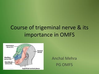
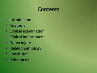
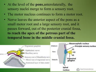
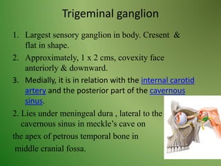
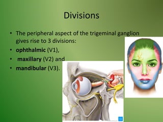
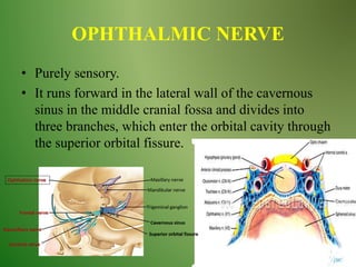
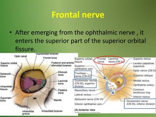
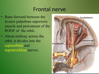
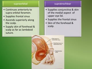
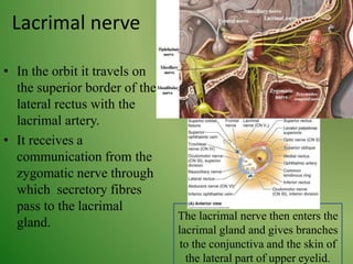
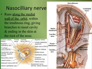
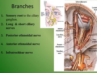
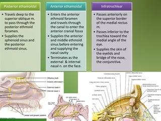
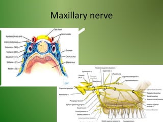
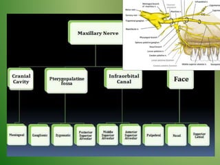
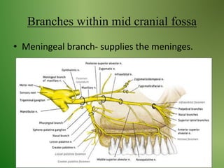
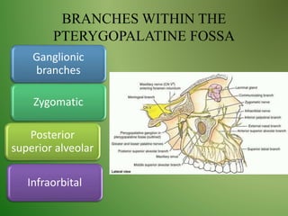
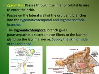
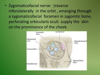
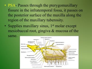
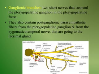
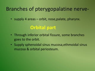
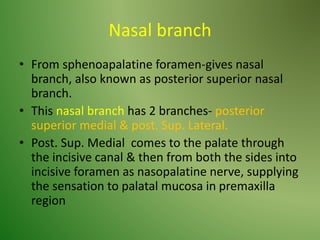
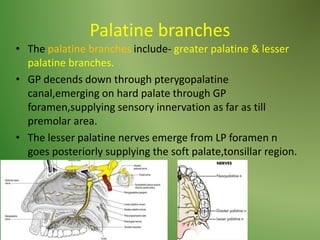
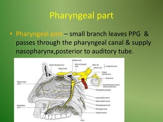
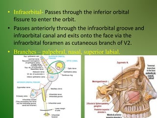
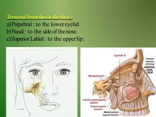
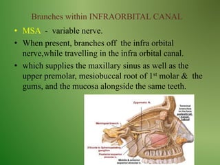
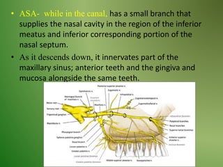
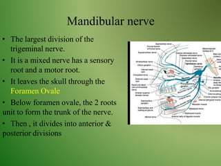
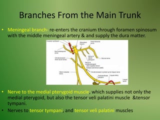
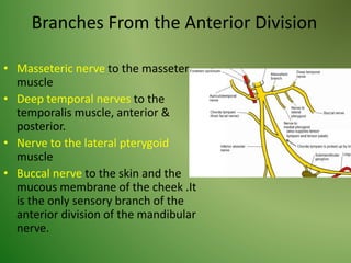
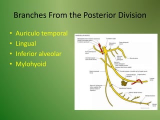
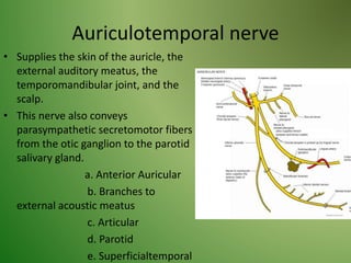
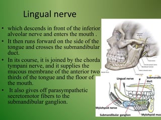
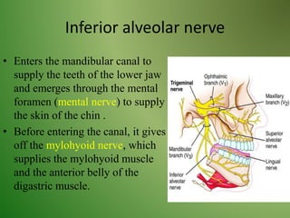
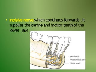
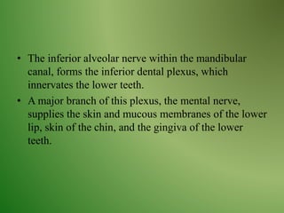
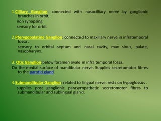
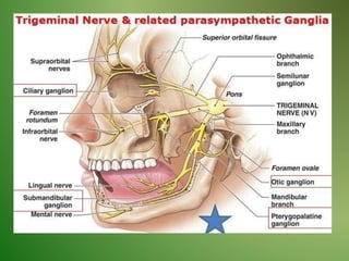
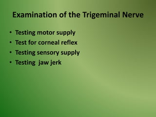
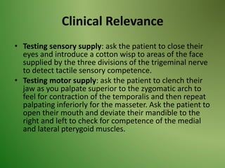
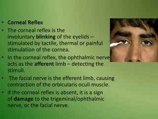
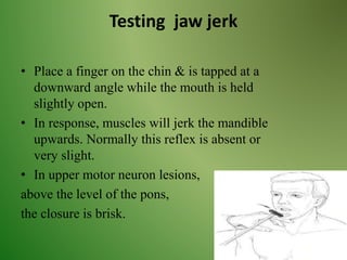
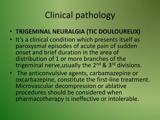
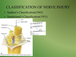
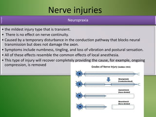
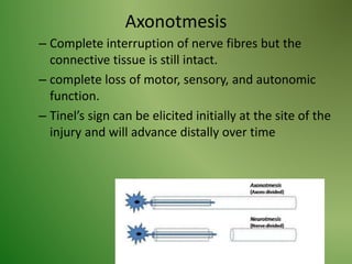
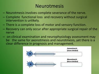
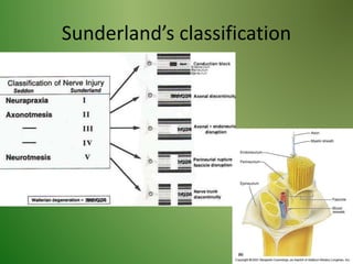
SlideShare a Scribd company logo
1 of 76
Course of trigeminal nerve & its
importance in OMFS
Anchal Mehra
PG OMFS
Contents
• Introduction
• Anatomy
• Clinical examination
• Clinical importance
• Nerve injury
• Related pathology
• Conclusion
• References
INTRODUCTION
It’s motor to the muscles of 1st
branchial arch & several small muscles.
It’s sensory to the head & face region.
• At the level of the pons,anterolaterally, the
sensory nuclei merge to form a sensory root.
• The motor nucleus continues to form a motor root.
• Nerve leaves the anterior aspect of the pons as a
small motor root and a large sensory root, and it
passes forward, out of the posterior cranial fossa,
to reach the apex of the petrous part of the
temporal bone in the middle cranial fossa.
Functional component
• Special visceral efferent (SVE) fibers- arise from
motor nucleus of CN5, & supply muscles of
mastication,mylohyoid,anterior belly of
diagastric,tensor veli palatini,tensor tympani.
• General somatic afferent (GSA)- they carry
proprioceptive sensation from
muscles of mastication, TMJ,
teeth & terminate in the motor
nucleus.
Exteroceptive sensation (i.e,
pain.touch & temperature ), from
skin of the head & face,,nasal
cavity,meninges etc. & terminate in
the principal nucleus of CN5
Sensory root
GSA- face,scalp,teeth,gingiva,oral & nasal
cavities, para nasal sinus, conjunctiva, cornea.
Motor root
CNS
Motor nucleus
Motor root
Mandibular nerve
Muscles of mastication, mylohyoid,tensor
tympani,tensor veli palatini.
Trigeminal ganglion
1. Largest sensory ganglion in body. Cresent &
flat in shape.
2. Approximately, 1 x 2 cms, covexity face
anteriorly & downward.
3. Medially, it is in relation with the internal carotid
artery and the posterior part of the cavernous
sinus.
2. Lies under meningeal dura , lateral to the
cavernous sinus in meckle’s cave on
the apex of petrous temporal bone in
middle cranial fossa.
Divisions
• The peripheral aspect of the trigeminal ganglion
gives rise to 3 divisions:
• ophthalmic (V1),
• maxillary (V2) and
• mandibular (V3).
OPHTHALMIC NERVE
• Purely sensory.
• It runs forward in the lateral wall of the cavernous
sinus in the middle cranial fossa and divides into
three branches, which enter the orbital cavity through
the superior orbital fissure.
Branches
Tentorial
branch
Frontal
Supraorbital
Supratrochlear
Lacrimal
Nasocilliary
Frontal nerve
• After emerging from the ophthalmic nerve , it
enters the superior part of the superior orbital
fissure.
Frontal nerve
• Runs forward between the
levator palpebrae superioris
muscle and periosteum of the
ROOF of the orbit.
• About midway across the
orbit, it divides into the
supraorbital and
supratrochlear nerves.
supraorbital
• Continues anteriorly to
supra orbital foramen.
• Supplies frontal sinus
• Ascends superiorly along
the scalp.
• Supply skin of forehead &
scalp as far as lambdoid
suture.
supratrochlear
• Supplies conjunctiva & skin
of the medial aspect of
upper eye lid.
• Supplies the frontal sinus
• Skin of the forehead &
scalp.
Lacrimal nerve
• In the orbit it travels on
the superior border of the
lateral rectus with the
lacrimal artery.
• It receives a
communication from the
zygomatic nerve through
which secretory fibres
pass to the lacrimal
gland.
The lacrimal nerve then enters the
lacrimal gland and gives branches
to the conjunctiva and the skin of
the lateral part of upper eyelid.
Nasocilliary nerve
• Runs along the medial
wall of the orbit, within
the tendinous ring, giving
branches to nasal cavity
& ending in the skin at
the root of the nose.
Branches
1. Sensory root to the ciliary
ganglion.
2. Long & short ciliary
nerves
3. Posterior ethmoidal nerve
4. Anterior ethmoidal nerve
5. Infratrochlear nerve
Posterior ethamoidal
• Travels deep to the
superior oblique m.
to pass through the
posterior ethmoid
foramen.
• Supplies the
sphenoid sinus and
the posterior
ethmoid sinus.
Anterior ethamoidal
• Enters the anterior
ethmoid foramen
and travels through
the canal to enter the
anterior cranial fossa
• Supplies the anterior
and middle ethmoid
sinus before entering
and supplying the
nasal cavity
• Terminates as the
external & internal
nasal n. on the face.
Infratrochlear
• Passes anteriorly on
the superior border
of the medial rectus
m.
• Passes inferior to the
trochlea toward the
medial angle of the
eye.
• Supplies the skin of
the eyelids and
bridge of the nose,
the conjunctiva.
Maxillary nerve
Branches within mid cranial fossa
• Meningeal branch- supplies the meninges.
BRANCHES WITHIN THE
PTERYGOPALATINE FOSSA
Posterior
superior alveolar
Zygomatic
Ganglionic
branches
Infraorbital
• Zygomatic -Passes through the inferior orbital fissure
to enter the orbit.
• Passes on the lateral wall of the orbit and branches
into the zygomaticotemporal and zygomaticofacial
branches
• The zygomaticotemporal branch gives
parasympathetic secretomotor fibers to the lacrimal
gland via the lacrimal nerve. Supply the skin on side
of the forehead.
• Zygomaticofacial nerve- traverse
inferolaterally in the orbit , emerging through
a zygomaticofacial foramen in zygomtic bone,
perforating orbicularis oculi. supply the skin
on the prominence of the cheek.
• PSA- Passes through the pterygomaxillary
fissure in the infratemporal fossa, it passes on
the posterior surface of the maxilla along the
region of the maxillary tuberosity.
• Supplies maxillary sinus, 1st molar except
mesiobuccal root, gingiva & mucosa of the
same.
• Ganglionic branches- two short nerves that suspend
the pterygopalatine ganglion in the pterygopalatine
fossa.
• They also contain postganglionic parasympathetic
fibers from the pterygopalatine ganglion & from the
zygomaticotemporal nerve, that are going to the
lacrimal gland.
Branches of pterygopalatine nerve-
• supply 4 areas – orbit, nose,palate, pharynx.
• Through inferior orbital fissure, some branches
goes to the orbit.
• Supply sphenoidal sinus mucosa,ethmoidal sinus
mucosa & orbital periosteum.
Orbital part
Nasal branch
• From sphenoapalatine foramen-gives nasal
branch, also known as posterior superior nasal
branch.
• This nasal branch has 2 branches- posterior
superior medial & post. Sup. Lateral.
• Post. Sup. Medial comes to the palate through
the incisive canal & then from both the sides into
incisive foramen as nasopalatine nerve, supplying
the sensation to palatal mucosa in premaxilla
region
Palatine branches
• The palatine branches include- greater palatine & lesser
palatine branches.
• GP decends down through pterygopalatine
canal,emerging on hard palate through GP
foramen,supplying sensory innervation as far as till
premolar area.
• The lesser palatine nerves emerge from LP foramen n
goes posteriorly supplying the soft palate,tonsillar region.
Pharyngeal part
• Pharyngeal part – small branch leaves PPG &
passes through the pharyngeal canal & supply
nasopharynx,posterior to auditory tube.
• Infraorbital- Passes through the inferior orbital
fissure to enter the orbit.
• Passes anteriorly through the infraorbital groove and
infraorbital canal and exits onto the face via the
infraorbital foramen as cutaneous branch of V2.
• Branches – palpebral, nasal, superior labial.
Terminal branchesin the face :
a)Palpebral: to the lowereyelid.
b)Nasal: to the sideof thenose.
c)SuperiorLabial: to the upperlip.
Branches within INFRAORBITAL CANAL
• MSA - variable nerve.
• When present, branches off the infra orbital
nerve,while travelling in the infra orbital canal.
• which supplies the maxillary sinus as well as the
upper premolar, mesiobuccal root of 1st molar & the
gums, and the mucosa alongside the same teeth.
• ASA- while in the canal, has a small branch that
supplies the nasal cavity in the region of the inferior
meatus and inferior corresponding portion of the
nasal septum.
• As it descends down, it innervates part of the
maxillary sinus; anterior teeth and the gingiva and
mucosa alongside the same teeth.
Mandibular nerve
• The largest division of the
trigeminal nerve.
• It is a mixed nerve has a sensory
root and a motor root.
• It leaves the skull through the
Foramen Ovale
• Below foramen ovale, the 2 roots
unit to form the trunk of the nerve.
• Then , it divides into anterior &
posterior divisions
Branches From the Main Trunk
• Meningeal branch- re-enters the cranium through foramen spinosum
with the middle meningeal artery & and supply the dura matter.
• Nerve to the medial pterygoid muscle, which supplies not only the
medial pterygoid, but also the tensor veli palatini muscle &tensor
tympani.
• Nerves to tensor tympani, and tensor veli palatini muscles
Branches From the Anterior Division
• Masseteric nerve to the masseter
muscle
• Deep temporal nerves to the
temporalis muscle, anterior &
posterior.
• Nerve to the lateral pterygoid
muscle
• Buccal nerve to the skin and the
mucous membrane of the cheek .It
is the only sensory branch of the
anterior division of the mandibular
nerve.
Branches From the Posterior Division
• Auriculo temporal
• Lingual
• Inferior alveolar
• Mylohyoid
Auriculotemporal nerve
• Supplies the skin of the auricle, the
external auditory meatus, the
temporomandibular joint, and the
scalp.
• This nerve also conveys
parasympathetic secretomotor fibers
from the otic ganglion to the parotid
salivary gland.
a. Anterior Auricular
b. Branches to
external acoustic meatus
c. Articular
d. Parotid
e. Superficialtemporal
Lingual nerve
• which descends in front of the inferior
alveolar nerve and enters the mouth .
• It then runs forward on the side of the
tongue and crosses the submandibular
duct.
• In its course, it is joined by the chorda
tympani nerve, and it supplies the
mucous membrane of the anterior two
thirds of the tongue and the floor of
the mouth.
• It also gives off parasympathetic
secretomotor fibers to the
submandibular ganglion.
Inferior alveolar nerve
• Enters the mandibular canal to
supply the teeth of the lower jaw
and emerges through the mental
foramen (mental nerve) to supply
the skin of the chin .
• Before entering the canal, it gives
off the mylohyoid nerve, which
supplies the mylohyoid muscle
and the anterior belly of the
digastric muscle.
• Incisivenervewhich continues forwards .It
supplies the canine and incisor teeth of the
lower jaw.
• The inferior alveolar nerve within the mandibular
canal, forms the inferior dental plexus, which
innervates the lower teeth.
• A major branch of this plexus, the mental nerve,
supplies the skin and mucous membranes of the lower
lip, skin of the chin, and the gingiva of the lower
teeth.
1.Cilliary Ganglion: connected with nasocilliary nerve by ganglionic
branches in orbit,
non synapsing
sensory for orbit
2.Pterygopalatine Ganglion: connected to maxillary nerve in infratemporal
fossa .
sensory to orbital septum and nasal cavity, max sinus, palate,
nasopharynx.
3. Otic Ganglion:below foramen ovale in infra temporal fossa.
On the medial surface of mandibular nerve. Supplies secretomotor fibres
to the parotid gland.
4.Submandibular Ganglion: related to lingual nerve, rests on hypoglossus .
supplies post ganglionic parasympathetic secretomotor fibres to
submandibular and sublingual gland.
Examination of the Trigeminal Nerve
• Testing motor supply
• Test for corneal reflex
• Testing sensory supply
• Testing jaw jerk
Clinical Relevance
• Testing sensory supply: ask the patient to close their
eyes and introduce a cotton wisp to areas of the face
supplied by the three divisions of the trigeminal nerve
to detect tactile sensory competence.
• Testing motor supply: ask the patient to clench their
jaw as you palpate superior to the zygomatic arch to
feel for contraction of the temporalis and then repeat
palpating inferiorly for the masseter. Ask the patient to
open their mouth and deviate their mandible to the
right and left to check for competence of the medial
and lateral pterygoid muscles.
• Corneal Reflex
• The corneal reflex is the
involuntary blinking of the eyelids –
stimulated by tactile, thermal or painful
stimulation of the cornea.
• In the corneal reflex, the ophthalmic nerve
acts as the afferent limb – detecting the
stimuli.
• The facial nerve is the efferent limb, causing
contraction of the orbicularis oculi muscle.
• If the corneal reflex is absent, it is a sign
of damage to the trigeminal/ophthalmic
nerve, or the facial nerve.
Testing jaw jerk
• Place a finger on the chin & is tapped at a
downward angle while the mouth is held
slightly open.
• In response, muscles will jerk the mandible
upwards. Normally this reflex is absent or
very slight.
• In upper motor neuron lesions,
above the level of the pons,
the closure is brisk.
Clinical pathology
• TRIGEMINAL NEURALGIA (TIC DOULOUREUX)
• It’s a clinical condition which presents itself as
paroxysmal episodes of acute pain of sudden
onset and brief duration in the area of
distribution of 1 or more branches of the
trigeminal nerve,usually the 2nd & 3rd divisions.
• The anticonvulsive agents, carbamazepine or
oxcarbazepine, constitute the first-line treatment.
Microvascular decompression or ablative
procedures should be considered when
pharmacotherapy is ineffective or intolerable.
TCR
• TCR is a triad of bradycardia ,bradypnea and
gastric motility changes due to the efferent
activation of the vagal nerve in response to
the pressure distribution in the trigeminal
nerve.
• This unexpected phenomenon is usually seen
in orbital injuries and during surgical
manipulation of craniofacial structures in the
distribution of trigeminal nerve.
• Knowledge of the TCR is essential as it may
mimic a closed cranial injury or a cardiac
dysarrythmia in a post traumatic patient to
avoid unwarranted surgical intervention.
• A detailed ophthalmic examination in
maxillofacial injuries is essential.
LESIONS
• Lesion at foramen rotundum
or within infra orbital canal
involves maxillary nerve.
This results in paraesthesia
of the cheek & upper teeth.
• Loss of sneeze reflex as the
nerve supplies the afferent
limb of ‘’sneeze reflex’’.
•Lesion at the foramen ovale
involves mandibular nerve &
paraesthesia along the
mandible, lower teeth,& side
of the face.
•Paralysis of muscles of
mastication .
•Loss of the ‘’ jaw jerk
reflex ‘’, as mandibular nerve
supplies both the afferent &
efferent limbs for the jaw
jerk reflex.
Multiple scelrosis
• During periods of multiple sclerosis
(MS) activity, white blood cells are
drawn to regions of the white matter.
• These initiate and participate in the
inflammatory response.
• During the inflammatory phase, the
myelin surrounding the axons is
destroyed in a process known as
demyelination.
• Approximately 2 % of patients with
MS also have symptoms of trigeminal
neuralgia.
Herpes zoster of CN V
Trigeminal Schwannomas
• Arise from the trigeminal ganglion and cause
numbness and paresthesias are the most
common symptoms from these tumors, but
pain and crawling sensations are sometimes
present.
• Other tumors and perineurial spread of other
cancers in the region of the trigeminal
ganglion are rare, but can cause variable
clinical symptoms.
The superior orbital fissure syndrome
• Is a complex of impaired function of the cranial
nerves (III, IV, V, and VI) that enter the orbit
through the superior orbital fissure.
• Three major precipitating factors for SOFS are
trauma, tumor, and inflammation.
• Any unnatural narrowing of SOF due to trauma of
high impact to the upper and midface
(frontobasal skull, Le Fort II, III, and zygomatic
complex fractures) can precipitate this condition.
CLASSIFICATION OF NERVE INJURY
• Seddon’s Classification(1943)
• Sunderland’s Classification(1951)
Nerve injuries
Neuropraxia
• the mildest injury type that is transient.
• There is no effect on nerve continuity.
• Caused by a temporary disturbance in the conduction pathway that blocks neural
transmission but does not damage the axon.
• Symptoms include numbness, tingling, and loss of vibration and postural sensation.
• All of these effects resemble the common effects of local anesthesia.
• This type of injury will recover completely providing the cause, for example, ongoing
compression, is removed
Axonotmesis
– Complete interruption of nerve fibres but the
connective tissue is still intact.
– complete loss of motor, sensory, and autonomic
function.
– Tinel’s sign can be elicited initially at the site of the
injury and will advance distally over time
Neurotmesis
– Neurotmesis involves complete severance of the nerve.
– Complete functional loss and recovery without surgical
intervention is unlikely.
– There is a complete loss of motor and sensory function.
– Recovery can only occur after appropriate surgical repair of the
nerve
– on clinical examination and neurophysiology assessment may
be the same for axonotmesis and neurotmesis, yet there is a
clear difference in prognosis and management.
Sunderland’s classification
TRIGEMINAL NEUROPATHY
• Well recognized disorders characterized and manifesting as skin and mucosal
numbness in the region innervated by the trigeminal nerve.
• Facial numbness indicates trigeminal sensory alteration affecting the trigeminal
system
• They can be the result of traumatism, tumors, or diseases of the connective tissue,
infectious or demyelinating diseases, or may be of idiopathic origin.
• The clinical exploration reveals a loss of sensitivity in the cutaneous territory
corresponding to the affected nerve, which can be partial (hypoesthesia) or
complete (anesthesia).
Trigeminal nerve injuries
• CN5 may be injured by
trauma,tumors,aneurysms or meningeal
infections.
• The sensory & motor nuclei in the pons &
medulla may be destroyed by intermedullary
tumors or vascular lesions.
• An isolated lesion of the spinal trigeminal tract
also may occur with multiple sclerosis.
INJURY CAUSES THE FOLLOWING-
• Paralysis of muscle of mastication with
deviation of the mandible towards the side of
the lesion.
• Loss of the ability to appreciate soft
tactile,thermal or painful sensations in the
face.
• Loss of corneal reflex & sneezing reflex.
• Eg: involvement of the IAN in mandibular
fractures and infra -orbital in maxillary and
ZMC fractures
• Loss or diminished sensation around the lower
lips ,usually involving the lower lip and/or
tongue areas causing a mixture of pain,
numbness that may be present all the time or
intermittently.
• Lingual nerve lies in contact with lingual
gingiva medial to 3rd molar, may get injured
during removal of the tooth.
Conclusion
Bibliography
• Snell’s 8th edition
• Textbook of Local Anesthesia-Stanley F
Malamed
• Gray’s Anatomy
• Articles
• Internet sources
Thank you 
Applied anatomy
• Supraorbital injuries
Trauma to the supraorbital margin may damage the
supraorbital and/or supratrochlear nerves causing sensory
loss in the scalp.
• Ethmoid tumours
Malignant tumours of the mucous lining of the ethmoid air cells
may expand into the orbits, damaging branches of Va. This
may lead to displacement of the orbital contents causing
proptosis and squint, and sensory loss over the anterior nasal
skin.
• Nasal fractures
Trauma to the nose may damage the external nasal nerve as it
becomes superficial. Sensory loss of the skin down to the tip
of the nose.
• Infraorbital injuries: malar fractures
Trauma to infraorbital margin may cause sensory loss of infraorbital
skin.
• Maxillary sinus infections
Infections of the maxillary sinus may cause infraorbital pain or
may cause referred pain to other structures supplied by Vb .
• Maxillary teeth abscesses :The roots of the maxillary teeth
(especially the second molars)are intimately related to the maxillary
sinus. Root abscesses are painful.
• Maxillary antrumtumours
Malignant tumours of the mucous lining of the maxillary antrum may
expand into the orbit, damaging branches of Vb, particularly the
infraorbital. This may lead to anaesthesia over the facial skin. The
orbital contents may also be displaced causing proptosis
• TCR is actually endogenous physiological
protective mechanisms found in brain against
ischemia.
• It is one of the oxygen conserving reflexes.
• Within seconds of initiation of such reflex, there
is activation of sympathetic nerves which leads to
cerebro vascular vasodilatation.
• These responses are exaggerated and put the
patient at risk .
• During initial period of vagal stimulation,the
cardiac depression is at peak.
Management of nerve injury
1.Manipulation of the nerve (neurolysis)
2.Repair with a direct anastomosis or the use of
graft
3.Coaptation
4.Neurorrhaphy
5. Entubulation

More Related Content

What's hot

Mandibular nerve dental surgery
Mandibular nerve dental surgeryMandibular nerve dental surgery
Mandibular nerve dental surgeryDr-Faisal Al-Qahtani
 
Trigeminal Nerve and its applied aspects
Trigeminal Nerve and its applied aspectsTrigeminal Nerve and its applied aspects
Trigeminal Nerve and its applied aspectsAMBARKHAN4
 
Facial nerve and it's applied aspect
Facial nerve and it's applied aspectFacial nerve and it's applied aspect
Facial nerve and it's applied aspectPrachi Jha
 
Trigeminal nerve
Trigeminal nerve Trigeminal nerve
Trigeminal nerve AnuRaaga
 
Trigeminal nerve
Trigeminal nerveTrigeminal nerve
Trigeminal nervevinod panchal
 
Trigeminal nerve
Trigeminal nerveTrigeminal nerve
Trigeminal nerveashish25200
 
Opthalmic division of trigeminal nerve/ oral surgery courses  
Opthalmic division of trigeminal nerve/ oral surgery courses  Opthalmic division of trigeminal nerve/ oral surgery courses  
Opthalmic division of trigeminal nerve/ oral surgery courses  Indian dental academy
 
Pterygopalatine ganglion 1
Pterygopalatine ganglion 1Pterygopalatine ganglion 1
Pterygopalatine ganglion 1Omar Eraky
 
Anatomy of Soft palate
Anatomy of  Soft palateAnatomy of  Soft palate
Anatomy of Soft palateddert
 
External carotid artery, branches and ligation
External carotid artery, branches and ligationExternal carotid artery, branches and ligation
External carotid artery, branches and ligationbenjamin Emmanuel
 
Facial nerve
Facial nerve Facial nerve
Facial nerve Adarsh Nath
 
Maxilla anatomy, development & surgical anatomy
Maxilla  anatomy, development & surgical anatomyMaxilla  anatomy, development & surgical anatomy
Maxilla anatomy, development & surgical anatomyDr. SHEETAL KAPSE
 
SURICAL ANATOMY OF FACIAL NERVE
SURICAL ANATOMY OF FACIAL NERVESURICAL ANATOMY OF FACIAL NERVE
SURICAL ANATOMY OF FACIAL NERVEAmar Shinde
 
trigeminal nerve
trigeminal  nerve trigeminal  nerve
trigeminal nerve Zunaidahaneef
 

What's hot (20)

Mandibular nerve dental surgery
Mandibular nerve dental surgeryMandibular nerve dental surgery
Mandibular nerve dental surgery
 
Trigeminal Nerve and its applied aspects
Trigeminal Nerve and its applied aspectsTrigeminal Nerve and its applied aspects
Trigeminal Nerve and its applied aspects
 
Facial nerve and it's applied aspect
Facial nerve and it's applied aspectFacial nerve and it's applied aspect
Facial nerve and it's applied aspect
 
Trigeminal nerve
Trigeminal nerve Trigeminal nerve
Trigeminal nerve
 
Trigeminal nerve
Trigeminal nerveTrigeminal nerve
Trigeminal nerve
 
Facial nerve
Facial nerveFacial nerve
Facial nerve
 
Trigeminal nerve
Trigeminal nerveTrigeminal nerve
Trigeminal nerve
 
Opthalmic division of trigeminal nerve/ oral surgery courses  
Opthalmic division of trigeminal nerve/ oral surgery courses  Opthalmic division of trigeminal nerve/ oral surgery courses  
Opthalmic division of trigeminal nerve/ oral surgery courses  
 
Pterygopalatine ganglion 1
Pterygopalatine ganglion 1Pterygopalatine ganglion 1
Pterygopalatine ganglion 1
 
Anatomy of Soft palate
Anatomy of  Soft palateAnatomy of  Soft palate
Anatomy of Soft palate
 
External carotid artery, branches and ligation
External carotid artery, branches and ligationExternal carotid artery, branches and ligation
External carotid artery, branches and ligation
 
Mandibular nerve
Mandibular nerveMandibular nerve
Mandibular nerve
 
Facial nerve
Facial nerve Facial nerve
Facial nerve
 
Trigeminal nerve
Trigeminal nerveTrigeminal nerve
Trigeminal nerve
 
Maxilla anatomy, development & surgical anatomy
Maxilla  anatomy, development & surgical anatomyMaxilla  anatomy, development & surgical anatomy
Maxilla anatomy, development & surgical anatomy
 
SURICAL ANATOMY OF FACIAL NERVE
SURICAL ANATOMY OF FACIAL NERVESURICAL ANATOMY OF FACIAL NERVE
SURICAL ANATOMY OF FACIAL NERVE
 
trigeminal nerve
trigeminal  nerve trigeminal  nerve
trigeminal nerve
 
Facial artery
Facial arteryFacial artery
Facial artery
 
Mandibular nerve
Mandibular nerveMandibular nerve
Mandibular nerve
 
Trigeminal nerve
Trigeminal nerveTrigeminal nerve
Trigeminal nerve
 

Similar to Trigeminal nerve

Trigeminal Nerve Dr.AD.pptx
Trigeminal Nerve Dr.AD.pptxTrigeminal Nerve Dr.AD.pptx
Trigeminal Nerve Dr.AD.pptxArunCreations
 
Cranial Nerves
Cranial NervesCranial Nerves
Cranial NervesHadi Munib
 
Trigeminal and facial nerve.pptx
Trigeminal and facial nerve.pptxTrigeminal and facial nerve.pptx
Trigeminal and facial nerve.pptxAnkur Rathaur
 
Trigeminal and facial nerve.pptx
Trigeminal and facial nerve.pptxTrigeminal and facial nerve.pptx
Trigeminal and facial nerve.pptxAnkur Rathaur
 
trigeminal-neuralgia.pptx
trigeminal-neuralgia.pptxtrigeminal-neuralgia.pptx
trigeminal-neuralgia.pptxssuser20cec1
 
Temporal_and_infratemporal_fossa.ppt
Temporal_and_infratemporal_fossa.pptTemporal_and_infratemporal_fossa.ppt
Temporal_and_infratemporal_fossa.pptDr Ndayisaba Corneille
 
Anatomy of infratemporal region
Anatomy of infratemporal regionAnatomy of infratemporal region
Anatomy of infratemporal regionDr. Mohammad Mahmoud
 
MICROSURGICAL ANATOMY OF CRANIAL NERVES
MICROSURGICAL ANATOMY OF CRANIAL NERVESMICROSURGICAL ANATOMY OF CRANIAL NERVES
MICROSURGICAL ANATOMY OF CRANIAL NERVESpankaj patel
 
The cerebello pontine angle
The cerebello pontine angleThe cerebello pontine angle
The cerebello pontine angleDr Himanshu Soni
 
trigeminal-neuralgia (1).pptx
trigeminal-neuralgia (1).pptxtrigeminal-neuralgia (1).pptx
trigeminal-neuralgia (1).pptxAkshaySingh503850
 
cranial nerve 5.pptx
cranial nerve 5.pptxcranial nerve 5.pptx
cranial nerve 5.pptxDeepthinker14
 
Dental Anesthesia
Dental AnesthesiaDental Anesthesia
Dental AnesthesiaOzident
 
Anesth mouth
Anesth mouthAnesth mouth
Anesth mouthLE HAI TRIEU
 
Facialnerve 160502100010
Facialnerve 160502100010Facialnerve 160502100010
Facialnerve 160502100010Indhu Reddy
 
Nerve supply of face 1
Nerve supply of face 1Nerve supply of face 1
Nerve supply of face 1RenukaAjay
 
Temporal & infratemporal regions II
Temporal & infratemporal regions IITemporal & infratemporal regions II
Temporal & infratemporal regions IIPrabhakar Yadav
 

Similar to Trigeminal nerve (20)

Trigeminal Nerve Dr.AD.pptx
Trigeminal Nerve Dr.AD.pptxTrigeminal Nerve Dr.AD.pptx
Trigeminal Nerve Dr.AD.pptx
 
Cranial Nerves
Cranial NervesCranial Nerves
Cranial Nerves
 
Trigeminal and facial nerve.pptx
Trigeminal and facial nerve.pptxTrigeminal and facial nerve.pptx
Trigeminal and facial nerve.pptx
 
Trigeminal and facial nerve.pptx
Trigeminal and facial nerve.pptxTrigeminal and facial nerve.pptx
Trigeminal and facial nerve.pptx
 
trigeminal-neuralgia.pptx
trigeminal-neuralgia.pptxtrigeminal-neuralgia.pptx
trigeminal-neuralgia.pptx
 
Temporal_and_infratemporal_fossa.ppt
Temporal_and_infratemporal_fossa.pptTemporal_and_infratemporal_fossa.ppt
Temporal_and_infratemporal_fossa.ppt
 
Anatomy of infratemporal region
Anatomy of infratemporal regionAnatomy of infratemporal region
Anatomy of infratemporal region
 
MICROSURGICAL ANATOMY OF CRANIAL NERVES
MICROSURGICAL ANATOMY OF CRANIAL NERVESMICROSURGICAL ANATOMY OF CRANIAL NERVES
MICROSURGICAL ANATOMY OF CRANIAL NERVES
 
The cerebello pontine angle
The cerebello pontine angleThe cerebello pontine angle
The cerebello pontine angle
 
trigeminal-neuralgia (1).pptx
trigeminal-neuralgia (1).pptxtrigeminal-neuralgia (1).pptx
trigeminal-neuralgia (1).pptx
 
Facial nerve anatomy
Facial nerve anatomyFacial nerve anatomy
Facial nerve anatomy
 
cranial nerve 5.pptx
cranial nerve 5.pptxcranial nerve 5.pptx
cranial nerve 5.pptx
 
Dental Anesthesia
Dental AnesthesiaDental Anesthesia
Dental Anesthesia
 
Anesth mouth
Anesth mouthAnesth mouth
Anesth mouth
 
Facial nerve
Facial nerveFacial nerve
Facial nerve
 
Facialnerve 160502100010
Facialnerve 160502100010Facialnerve 160502100010
Facialnerve 160502100010
 
Nerve supply of face 1
Nerve supply of face 1Nerve supply of face 1
Nerve supply of face 1
 
FACIAL NERVE ANATOMY
FACIAL NERVE ANATOMYFACIAL NERVE ANATOMY
FACIAL NERVE ANATOMY
 
Trigeminal nerve
Trigeminal nerveTrigeminal nerve
Trigeminal nerve
 
Temporal & infratemporal regions II
Temporal & infratemporal regions IITemporal & infratemporal regions II
Temporal & infratemporal regions II
 

Recently uploaded

Hemostasis Physiology and Clinical correlations by Dr Faiza.pdf
Hemostasis Physiology and Clinical correlations by Dr Faiza.pdfHemostasis Physiology and Clinical correlations by Dr Faiza.pdf
Hemostasis Physiology and Clinical correlations by Dr Faiza.pdfMedicoseAcademics
 
Bangalore Call Girls Marathahalli 📞 9907093804 High Profile Service 100% Safe
Bangalore Call Girls Marathahalli 📞 9907093804 High Profile Service 100% SafeBangalore Call Girls Marathahalli 📞 9907093804 High Profile Service 100% Safe
Bangalore Call Girls Marathahalli 📞 9907093804 High Profile Service 100% Safenarwatsonia7
 
Artifacts in Nuclear Medicine with Identifying and resolving artifacts.
Artifacts in Nuclear Medicine with Identifying and resolving artifacts.Artifacts in Nuclear Medicine with Identifying and resolving artifacts.
Artifacts in Nuclear Medicine with Identifying and resolving artifacts.MiadAlsulami
 
Call Girls Hsr Layout Just Call 7001305949 Top Class Call Girl Service Available
Call Girls Hsr Layout Just Call 7001305949 Top Class Call Girl Service AvailableCall Girls Hsr Layout Just Call 7001305949 Top Class Call Girl Service Available
Call Girls Hsr Layout Just Call 7001305949 Top Class Call Girl Service Availablenarwatsonia7
 
Call Girls Electronic City Just Call 7001305949 Top Class Call Girl Service A...
Call Girls Electronic City Just Call 7001305949 Top Class Call Girl Service A...Call Girls Electronic City Just Call 7001305949 Top Class Call Girl Service A...
Call Girls Electronic City Just Call 7001305949 Top Class Call Girl Service A...narwatsonia7
 
Call Girls Service In Shyam Nagar Whatsapp 8445551418 Independent Escort Service
Call Girls Service In Shyam Nagar Whatsapp 8445551418 Independent Escort ServiceCall Girls Service In Shyam Nagar Whatsapp 8445551418 Independent Escort Service
Call Girls Service In Shyam Nagar Whatsapp 8445551418 Independent Escort Serviceparulsinha
 
Call Girls Jayanagar Just Call 7001305949 Top Class Call Girl Service Available
Call Girls Jayanagar Just Call 7001305949 Top Class Call Girl Service AvailableCall Girls Jayanagar Just Call 7001305949 Top Class Call Girl Service Available
Call Girls Jayanagar Just Call 7001305949 Top Class Call Girl Service Availablenarwatsonia7
 
Call Girls Frazer Town Just Call 7001305949 Top Class Call Girl Service Avail...
Call Girls Frazer Town Just Call 7001305949 Top Class Call Girl Service Avail...Call Girls Frazer Town Just Call 7001305949 Top Class Call Girl Service Avail...
Call Girls Frazer Town Just Call 7001305949 Top Class Call Girl Service Avail...narwatsonia7
 
Aspirin presentation slides by Dr. Rewas Ali
Aspirin presentation slides by Dr. Rewas AliAspirin presentation slides by Dr. Rewas Ali
Aspirin presentation slides by Dr. Rewas AliRewAs ALI
 
Housewife Call Girls Hoskote | 7001305949 At Low Cost Cash Payment Booking
Housewife Call Girls Hoskote | 7001305949 At Low Cost Cash Payment BookingHousewife Call Girls Hoskote | 7001305949 At Low Cost Cash Payment Booking
Housewife Call Girls Hoskote | 7001305949 At Low Cost Cash Payment Bookingnarwatsonia7
 
Call Girls In Andheri East Call 9920874524 Book Hot And Sexy Girls
Call Girls In Andheri East Call 9920874524 Book Hot And Sexy GirlsCall Girls In Andheri East Call 9920874524 Book Hot And Sexy Girls
Call Girls In Andheri East Call 9920874524 Book Hot And Sexy Girlsnehamumbai
 
High Profile Call Girls Jaipur Vani 8445551418 Independent Escort Service Jaipur
High Profile Call Girls Jaipur Vani 8445551418 Independent Escort Service JaipurHigh Profile Call Girls Jaipur Vani 8445551418 Independent Escort Service Jaipur
High Profile Call Girls Jaipur Vani 8445551418 Independent Escort Service Jaipurparulsinha
 
Low Rate Call Girls Mumbai Suman 9910780858 Independent Escort Service Mumbai
Low Rate Call Girls Mumbai Suman 9910780858 Independent Escort Service MumbaiLow Rate Call Girls Mumbai Suman 9910780858 Independent Escort Service Mumbai
Low Rate Call Girls Mumbai Suman 9910780858 Independent Escort Service Mumbaisonalikaur4
 
Bangalore Call Girls Majestic 📞 9907093804 High Profile Service 100% Safe
Bangalore Call Girls Majestic 📞 9907093804 High Profile Service 100% SafeBangalore Call Girls Majestic 📞 9907093804 High Profile Service 100% Safe
Bangalore Call Girls Majestic 📞 9907093804 High Profile Service 100% Safenarwatsonia7
 
Russian Call Girl Brookfield - 7001305949 Escorts Service 50% Off with Cash O...
Russian Call Girl Brookfield - 7001305949 Escorts Service 50% Off with Cash O...Russian Call Girl Brookfield - 7001305949 Escorts Service 50% Off with Cash O...
Russian Call Girl Brookfield - 7001305949 Escorts Service 50% Off with Cash O...narwatsonia7
 
Vip Call Girls Anna Salai Chennai 👉 8250192130 ❣️💯 Top Class Girls Available
Vip Call Girls Anna Salai Chennai 👉 8250192130 ❣️💯 Top Class Girls AvailableVip Call Girls Anna Salai Chennai 👉 8250192130 ❣️💯 Top Class Girls Available
Vip Call Girls Anna Salai Chennai 👉 8250192130 ❣️💯 Top Class Girls AvailableNehru place Escorts
 
Call Girls Service Chennai Jiya 7001305949 Independent Escort Service Chennai
Call Girls Service Chennai Jiya 7001305949 Independent Escort Service ChennaiCall Girls Service Chennai Jiya 7001305949 Independent Escort Service Chennai
Call Girls Service Chennai Jiya 7001305949 Independent Escort Service ChennaiNehru place Escorts
 
Book Call Girls in Kasavanahalli - 7001305949 with real photos and phone numbers
Book Call Girls in Kasavanahalli - 7001305949 with real photos and phone numbersBook Call Girls in Kasavanahalli - 7001305949 with real photos and phone numbers
Book Call Girls in Kasavanahalli - 7001305949 with real photos and phone numbersnarwatsonia7
 

Recently uploaded (20)

Hemostasis Physiology and Clinical correlations by Dr Faiza.pdf
Hemostasis Physiology and Clinical correlations by Dr Faiza.pdfHemostasis Physiology and Clinical correlations by Dr Faiza.pdf
Hemostasis Physiology and Clinical correlations by Dr Faiza.pdf
 
Bangalore Call Girls Marathahalli 📞 9907093804 High Profile Service 100% Safe
Bangalore Call Girls Marathahalli 📞 9907093804 High Profile Service 100% SafeBangalore Call Girls Marathahalli 📞 9907093804 High Profile Service 100% Safe
Bangalore Call Girls Marathahalli 📞 9907093804 High Profile Service 100% Safe
 
Artifacts in Nuclear Medicine with Identifying and resolving artifacts.
Artifacts in Nuclear Medicine with Identifying and resolving artifacts.Artifacts in Nuclear Medicine with Identifying and resolving artifacts.
Artifacts in Nuclear Medicine with Identifying and resolving artifacts.
 
Call Girls Hsr Layout Just Call 7001305949 Top Class Call Girl Service Available
Call Girls Hsr Layout Just Call 7001305949 Top Class Call Girl Service AvailableCall Girls Hsr Layout Just Call 7001305949 Top Class Call Girl Service Available
Call Girls Hsr Layout Just Call 7001305949 Top Class Call Girl Service Available
 
Call Girls Electronic City Just Call 7001305949 Top Class Call Girl Service A...
Call Girls Electronic City Just Call 7001305949 Top Class Call Girl Service A...Call Girls Electronic City Just Call 7001305949 Top Class Call Girl Service A...
Call Girls Electronic City Just Call 7001305949 Top Class Call Girl Service A...
 
Call Girls Service In Shyam Nagar Whatsapp 8445551418 Independent Escort Service
Call Girls Service In Shyam Nagar Whatsapp 8445551418 Independent Escort ServiceCall Girls Service In Shyam Nagar Whatsapp 8445551418 Independent Escort Service
Call Girls Service In Shyam Nagar Whatsapp 8445551418 Independent Escort Service
 
Call Girls Jayanagar Just Call 7001305949 Top Class Call Girl Service Available
Call Girls Jayanagar Just Call 7001305949 Top Class Call Girl Service AvailableCall Girls Jayanagar Just Call 7001305949 Top Class Call Girl Service Available
Call Girls Jayanagar Just Call 7001305949 Top Class Call Girl Service Available
 
Russian Call Girls in Delhi Tanvi ➡️ 9711199012 💋📞 Independent Escort Service...
Russian Call Girls in Delhi Tanvi ➡️ 9711199012 💋📞 Independent Escort Service...Russian Call Girls in Delhi Tanvi ➡️ 9711199012 💋📞 Independent Escort Service...
Russian Call Girls in Delhi Tanvi ➡️ 9711199012 💋📞 Independent Escort Service...
 
Call Girls Frazer Town Just Call 7001305949 Top Class Call Girl Service Avail...
Call Girls Frazer Town Just Call 7001305949 Top Class Call Girl Service Avail...Call Girls Frazer Town Just Call 7001305949 Top Class Call Girl Service Avail...
Call Girls Frazer Town Just Call 7001305949 Top Class Call Girl Service Avail...
 
Aspirin presentation slides by Dr. Rewas Ali
Aspirin presentation slides by Dr. Rewas AliAspirin presentation slides by Dr. Rewas Ali
Aspirin presentation slides by Dr. Rewas Ali
 
Housewife Call Girls Hoskote | 7001305949 At Low Cost Cash Payment Booking
Housewife Call Girls Hoskote | 7001305949 At Low Cost Cash Payment BookingHousewife Call Girls Hoskote | 7001305949 At Low Cost Cash Payment Booking
Housewife Call Girls Hoskote | 7001305949 At Low Cost Cash Payment Booking
 
Call Girls In Andheri East Call 9920874524 Book Hot And Sexy Girls
Call Girls In Andheri East Call 9920874524 Book Hot And Sexy GirlsCall Girls In Andheri East Call 9920874524 Book Hot And Sexy Girls
Call Girls In Andheri East Call 9920874524 Book Hot And Sexy Girls
 
High Profile Call Girls Jaipur Vani 8445551418 Independent Escort Service Jaipur
High Profile Call Girls Jaipur Vani 8445551418 Independent Escort Service JaipurHigh Profile Call Girls Jaipur Vani 8445551418 Independent Escort Service Jaipur
High Profile Call Girls Jaipur Vani 8445551418 Independent Escort Service Jaipur
 
Low Rate Call Girls Mumbai Suman 9910780858 Independent Escort Service Mumbai
Low Rate Call Girls Mumbai Suman 9910780858 Independent Escort Service MumbaiLow Rate Call Girls Mumbai Suman 9910780858 Independent Escort Service Mumbai
Low Rate Call Girls Mumbai Suman 9910780858 Independent Escort Service Mumbai
 
Bangalore Call Girls Majestic 📞 9907093804 High Profile Service 100% Safe
Bangalore Call Girls Majestic 📞 9907093804 High Profile Service 100% SafeBangalore Call Girls Majestic 📞 9907093804 High Profile Service 100% Safe
Bangalore Call Girls Majestic 📞 9907093804 High Profile Service 100% Safe
 
Russian Call Girl Brookfield - 7001305949 Escorts Service 50% Off with Cash O...
Russian Call Girl Brookfield - 7001305949 Escorts Service 50% Off with Cash O...Russian Call Girl Brookfield - 7001305949 Escorts Service 50% Off with Cash O...
Russian Call Girl Brookfield - 7001305949 Escorts Service 50% Off with Cash O...
 
Vip Call Girls Anna Salai Chennai 👉 8250192130 ❣️💯 Top Class Girls Available
Vip Call Girls Anna Salai Chennai 👉 8250192130 ❣️💯 Top Class Girls AvailableVip Call Girls Anna Salai Chennai 👉 8250192130 ❣️💯 Top Class Girls Available
Vip Call Girls Anna Salai Chennai 👉 8250192130 ❣️💯 Top Class Girls Available
 
sauth delhi call girls in Bhajanpura 🔝 9953056974 🔝 escort Service
sauth delhi call girls in Bhajanpura 🔝 9953056974 🔝 escort Servicesauth delhi call girls in Bhajanpura 🔝 9953056974 🔝 escort Service
sauth delhi call girls in Bhajanpura 🔝 9953056974 🔝 escort Service
 
Call Girls Service Chennai Jiya 7001305949 Independent Escort Service Chennai
Call Girls Service Chennai Jiya 7001305949 Independent Escort Service ChennaiCall Girls Service Chennai Jiya 7001305949 Independent Escort Service Chennai
Call Girls Service Chennai Jiya 7001305949 Independent Escort Service Chennai
 
Book Call Girls in Kasavanahalli - 7001305949 with real photos and phone numbers
Book Call Girls in Kasavanahalli - 7001305949 with real photos and phone numbersBook Call Girls in Kasavanahalli - 7001305949 with real photos and phone numbers
Book Call Girls in Kasavanahalli - 7001305949 with real photos and phone numbers
 

Trigeminal nerve

  • 1. Course of trigeminal nerve & its importance in OMFS Anchal Mehra PG OMFS
  • 2. Contents • Introduction • Anatomy • Clinical examination • Clinical importance • Nerve injury • Related pathology • Conclusion • References
  • 3. INTRODUCTION It’s motor to the muscles of 1st branchial arch & several small muscles. It’s sensory to the head & face region.
  • 4. • At the level of the pons,anterolaterally, the sensory nuclei merge to form a sensory root. • The motor nucleus continues to form a motor root. • Nerve leaves the anterior aspect of the pons as a small motor root and a large sensory root, and it passes forward, out of the posterior cranial fossa, to reach the apex of the petrous part of the temporal bone in the middle cranial fossa.
  • 5. Functional component • Special visceral efferent (SVE) fibers- arise from motor nucleus of CN5, & supply muscles of mastication,mylohyoid,anterior belly of diagastric,tensor veli palatini,tensor tympani. • General somatic afferent (GSA)- they carry proprioceptive sensation from muscles of mastication, TMJ, teeth & terminate in the motor nucleus. Exteroceptive sensation (i.e, pain.touch & temperature ), from skin of the head & face,,nasal cavity,meninges etc. & terminate in the principal nucleus of CN5
  • 6. Sensory root GSA- face,scalp,teeth,gingiva,oral & nasal cavities, para nasal sinus, conjunctiva, cornea.
  • 7. Motor root CNS Motor nucleus Motor root Mandibular nerve Muscles of mastication, mylohyoid,tensor tympani,tensor veli palatini.
  • 8. Trigeminal ganglion 1. Largest sensory ganglion in body. Cresent & flat in shape. 2. Approximately, 1 x 2 cms, covexity face anteriorly & downward. 3. Medially, it is in relation with the internal carotid artery and the posterior part of the cavernous sinus. 2. Lies under meningeal dura , lateral to the cavernous sinus in meckle’s cave on the apex of petrous temporal bone in middle cranial fossa.
  • 9. Divisions • The peripheral aspect of the trigeminal ganglion gives rise to 3 divisions: • ophthalmic (V1), • maxillary (V2) and • mandibular (V3).
  • 10. OPHTHALMIC NERVE • Purely sensory. • It runs forward in the lateral wall of the cavernous sinus in the middle cranial fossa and divides into three branches, which enter the orbital cavity through the superior orbital fissure.
  • 12. Frontal nerve • After emerging from the ophthalmic nerve , it enters the superior part of the superior orbital fissure.
  • 13. Frontal nerve • Runs forward between the levator palpebrae superioris muscle and periosteum of the ROOF of the orbit. • About midway across the orbit, it divides into the supraorbital and supratrochlear nerves.
  • 14. supraorbital • Continues anteriorly to supra orbital foramen. • Supplies frontal sinus • Ascends superiorly along the scalp. • Supply skin of forehead & scalp as far as lambdoid suture. supratrochlear • Supplies conjunctiva & skin of the medial aspect of upper eye lid. • Supplies the frontal sinus • Skin of the forehead & scalp.
  • 15. Lacrimal nerve • In the orbit it travels on the superior border of the lateral rectus with the lacrimal artery. • It receives a communication from the zygomatic nerve through which secretory fibres pass to the lacrimal gland. The lacrimal nerve then enters the lacrimal gland and gives branches to the conjunctiva and the skin of the lateral part of upper eyelid.
  • 16. Nasocilliary nerve • Runs along the medial wall of the orbit, within the tendinous ring, giving branches to nasal cavity & ending in the skin at the root of the nose.
  • 17. Branches 1. Sensory root to the ciliary ganglion. 2. Long & short ciliary nerves 3. Posterior ethmoidal nerve 4. Anterior ethmoidal nerve 5. Infratrochlear nerve
  • 18. Posterior ethamoidal • Travels deep to the superior oblique m. to pass through the posterior ethmoid foramen. • Supplies the sphenoid sinus and the posterior ethmoid sinus. Anterior ethamoidal • Enters the anterior ethmoid foramen and travels through the canal to enter the anterior cranial fossa • Supplies the anterior and middle ethmoid sinus before entering and supplying the nasal cavity • Terminates as the external & internal nasal n. on the face. Infratrochlear • Passes anteriorly on the superior border of the medial rectus m. • Passes inferior to the trochlea toward the medial angle of the eye. • Supplies the skin of the eyelids and bridge of the nose, the conjunctiva.
  • 20.
  • 21. Branches within mid cranial fossa • Meningeal branch- supplies the meninges.
  • 22. BRANCHES WITHIN THE PTERYGOPALATINE FOSSA Posterior superior alveolar Zygomatic Ganglionic branches Infraorbital
  • 23. • Zygomatic -Passes through the inferior orbital fissure to enter the orbit. • Passes on the lateral wall of the orbit and branches into the zygomaticotemporal and zygomaticofacial branches • The zygomaticotemporal branch gives parasympathetic secretomotor fibers to the lacrimal gland via the lacrimal nerve. Supply the skin on side of the forehead.
  • 24. • Zygomaticofacial nerve- traverse inferolaterally in the orbit , emerging through a zygomaticofacial foramen in zygomtic bone, perforating orbicularis oculi. supply the skin on the prominence of the cheek.
  • 25. • PSA- Passes through the pterygomaxillary fissure in the infratemporal fossa, it passes on the posterior surface of the maxilla along the region of the maxillary tuberosity. • Supplies maxillary sinus, 1st molar except mesiobuccal root, gingiva & mucosa of the same.
  • 26. • Ganglionic branches- two short nerves that suspend the pterygopalatine ganglion in the pterygopalatine fossa. • They also contain postganglionic parasympathetic fibers from the pterygopalatine ganglion & from the zygomaticotemporal nerve, that are going to the lacrimal gland.
  • 27. Branches of pterygopalatine nerve- • supply 4 areas – orbit, nose,palate, pharynx. • Through inferior orbital fissure, some branches goes to the orbit. • Supply sphenoidal sinus mucosa,ethmoidal sinus mucosa & orbital periosteum. Orbital part
  • 28. Nasal branch • From sphenoapalatine foramen-gives nasal branch, also known as posterior superior nasal branch. • This nasal branch has 2 branches- posterior superior medial & post. Sup. Lateral. • Post. Sup. Medial comes to the palate through the incisive canal & then from both the sides into incisive foramen as nasopalatine nerve, supplying the sensation to palatal mucosa in premaxilla region
  • 29. Palatine branches • The palatine branches include- greater palatine & lesser palatine branches. • GP decends down through pterygopalatine canal,emerging on hard palate through GP foramen,supplying sensory innervation as far as till premolar area. • The lesser palatine nerves emerge from LP foramen n goes posteriorly supplying the soft palate,tonsillar region.
  • 30. Pharyngeal part • Pharyngeal part – small branch leaves PPG & passes through the pharyngeal canal & supply nasopharynx,posterior to auditory tube.
  • 31. • Infraorbital- Passes through the inferior orbital fissure to enter the orbit. • Passes anteriorly through the infraorbital groove and infraorbital canal and exits onto the face via the infraorbital foramen as cutaneous branch of V2. • Branches – palpebral, nasal, superior labial.
  • 32. Terminal branchesin the face : a)Palpebral: to the lowereyelid. b)Nasal: to the sideof thenose. c)SuperiorLabial: to the upperlip.
  • 33. Branches within INFRAORBITAL CANAL • MSA - variable nerve. • When present, branches off the infra orbital nerve,while travelling in the infra orbital canal. • which supplies the maxillary sinus as well as the upper premolar, mesiobuccal root of 1st molar & the gums, and the mucosa alongside the same teeth.
  • 34. • ASA- while in the canal, has a small branch that supplies the nasal cavity in the region of the inferior meatus and inferior corresponding portion of the nasal septum. • As it descends down, it innervates part of the maxillary sinus; anterior teeth and the gingiva and mucosa alongside the same teeth.
  • 35. Mandibular nerve • The largest division of the trigeminal nerve. • It is a mixed nerve has a sensory root and a motor root. • It leaves the skull through the Foramen Ovale • Below foramen ovale, the 2 roots unit to form the trunk of the nerve. • Then , it divides into anterior & posterior divisions
  • 36. Branches From the Main Trunk • Meningeal branch- re-enters the cranium through foramen spinosum with the middle meningeal artery & and supply the dura matter. • Nerve to the medial pterygoid muscle, which supplies not only the medial pterygoid, but also the tensor veli palatini muscle &tensor tympani. • Nerves to tensor tympani, and tensor veli palatini muscles
  • 37. Branches From the Anterior Division • Masseteric nerve to the masseter muscle • Deep temporal nerves to the temporalis muscle, anterior & posterior. • Nerve to the lateral pterygoid muscle • Buccal nerve to the skin and the mucous membrane of the cheek .It is the only sensory branch of the anterior division of the mandibular nerve.
  • 38. Branches From the Posterior Division • Auriculo temporal • Lingual • Inferior alveolar • Mylohyoid
  • 39. Auriculotemporal nerve • Supplies the skin of the auricle, the external auditory meatus, the temporomandibular joint, and the scalp. • This nerve also conveys parasympathetic secretomotor fibers from the otic ganglion to the parotid salivary gland. a. Anterior Auricular b. Branches to external acoustic meatus c. Articular d. Parotid e. Superficialtemporal
  • 40. Lingual nerve • which descends in front of the inferior alveolar nerve and enters the mouth . • It then runs forward on the side of the tongue and crosses the submandibular duct. • In its course, it is joined by the chorda tympani nerve, and it supplies the mucous membrane of the anterior two thirds of the tongue and the floor of the mouth. • It also gives off parasympathetic secretomotor fibers to the submandibular ganglion.
  • 41. Inferior alveolar nerve • Enters the mandibular canal to supply the teeth of the lower jaw and emerges through the mental foramen (mental nerve) to supply the skin of the chin . • Before entering the canal, it gives off the mylohyoid nerve, which supplies the mylohyoid muscle and the anterior belly of the digastric muscle.
  • 42. • Incisivenervewhich continues forwards .It supplies the canine and incisor teeth of the lower jaw.
  • 43. • The inferior alveolar nerve within the mandibular canal, forms the inferior dental plexus, which innervates the lower teeth. • A major branch of this plexus, the mental nerve, supplies the skin and mucous membranes of the lower lip, skin of the chin, and the gingiva of the lower teeth.
  • 44. 1.Cilliary Ganglion: connected with nasocilliary nerve by ganglionic branches in orbit, non synapsing sensory for orbit 2.Pterygopalatine Ganglion: connected to maxillary nerve in infratemporal fossa . sensory to orbital septum and nasal cavity, max sinus, palate, nasopharynx. 3. Otic Ganglion:below foramen ovale in infra temporal fossa. On the medial surface of mandibular nerve. Supplies secretomotor fibres to the parotid gland. 4.Submandibular Ganglion: related to lingual nerve, rests on hypoglossus . supplies post ganglionic parasympathetic secretomotor fibres to submandibular and sublingual gland.
  • 45.
  • 46. Examination of the Trigeminal Nerve • Testing motor supply • Test for corneal reflex • Testing sensory supply • Testing jaw jerk
  • 47. Clinical Relevance • Testing sensory supply: ask the patient to close their eyes and introduce a cotton wisp to areas of the face supplied by the three divisions of the trigeminal nerve to detect tactile sensory competence. • Testing motor supply: ask the patient to clench their jaw as you palpate superior to the zygomatic arch to feel for contraction of the temporalis and then repeat palpating inferiorly for the masseter. Ask the patient to open their mouth and deviate their mandible to the right and left to check for competence of the medial and lateral pterygoid muscles.
  • 48. • Corneal Reflex • The corneal reflex is the involuntary blinking of the eyelids – stimulated by tactile, thermal or painful stimulation of the cornea. • In the corneal reflex, the ophthalmic nerve acts as the afferent limb – detecting the stimuli. • The facial nerve is the efferent limb, causing contraction of the orbicularis oculi muscle. • If the corneal reflex is absent, it is a sign of damage to the trigeminal/ophthalmic nerve, or the facial nerve.
  • 49. Testing jaw jerk • Place a finger on the chin & is tapped at a downward angle while the mouth is held slightly open. • In response, muscles will jerk the mandible upwards. Normally this reflex is absent or very slight. • In upper motor neuron lesions, above the level of the pons, the closure is brisk.
  • 50. Clinical pathology • TRIGEMINAL NEURALGIA (TIC DOULOUREUX) • It’s a clinical condition which presents itself as paroxysmal episodes of acute pain of sudden onset and brief duration in the area of distribution of 1 or more branches of the trigeminal nerve,usually the 2nd & 3rd divisions. • The anticonvulsive agents, carbamazepine or oxcarbazepine, constitute the first-line treatment. Microvascular decompression or ablative procedures should be considered when pharmacotherapy is ineffective or intolerable.
  • 51.
  • 52. TCR • TCR is a triad of bradycardia ,bradypnea and gastric motility changes due to the efferent activation of the vagal nerve in response to the pressure distribution in the trigeminal nerve. • This unexpected phenomenon is usually seen in orbital injuries and during surgical manipulation of craniofacial structures in the distribution of trigeminal nerve.
  • 53. • Knowledge of the TCR is essential as it may mimic a closed cranial injury or a cardiac dysarrythmia in a post traumatic patient to avoid unwarranted surgical intervention. • A detailed ophthalmic examination in maxillofacial injuries is essential.
  • 54.
  • 55. LESIONS • Lesion at foramen rotundum or within infra orbital canal involves maxillary nerve. This results in paraesthesia of the cheek & upper teeth. • Loss of sneeze reflex as the nerve supplies the afferent limb of ‘’sneeze reflex’’. •Lesion at the foramen ovale involves mandibular nerve & paraesthesia along the mandible, lower teeth,& side of the face. •Paralysis of muscles of mastication . •Loss of the ‘’ jaw jerk reflex ‘’, as mandibular nerve supplies both the afferent & efferent limbs for the jaw jerk reflex.
  • 56. Multiple scelrosis • During periods of multiple sclerosis (MS) activity, white blood cells are drawn to regions of the white matter. • These initiate and participate in the inflammatory response. • During the inflammatory phase, the myelin surrounding the axons is destroyed in a process known as demyelination. • Approximately 2 % of patients with MS also have symptoms of trigeminal neuralgia.
  • 58. Trigeminal Schwannomas • Arise from the trigeminal ganglion and cause numbness and paresthesias are the most common symptoms from these tumors, but pain and crawling sensations are sometimes present. • Other tumors and perineurial spread of other cancers in the region of the trigeminal ganglion are rare, but can cause variable clinical symptoms.
  • 59. The superior orbital fissure syndrome • Is a complex of impaired function of the cranial nerves (III, IV, V, and VI) that enter the orbit through the superior orbital fissure. • Three major precipitating factors for SOFS are trauma, tumor, and inflammation. • Any unnatural narrowing of SOF due to trauma of high impact to the upper and midface (frontobasal skull, Le Fort II, III, and zygomatic complex fractures) can precipitate this condition.
  • 60. CLASSIFICATION OF NERVE INJURY • Seddon’s Classification(1943) • Sunderland’s Classification(1951)
  • 61. Nerve injuries Neuropraxia • the mildest injury type that is transient. • There is no effect on nerve continuity. • Caused by a temporary disturbance in the conduction pathway that blocks neural transmission but does not damage the axon. • Symptoms include numbness, tingling, and loss of vibration and postural sensation. • All of these effects resemble the common effects of local anesthesia. • This type of injury will recover completely providing the cause, for example, ongoing compression, is removed
  • 62. Axonotmesis – Complete interruption of nerve fibres but the connective tissue is still intact. – complete loss of motor, sensory, and autonomic function. – Tinel’s sign can be elicited initially at the site of the injury and will advance distally over time
  • 63. Neurotmesis – Neurotmesis involves complete severance of the nerve. – Complete functional loss and recovery without surgical intervention is unlikely. – There is a complete loss of motor and sensory function. – Recovery can only occur after appropriate surgical repair of the nerve – on clinical examination and neurophysiology assessment may be the same for axonotmesis and neurotmesis, yet there is a clear difference in prognosis and management.
  • 65. TRIGEMINAL NEUROPATHY • Well recognized disorders characterized and manifesting as skin and mucosal numbness in the region innervated by the trigeminal nerve. • Facial numbness indicates trigeminal sensory alteration affecting the trigeminal system • They can be the result of traumatism, tumors, or diseases of the connective tissue, infectious or demyelinating diseases, or may be of idiopathic origin. • The clinical exploration reveals a loss of sensitivity in the cutaneous territory corresponding to the affected nerve, which can be partial (hypoesthesia) or complete (anesthesia).
  • 66. Trigeminal nerve injuries • CN5 may be injured by trauma,tumors,aneurysms or meningeal infections. • The sensory & motor nuclei in the pons & medulla may be destroyed by intermedullary tumors or vascular lesions. • An isolated lesion of the spinal trigeminal tract also may occur with multiple sclerosis.
  • 67. INJURY CAUSES THE FOLLOWING- • Paralysis of muscle of mastication with deviation of the mandible towards the side of the lesion. • Loss of the ability to appreciate soft tactile,thermal or painful sensations in the face. • Loss of corneal reflex & sneezing reflex.
  • 68. • Eg: involvement of the IAN in mandibular fractures and infra -orbital in maxillary and ZMC fractures • Loss or diminished sensation around the lower lips ,usually involving the lower lip and/or tongue areas causing a mixture of pain, numbness that may be present all the time or intermittently.
  • 69. • Lingual nerve lies in contact with lingual gingiva medial to 3rd molar, may get injured during removal of the tooth.
  • 71. Bibliography • Snell’s 8th edition • Textbook of Local Anesthesia-Stanley F Malamed • Gray’s Anatomy • Articles • Internet sources
  • 73. Applied anatomy • Supraorbital injuries Trauma to the supraorbital margin may damage the supraorbital and/or supratrochlear nerves causing sensory loss in the scalp. • Ethmoid tumours Malignant tumours of the mucous lining of the ethmoid air cells may expand into the orbits, damaging branches of Va. This may lead to displacement of the orbital contents causing proptosis and squint, and sensory loss over the anterior nasal skin. • Nasal fractures Trauma to the nose may damage the external nasal nerve as it becomes superficial. Sensory loss of the skin down to the tip of the nose.
  • 74. • Infraorbital injuries: malar fractures Trauma to infraorbital margin may cause sensory loss of infraorbital skin. • Maxillary sinus infections Infections of the maxillary sinus may cause infraorbital pain or may cause referred pain to other structures supplied by Vb . • Maxillary teeth abscesses :The roots of the maxillary teeth (especially the second molars)are intimately related to the maxillary sinus. Root abscesses are painful. • Maxillary antrumtumours Malignant tumours of the mucous lining of the maxillary antrum may expand into the orbit, damaging branches of Vb, particularly the infraorbital. This may lead to anaesthesia over the facial skin. The orbital contents may also be displaced causing proptosis
  • 75. • TCR is actually endogenous physiological protective mechanisms found in brain against ischemia. • It is one of the oxygen conserving reflexes. • Within seconds of initiation of such reflex, there is activation of sympathetic nerves which leads to cerebro vascular vasodilatation. • These responses are exaggerated and put the patient at risk . • During initial period of vagal stimulation,the cardiac depression is at peak.
  • 76. Management of nerve injury 1.Manipulation of the nerve (neurolysis) 2.Repair with a direct anastomosis or the use of graft 3.Coaptation 4.Neurorrhaphy 5. Entubulation