
Recommended
More Related Content
What's hot
What's hot (19)
Similar to Trigeminal
Similar to Trigeminal (20)
Recently uploaded
Recently uploaded (20)
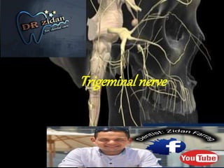
Trigeminal
- 2. Trigeminal nerve: 1-it is one of cranial nerves 2- it is the nerve NO.5 3- consist of three branches 1- ophthalmic 2- maxillary 3 - mandibular 4- it is mixed nerve (sensory and motor)
- 3. Ophthalmic nerve: it is the smallest devision of trigeminal nerve passes forwards in the lateral wall of cavernous sinus and then it divides beforethe superior orbital fissure (SOF) into 3 branches that enter the orbit through the fissure, they are: 1. Lacrimal Nerve: The smallest branch, which enters the orbit through the lateral end of SOF and supplies the lacrimal gland and skin of the lateral 1/3 of upper eyelid. 2. Frontal Nerve: The largest branch which enters the orbit through SOF and passes forwards below the middle of the roof of the orbit and divides into 2 terminal branches; supra-trochlear and supra-orbital branches which supply the skin of the anterior part of the scalp and upper eye lid. 3. Nasociliary Nerve: It enters the orbit through the medial part of SOF. It crosses optic nerve and passes forwards along the medial wall of the orbit to give the following branches: a) Anterior and posterior ethmoidal nerves. b) Long ciliary nerve (to the eyeball). c) Infratrochlear nerve: Reaches the face and supplies the skin of the medial parts of eyelids and root of nose. d) Communicating branch to the ciliary ganglion.
- 4. Ophthalmic nerve (CN V1). (1, gasserian [trigeminal, semilunar] ganglion; 2, ophthalmic nerve; 3, nasociliary nerve; 4, posterior ethmoidal nerve; 5, frontal nerve; 6, lacrimal nerve; 7, anterior ethmoidal nerve; 8, lacrimal gland; 9, supraorbital nerve; 10, supratrochlear nerve; 11, infratrochlear nerve; 12, long ciliary nerves; 13, ciliary ganglion.)
- 5. Maxillary Nerve purely sensory nerve, which is intermediate in position and size between ophthalmic and mandibular nerve Course: it leaves the middle cranial fossa through foramen rotundum to enter the pterygo- palatine fossa enters the orbit through the inferior orbital fissure. It is now called infra-orbital nerve, which passes in the infralorbital groove, infra-orbital canal and finally passes through the infra- orbital foramen to reach the face and terminates by dividing into 3 branches: Palpebral, nasal and labial.
- 7. In Pterygopalatine Fossa, Branches Into 1. Infraorbital nerve. Enters orbit via inferior orbital fissure (IOF). Structures in IOF: infraorbital nerve, zygomatic nerve, infraorbital artery and vein, and inferior ophthalmic vein. Then infraorbital nerve travels under orbital periosteum; then enters and traverses the infraorbital canal and exits via the infraorbital foramen to innervate the midportion of the face. Along its course it gives off • Posterior superior alveolar nerves. To maxillary sinus, molar teeth of maxilla, and adjacent gums and cheek. • Middle superior alveolar nerve. To maxillary premolar teeth. • Anterior superior alveolar nerves. To the maxillary incisor and canine teeth. • Inferior palpebral branches to lower lid skin and conjunctiva, external nasal branches to side of nose, and superior labial branches to upper lip
- 8. Maxillary nerve (CN V2). (1, maxillary nerve; 2, foramen rotundum; 3, infraorbital nerve; 4, zygomaticotemporal nerve; 5, zygomaticofacial nerve; 6, infraorbital foramen; 7, anterior superior alveolar nerves; 8, zygomatic nerve; 9, middle superior alveolar nerve; 10, posterior superior alveolar nerves; 11, ganglionic branches [fine filaments running to the pterygopalatine ganglion]; 12, gasserian [trigeminal, semilunar] ganglion.)
- 9. Sphenopalatine Ganglion: Found in Pterygopalatine fossa. Branches: Orbital Nasal: Short Sphenopalatine. Long Sphenopalatine (Nasopalatine): comes out from the incisive foramen & runs backward, it supplies the mucoperiosteum of the anterior part of the palate as far as the canine. Pharyngeal Palatine: Greater palatine: descends through the greater palatine canal, emerges on the hard palate from the greater palatine foramen and runs forwards in a groove on the inferior surface of the bony palate, it supplies the mucoperiosteum of the posterior part of the palate as far as the canine. Lesser palatine: comes out from the lesser palatine foramen & runs backward, it supplies the mucosa of the soft palate.
- 10. Mandibular Division • Sensory and motor. • Largest of the three divisions. Course: The two roots leave the skull through the foramen oval where they unite together just beyond the foramen forming mandibular nerve trunk, the trunk is one cm long, it divides into anterior division (small and mainly motor) and posterior division (large and mainly sensory). Branches: 1) From the trunk Motor: Nerve to medial pterygoid, it gives 3 branches for 3 muscles: Medial pterygoid muscle. Tensor palati muscle. Tensor tympani muscle. Sensory: Nervus spinosus: It reaches the middle cranial fossa through foramen spinosum to supply its dura mater
- 11. 2) From the anterior division: All its branches are motor except one sensory branch, they are: Motor branches: Two branches for temporalis, called deep temporal nerves. Branch for masseter, passes through mandibular notch. Branch for lateral pterygoid. Sensory branch: Long Buccal nerve: It is the continuation of the anterior division of the mandibular nerve. It emerges between the 2 heads of lateral pterygoid muscle then descends on its lower head to reach and supply the skin covering and the mucosa lining the buccinator muscle.
- 12. 3) From the posterior division: It gives off auriculo-temporal aurcular nerve Temporal nerve Glandular nerve Articular nerve l then divides into lingual nerve Pre ganglion parathympathatic give sub maxillary gland and sub lingual through chordatympani Ganglion parathympathatic To sub mandibular gland Post ganglion parathympathatic Give sub lingual gland Sensory branche two anterior 2/3 of tongue Floor of mouth and lingual aspect of gingiva Taste sensation anterior 2/3 of tongue via chordatympani
- 13. & inferior alveolar nerves It is the largest branch of posterior division of mandibular nervemuscle, posterior to the lingual nerve to enter the mandibular foramen. It runs in the mandibular canal and divides at the mental foramen into two terminal branches; mental and incisive. Prior entering the mandibular canal the mylohyoid nerve (motor) arises from it. The inferior alveolar nerve gives: Dental branches: They arise in the mandibular canal supplying the lower teeth, and alveolar bone. Branches to the canine and incisor teeth arise from incisive nerve while branches to the remaining teeth arise from inferior alveolar nerve itself. Mental nerve: It comes out from the mental foramen to supply the skin of chin and lower lip Motor branch: Nerve to mylohyoid (Motor nerve from posterior division): It arises from the inferior alveolar nerve near the mandibular foramen, runs in the mylohyoid groove supplying the mylohyoid and anterior belly of digastric muscles.
- 14. Mandibular nerve (CN V3). (1, gasserian [trigeminal, semilunar] ganglion; 2, deep temporal nerves; 3, pterygoid nerves; 4, buccal nerve; 5, buccinator muscle; 6, mental foramen; 7, inferior dental branches of inferior alveolar nerve; 8, masseteric nerve; 9, inferior alveolar nerve; 10, lingual nerve; 11, auriculotemporal nerve; 12, recurrent meningeal branch; 13, temporalis muscle; 14, pterygoid muscles; 15, masseter muscle.)
- 16. Thank You Don’t Forget Subscribe , Like And Share