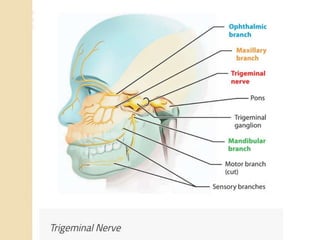
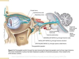
1. Seminar On : Opthalmic
Division Of Trigeminal Nerve
MODERATED BY-
DR. HIMANSHU CHAUHAN
SENIOR LECTURER
(Dept. Of OMFS)
PRESENTED BY-
Dr. MRINALINI MATHUR
3. Types Of Fibers-
a.) Afferent Fibers b.)Efferent Fibers
Afferent Fibers Are-
1- General Somatic Afferent: Carries touch, pain and
temperature from the surface of skin.
2- General Visceral Afferent: Carries touch, pain and
temperature from internal organs, glands and blood vessels.
4. 3- Special Somatic Afferent: Afferent fibers that carry
information regarding vision, hearing and balance. The cranial
nerves having these fibers are optic nerve and vestibulocochlear
nerve.
4- Special Visceral Afferent: These are the fibers associated
with gastro-intestinal tract. They carry special senses of smell
and taste. The cranial nerves having these fibers are
glossopharyngeal nerve and vagus nerve.
5. Efferent Fibers Are-
1- General Somatic Efferent- Provides motor impulse to the
skeletal muscles.
2- General Visceral Efferent- Provides motor innervation to the
smooth muscles, cardiac muscles and glands.
3- Special Visceral Efferent- Provides motor innervation to the
muscles associated with the pharyngeal arches.
7. -The trigeminal nerve, CN V, is the fifth paired cranial nerve.
-It is also the largest cranial nerve.
-It is a mixed nerve.
-It is composed of a small motor root and a considerably larger
sensory root.
INTRODUCTION
8.
9. -During the development of embryo, the pharyngeal arches appear
in the fourth and fifth week.
-The trigeminal nerve is derived from the 1st pharyngeal arch.
EMBRYOLOGY
10. Trigeminal nerve has got 4 nuclei:
1) Mesencephalic nuclei
2) Principal sensory nuclei SENSORY NUCLEUS
3) Spinal nuclei
4) Motor nuclei
NUCLEI OF TRIGEMINAL NERVE
11.
12. 1. Mesencephalic Nucleus
-Situated in midbrain.
-Receives general somatic afferent fibers.
-Relay proprioception from:
muscles of mastication
facial muscles
eye
SENSORY NUCLEI:
13. 2. Principal Sensory Nucleus
- Situated in upper part of pons lateral to motor nucleus.
- Receives general somatic afferent fibers.
- Relays impulse of touch and pressure from skin and mucous
membrane of facial region.
14. 3. The Spinal Nucleus
-It extends from caudal end of principal sensory nucleus in the
pons to 2nd or 3rd spinal segment where it continues with sub.
Gelatinosa.
-It is divided into 3 parts:-
1. Subnucleus oralis
2. Subnucleus interpolaris
3. Subnucleus caudalis
- It receives general somatic afferent fibres.
- Relays the impulses of pain and temperature of face.
15. 4. The Motor Nucleus
- It is situated in upper pons medial to principal sensory nucleus.
`
- Contains efferent fibers.
- Innervates muscles of mastication , tensor tympani ,tensor
palatini, mylohyoid and anterior belly of digastric.
17. -The motor root originates from the motor nucleus within the pons
and medulla oblongata and travel anteriorly to the
trigeminal/gasserian/semilunar ganglion.
- At the ganglion, the motor root passes in a lateral and inferior
direction under the ganglion towards foramen ovale, through which
it leaves the middle cranial fossa along with the third division of
sensory root, the mandibular nerve. So the mandibular nerve has
both sensory and motor fibers.
MOTOR ROOT
18. -The trigeminal ganglion is located in Meckel’s cavity, on the
anterior surface of the petrous portion of the temporal bone.
-Sensory root fibers enter the concave portion of the crescent,
and the three sensory divisions of the trigeminal nerve exit from
the convexity:
1. The opthalmic division (V1) travels anteriorly in the lateral
wall of the cavernous sinus(below the trochlear nerve) to the
medial part of the superior orbital fissure, through which it
exits the skull into the orbit.
SENSORY ROOT
19. 2. The maxillary division (V2) travels anteriorly and downward
to exit the cranium through the foramen rotundum into the upper
portion of the pterygopalatine fossa.
3. The mandibular division(V3) travels almost directly
downward to exit the skull along with the motor root, through the
foramen ovale.
22. - It is the first branch of the trigeminal nerve.
-It is purely sensory and is the smallest of the three divisions.
- It arises from the anteromedial end of the trigeminal ganglion.
Then it passes forward in the lateral wall of cavernous sinus,
below the oculomotor and trochlear nerves.
-It leaves the cranium and enters the orbit through the superior
orbital fissure.
-The nerve trunk is approximately 2.5cm long.
INTRODUCTION
23. -In the middle cranial fossa, the nervus tentori branches from the
opthalmic division to supply the dura.
-The opthalmic division also gives off communication branches
to the oculomotor, trochlear, and abducent cranial nerves.
-Just before the opthalmic nerve passes through the superior
orbital fissure, it divides into three main branches:
lacrimal nerve
frontal nerve
nasociliary nerve
24.
25. LACRIMAL NERVE
It is the smallest of the three
branches.
-It passes into the orbit at the
lateral angle of the superior
orbital fissure.
-It then courses in an anterolateral
direction to reach the lacrimal
gland. Here it supplies sensory
fibers to the gland and adjacent
conjunctiva.
- In the orbit, postganglionic
secretory fibers from the
pterygopalatine ganglion meet
and travel along with lacrimal
nerve. These fibers are from the
zygomatic nerve
27. -It enters the orbit through the superior orbital fissure.
- It is the largest nerve.
-At about middle of the orbit , between the base and apex the
frontal nerve divides into-
supraorbital nerve
supratrochlear nerve
FRONTAL NERVE
28. 1. SUPRAORBITAL NERVE:
- It is the largest branch of
frontal nerve.
- It passes forward between the
levator palpabrae superioris and
the orbital floor and leaves the
orbit through supraorbital
foramen, or notch, to supply the
skin of
- upper eyelid
- forehead
- anterior scalp region to
the vertex of skull
29. 2. SUPRATROCHLEAR NERVE:
-It is the smallest branch of
frontal nerve.
-It passes towards the upper
medial angle of the orbit.
- Then it emerges between
the trochlea and the
supraorbital foramen.
-There it pierces the fascia
of the upper eyelid to supply
the skin of the upper eyelid
and lower medial portion of
the forehead.
30. -It is the third main division of the opthalmic nerve.
-It enters the orbit through the superior orbital fissure.
- It has branches in the nasal cavity and the orbit .
NASOCILIARY NERVE
31. Branches Are-
1. LONG CILIARY NERVE: The long, or sensory, root
arises from the nasociliary nerve. It contains sensory
fibers, which pass through the ganglion without synapsing
and continue on to the eyeball by means of short ciliary
nerves.
32. 2. SENSORY ROOT TO THE CILIARY GANGLION: It is
given by the nasociliary nerve just before crossing the optic
nerve.
3. POSTERIOR ETHMOIDAL NERVE: The posterior ethmoidal
nerve enters the posterior ethmoidal canal to be distributed to the
mucous membrane lining the posterior ethmoidal cells and sphenoid
sinus
33. 4. ANTERIOR ETHMOIDAL NERVE:
The nasociliary nerve
continues anteriorly along the
medial wall of the orbit. In its
course it gives off filaments
that supplies the mucous
membrane of the anterior
ethmoid cells and frontal
sinus.
In the upper part of the nasal
cavity, the ethmoid nerve
divides into two sets of
anterior nasal branches-
internal nasal branch
external nasal branch
34. a) Internal Nasal Branches-
(1) Medial Or Septal
Branches- It travels downwards
to supply sensory innervation
to the mucous membrane of
that area.
(2) Lateral Branches –
These branches supply the
mucous membrane of the
anterior ends of the superior
and middle nasal conchae and
to the anterior lateral nasal
wall.
b) External Nasal Branches- It
supplies the skin over the tip of
nose and the skin over ala of
the nose.
35. 5. INFRATROCHLEAR NERVE -The infratrochlear nerve
extends forward and inferiorly to the trochlea, travels towards the
superior medial angle of the orbit, where it sends its terminal
branches for the innervation of the skin of the medial portion of
the upper eyelid and conjunctiva.
36.
37. - It belongs to the autonomic nervous system, and is functionally
added to the opthalmic nerve.
-It is placed in the orbit, on the lateral side of the optic nerve.
-Like any other autonomic ganglion, it has preganglionic and
postganglionic fibers.
-Preganglionic fibers are sensory, sympathetic and
parasympathetic. These are:
.Sensory fibers from the sensory root of trigeminal nerve.
.Sympathetic fibers originate from the sympathetic internal
carotid plexus and its anastomosis with opthalmic nerve.
.Oculomotor nerve provides the parasympathetic fibers.
CILIARY GANGLION
38. - Postganglionic fibers are the short ciliary nerves . They
penetrate the posterior part of the sclera and enter the eyeball to
which they are sensory.
- Parasympathetic fibers innervate the ciliary muscle and pupil
sphincter muscle, so the pupils are narrow in relaxed
state(domination of the parasympathetic nervous system).
- Sympathetic fibers innervate the pupil dilator muscle , so the
pupils dilate during stress(domination of the sympathetic
nervous system).
Both dilatation and constriction of the pupil are the
mechanisms take part in the eye accomodation.
40. 1. CORNEAL REFLEX-
It is the involuntary blinking of the eyelids stimulated by tactile,
thermal or painful stimulation of the cornea.
In the corneal reflex, the ophthalmic nerve acts as the afferent
limb –detecting the stimuli. The facial nerve is the efferent limb,
causing contraction of the orbicularis oculi muscle.
If the corneal reflex is absent, it is a sign of damage to the
opthalmic nerve or the facial nerve.
CLINICAL ASPECTS
41.
42. 2. HERPES ZOSTER OPTHALMICUS-
- Caused by varicella zoster.
- Predilection for nasociliary branch of opthalmic nerve.
Clinical Features-
Cutaneous Lesions-
Rash
Vesicle
Pustule crust permanent scar
44. Treatment:
- Acyclovir 800mg 5 times/day within 4 days of onset of rash
- Analgesics
-Systemic steroids
-Corneal grafting
45. 3. SUPRAORBITAL NEURALGIA-
The pain of supraorbital neuralgia is characterized as persistent
pain in the supraorbital region and forehead with occasional
sudden, shock like paresthesias in the distribution of the
supraorbital nerves. Occasionally, a patient suffering from
supraorbital neuralgia complains that the hair on the front of the
head “hurts”. Supraorbital nerve block is useful in the diagnosis
and treatment of supraorbital neuralgia.
46. 4. SUPRAORBITAL NERVE BLOCK-
-The supraorbital nerve is blocked by injecting a small amount
of local anesthetic into the supraorbital foramen.
-A 22 to 25 gauge needle is used.
-Foramen is palpated .
-The needle is guided towards the foramen , and once the
periosteum is contacted, the needle is slid slightly medially so
that its tip is abutting the rim of foramen.
-Following negative aspiration, 3 to 4ml of local anesthetic
solution is deposited.
NOTE:- The supraorbital nerve is usually located 2.7cm from
the midline.
47. -The supratrochlear nerve exits the orbit between the trochlea
and the supraorbital foramen.
- Supratrochlear nerve block is performed the same way as
supraorbital nerve block just at a site little more medial to the
insertion site described for supraorbital nerve block.
NOTE- Supratrochlear nerve is located 1.7cm from the midline.
48. 5. ORBITAL APEX SYNDROME-
Orbital apex syndrome involves damage to
Oculomotor nerve(III)
Trochlear nerve(IV)
Abducens nerve(VI)
Opthalmic branch of trigeminal nerve(V1)
Optic nerve(II)
Clinical Presentation-
Vision Loss Ptosis
Monocular Diplopia Ocular Deviation
Opthalmoplegia Headache
Periorbital Pain
Proptosis
49. 6. SUPERIOR ORBITAL FISSURE SYNDROME-
Applies to lesions located immediately anterior to the orbital
apex, including the structures exiting the annulus of Zinn and
often those external to the annulus as multiple cranial nerve
palsies maybe seen in the absence of optic nerve pathology.
Clinical Presentation-
Opthalmoplegia
Upper Eyelid Ptosis
Nonreactive Dilated Pupil
Anesthesia Over The Ipsilateral Forehead
Loss Of Corneal Reflex
Orbital Pain
Proptosis
50. 7. CAVERNOUS SINUS SYNDROME-
- It involves cranial nerves III, IV, V1,V2 and V1
Clinical Presentation-
Sensory Deficits Of The Nerves Involved
Vascular Congestion
Proptosis
Chemosis
Opthalmoplegia
Elevated Intraocular Pressure
51. REFERENCES-
1- BD Chaurasia, Human Anatomy
2- Stanley F. Malamed, Handbook Of Local Anesthesia
3- Monheim’s, Local Anesthesia And Pain Control In Dental
Practice
4- Vishram Singh, Textbook Of Anatomy