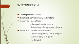
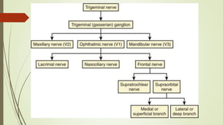
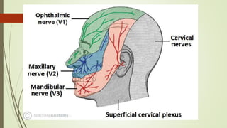
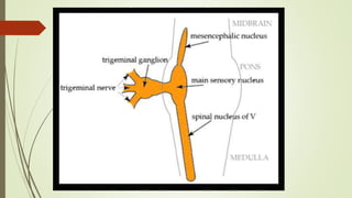
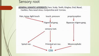
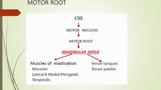
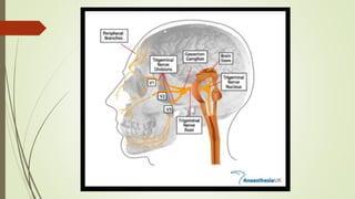
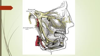
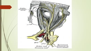
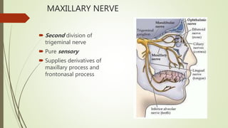
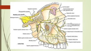
THE TRIGEMINAL NERVE
BY Dr A K PANDEY
RESIDENT OPHTHAL
INTRODUCTION
 The largest cranial nerve
 It is mixed nerve ( sensory and motor )
 Sensory to – Skin of face
-Mucosa of cranial viscera
-Except base of tongue and pharynx
 Motor to – Muscles of Mastication
-Tensor veli palatini, Tensor tympani
-Anterior belly of digastric
-Mylohyoid
TRIGEMINAL NUCLEI
A cranial nerve nucleus is a collection of neurons
(gray matter) in the brain stem that is associated
with one or more cranial nerves.
Axons carrying information to and from the cranial
nerves form a synapse first at these nuclei.
Lesions occurring at these nuclei can lead to effects
resembling those seen by the severing of nerve(s)
they are associated with.
Sensory Nuclei – mesencephalic nucleus in
midbrain
- primary sensory nucleus in pons
- spinal nucleus in medulla and
spinal cord
Motor nucleus - one motor nucleus in pons
SENSORY NUCLEI
MESENCEPHALIC
NUCLEUS
 Cell body of
Pseudounipolar neuron
 Relay proprioception from
muscles of mastication,
Extra ocular Muscles, Facial
muscles.
 Situated in Midbrain just
latetral to Aqueduct
Principal sensory
nucleus
Lies in Pons lateral to
Motor nucleus
Relays touch sensation
Spinal nucleus  Extends from caudal end of
principal sensory Nucles in
pons to 2nd or 3rd spinal
segment
 It relays Pain and
Temperature
MOTOR NUCLEUS
Innervates muscles of mastication and tensor
tympani and tensor palatini
Derived from first branchial arch.
Located in pons medial to principle sensory nucleus.
FUNCTIONAL COMPONENT
Sensory root
Motor root
Sensory root
MOTOR ROOT
COURSE AND DISTRIBUTION
Both motor and sensory root are attached vertically
to junction of pons and middle cerebellar peduncle
with motor root lying ventromedially to sensory root
Pass anteriorly in middle cranial fossa to lie below
tentorium cerebelli in cavum trigeminale, here motor
root lies inferior to sensory root
Sensory root connected to posteromedial concave
border of the trigeminal ganglion
Convex anterolateral margin of the ganglion gives
attachment to the 3 div of trigeminal nerve.
Motor root turns further inferior with sensory
component of V3 to emerge out of foramen Ovale as
Mandibular nerve.
Ophthalmic and Maxillary division emerges through
Superior orbital fissure and foramen Rotundum
respectively.
TRIGEMINAL GANGLION
 SEMILUNAR OR GASSERIAN GANGLION.
 Cresentric in shape with convexity anterolaterally.
 Contains cell bodies of pseudounipolar neurons.
 LOCATION: lies in a bony fossa at apex of the petrous
temporal bone on floor of middle cranial fossa, just lateral to
posterior part of lateral wall of the cavernous sinus.
 COVERINGS: covered by dural pouch = MECKLES CAVE or
CAVUM TRIGEMINALE. cave lined by pia and arachnoid thus
the ganglion is bathed in CSF.
 ARTERIAL SUPPLY: Ganglionic branches of Internal Carotid
Artery, middle meningeal artery and accessory meningeal
artery.
RELATIONS
 SUPERIORLY: uncus and temporal lobe
 INFERIORLY: *motor root *greater petrosal nerve *petrous
apex *foramen lacerum
 MEDIALLY: *posterior part of lateral wall of cavernous sinus
*Internal Carotid Artery with its sympathetic plexus
 LATERALLY: *middle meningeal artery and vein *nervous
spinosum
DIVISION OF TRIGEMINAL NERVE
1. Ophthalmic nerve
2. Maxillary nerve
3. Mandibular nerve
OPHTHALMIC NERVE
Smallest division.
Sensory only
Supplies : eyeballs, conjunctiva, lacrimal gland,
mucosa of nose and paranasal sinus, skin of forehead
eyelid and nose
COURSE
BRANCHES OF OPHTHALMIC NERVE
 Lacrimal nerve
 Frontal nerve
 Nasociliary nerve
LACRIMAL NERVE
Smallest
Passes into orbit through lateral compartment of the
Superior orbital fissure outside the tendinous ring.
Receives communicating branch from Trochlear
nerve
Receives branch from Zygomaticotemporal nerve
(secretomotor), branch of maxillary
Sensory to lateral conjunctiva, Upper Lid, lacrimal
gland
Post synaptic parasympathetic fibers from
pterygopalatine ganglion to lacrimal gland i.e
secretomotor
FRONTAL NERVE
 Largest
 Enters orbit through lateral part of superior orbital fissure
outside tendinous ring
 Passes forward between roof of orbit and Levator Palpebral
Superioris
 Divides midway into :
1. Supraorbital Nerve
2. Supratrochlear Nerve
SUPRATROCHLEAR NERVE
 Smaller nerve
 Medial branch
 Receives communication
branches from infratrochlear
nerve
 Curves around superomedial
margin of orbit
SUPRAORBITAL NERVE
 Larger nerve
 lateral branch
 Passes through supraorbital
notch
 Divides in medial and lateral
branches
 supplies: median
conjunctiva, Upper Lid and
lower part of forehead
 Lies between frontalis and
corrugator supercilliary
muscles
 Lies beneath frontalis
muscle
 Supplies: conjunctiva, scalp
upto vertex , mucous
membrane of frontal sinus
NASOCILLIARY NERVE
 Purely Sensory
 Passes through middle part of superior
orbital fissure within the tendenious
ring .
 Runs along medial wall of orbit
between Superior Oblique and Medial
Rectus
 Divides into Anterior Ethmoidal and
External Nasal
 5 branches in orbit.
 Short Clliary Nerves: Fibers reaches eyeball and also
contains fibers from Cilliary Ganglion
 Long Cilliary Nerves : 2 or 3 in no. supply to Iris and Cornea.
 Post Ethmoidal Nerve: passes through posterior ethmoidal
foramen to supply the Ethmoid and Sphenoid PNS.
 Infratrochlear Nerve: appears on face above medial angle of
the eye. Supplies to skin of lacrimal sac and caruncle
MAXILLARY NERVE
 Second division of
trigeminal nerve
 Pure sensory
 Supplies derivatives of
maxillary process and
frontonasal process
COURSE
After leaving foramen rotundum it moves anteriorly
in the uppermost part of pterygopalatine fossa.
As it passes through pterygopalatine fossa it also
gives branches to sphenopalatine ganglion, posterior
superior alveolar nerve and zygomatic branches
It then moves laterally and moves in a groove on
posterior surface of maxilla.
Then enters orbit through infra orbital fissure and
moves through infra orbital groove where it is called
as Infraorbital nerve and emerges on face from infra
orbital foramen
BRANCHES
 Meningeal branch – travels along the MMA and provides sensory innervation
to cranial dura matter
 IN PTERIGOPALATINE FOSSA:
1. Ganglionic branches-
Arises as 2 trunks. Trunks join to form single root within pterygopalatine
ganglion.
Gives Orital branches, Palatine branches, Pharyngeal branches,
Nasal branches
Gives postganglionic secretomotor fibers to lacrimal gland via
zygomaticotemporal and lacrimal
2.Orbital branch: Supplies periosteum of orbit
3.Nasal branch: Supplies to mucosa of superior and inferior conchae,
posterior ethmiodal sinus and posterior portion of nasal septum. It also
includes Nasopalatine branch.
-It passes across roof of nasal cavity downwards and forwards lying
between mucosa and periosteum of nasal septum.
-Reaches to floor of nasal cavity and give branch to anterior part of
nasal septum and floor of nasal cavity.
-Enters Incisor canal and enters oral cavity through insicisor foramen
-It provides sensation to palatal mucosa of premaxilla region.
4. Palatine branch: Arise as greater palatine (anterior) and
lesser palatine (middle and posterior)
-Greater palatine nerve descends through pterygopalatine
canal from the ganglion and emerges from greater palatine
foramen of hard palate.
-Then moves anteriorly between mucoperiostem and hard
palate upto 1st premolar supplying sensory innervation to
palatal soft tissue and bone.
-Middle palatine and posterior palatine emerges from lesser
palatine foramen and supply soft palate and tonsilar region
respectively
5. Pharyngeal branch: It leaves the posterior part
of pterygopalatine ganglion and passes through the
pharyngeal canal
It is distributed to the mucous membrane of the
nasal part of pharynx, posterior to eustachian
tube
6. POSTERIOR SUPERIOR ALVEOLAR NERVE
 It arises from the main trunk of maxillary nerve in the
petrygopalatine fossa just before the nerve enters the inferior
orbital canal
 Usually arises as 2 trunks.
 Passes downwards and crosses the pterygoplatine fossa reaching
infratemporal surface of maxilla.
 1st trunk continues downwards on posterior surface of maxilla and
provide sensory innervation to buccal gingiva in maxillary molar
region and adjacent facial mucosal surface
2nd trunk enters maxilla through PSA canal to travel
to posterolateral wall of maxillary sinus providing
sensory innervation to sinus mucosa. Continuing
downwards this also provides sensory innervation to
alveoli, pulp of molar tooth.
Branches In Infraorbital canal
Middle superior alveolar nerve- runs along the lateral
wall of maxilla and participate in superior dental
plexus. Supplies premolars
Anterior superior alveolar nerve – runs in canal in
anterior wall of maxilla and gives dental and nasal
branches
Facial branches – palpebral , nasal and superior labial
nerve
MANDIBULAR NERVE
Formed by union of two trunks, the large sensory
trunk and small motor trunk
These two trunks pass through the foramen ovale
and unite to form one trunk
Motor root supplies the muscles of mastication, the
tensor tympani, the tensor veli palatine, mylohyoid,
anterior belly of digastric
APPLIED ANATOMY
Trigeminal neuralgia – sudden, usually unilateral
severe, brief stabbing, lancinating, recurring pain in
the distribution of one or more branches of the 5th
nerve
Trigeminal neuropathy- sensory loss of face or
weakness of the jaw muscles, caused due to Sjogren
syndrome, herpes zoster, leprosy, meningioma and
schwanomma
HZO – recurrent infection of ophthalmic div of
trigeminal dermatome most frequently affecting
nasociliary nerve
 Cavernous sinus syndrome- multiple cranial neuropathies,
exophthalmos, ocular motor defects, sensory loss in V1 and/or
V2 . Pupils may be spared
 Gradenigos syndrome- petrous bone osteitis due to otitis
media. Characterized by ipsilateral trigeminal nerve palsy,
retro orbital pain and ipsilateral sixth nerve palsy
 Neuroparalytic keratitis – due to involvement of trigeminal
nerve or section of its sensory root for neuralgia
Trigeminal nerve

Trigeminal nerve

  • 1.
    THE TRIGEMINAL NERVE BYDr A K PANDEY RESIDENT OPHTHAL
  • 2.
    INTRODUCTION  The largestcranial nerve  It is mixed nerve ( sensory and motor )  Sensory to – Skin of face -Mucosa of cranial viscera -Except base of tongue and pharynx  Motor to – Muscles of Mastication -Tensor veli palatini, Tensor tympani -Anterior belly of digastric -Mylohyoid
  • 5.
    TRIGEMINAL NUCLEI A cranialnerve nucleus is a collection of neurons (gray matter) in the brain stem that is associated with one or more cranial nerves. Axons carrying information to and from the cranial nerves form a synapse first at these nuclei. Lesions occurring at these nuclei can lead to effects resembling those seen by the severing of nerve(s) they are associated with.
  • 6.
    Sensory Nuclei –mesencephalic nucleus in midbrain - primary sensory nucleus in pons - spinal nucleus in medulla and spinal cord Motor nucleus - one motor nucleus in pons
  • 8.
    SENSORY NUCLEI MESENCEPHALIC NUCLEUS  Cellbody of Pseudounipolar neuron  Relay proprioception from muscles of mastication, Extra ocular Muscles, Facial muscles.  Situated in Midbrain just latetral to Aqueduct
  • 9.
    Principal sensory nucleus Lies inPons lateral to Motor nucleus Relays touch sensation
  • 10.
    Spinal nucleus Extends from caudal end of principal sensory Nucles in pons to 2nd or 3rd spinal segment  It relays Pain and Temperature
  • 11.
    MOTOR NUCLEUS Innervates musclesof mastication and tensor tympani and tensor palatini Derived from first branchial arch. Located in pons medial to principle sensory nucleus.
  • 13.
  • 14.
  • 15.
  • 17.
    COURSE AND DISTRIBUTION Bothmotor and sensory root are attached vertically to junction of pons and middle cerebellar peduncle with motor root lying ventromedially to sensory root Pass anteriorly in middle cranial fossa to lie below tentorium cerebelli in cavum trigeminale, here motor root lies inferior to sensory root
  • 18.
    Sensory root connectedto posteromedial concave border of the trigeminal ganglion Convex anterolateral margin of the ganglion gives attachment to the 3 div of trigeminal nerve.
  • 19.
    Motor root turnsfurther inferior with sensory component of V3 to emerge out of foramen Ovale as Mandibular nerve. Ophthalmic and Maxillary division emerges through Superior orbital fissure and foramen Rotundum respectively.
  • 21.
    TRIGEMINAL GANGLION  SEMILUNAROR GASSERIAN GANGLION.  Cresentric in shape with convexity anterolaterally.  Contains cell bodies of pseudounipolar neurons.  LOCATION: lies in a bony fossa at apex of the petrous temporal bone on floor of middle cranial fossa, just lateral to posterior part of lateral wall of the cavernous sinus.
  • 22.
     COVERINGS: coveredby dural pouch = MECKLES CAVE or CAVUM TRIGEMINALE. cave lined by pia and arachnoid thus the ganglion is bathed in CSF.  ARTERIAL SUPPLY: Ganglionic branches of Internal Carotid Artery, middle meningeal artery and accessory meningeal artery.
  • 24.
    RELATIONS  SUPERIORLY: uncusand temporal lobe  INFERIORLY: *motor root *greater petrosal nerve *petrous apex *foramen lacerum  MEDIALLY: *posterior part of lateral wall of cavernous sinus *Internal Carotid Artery with its sympathetic plexus  LATERALLY: *middle meningeal artery and vein *nervous spinosum
  • 26.
    DIVISION OF TRIGEMINALNERVE 1. Ophthalmic nerve 2. Maxillary nerve 3. Mandibular nerve
  • 28.
    OPHTHALMIC NERVE Smallest division. Sensoryonly Supplies : eyeballs, conjunctiva, lacrimal gland, mucosa of nose and paranasal sinus, skin of forehead eyelid and nose
  • 29.
  • 31.
    BRANCHES OF OPHTHALMICNERVE  Lacrimal nerve  Frontal nerve  Nasociliary nerve
  • 33.
    LACRIMAL NERVE Smallest Passes intoorbit through lateral compartment of the Superior orbital fissure outside the tendinous ring. Receives communicating branch from Trochlear nerve
  • 34.
    Receives branch fromZygomaticotemporal nerve (secretomotor), branch of maxillary Sensory to lateral conjunctiva, Upper Lid, lacrimal gland Post synaptic parasympathetic fibers from pterygopalatine ganglion to lacrimal gland i.e secretomotor
  • 36.
    FRONTAL NERVE  Largest Enters orbit through lateral part of superior orbital fissure outside tendinous ring  Passes forward between roof of orbit and Levator Palpebral Superioris  Divides midway into : 1. Supraorbital Nerve 2. Supratrochlear Nerve
  • 38.
    SUPRATROCHLEAR NERVE  Smallernerve  Medial branch  Receives communication branches from infratrochlear nerve  Curves around superomedial margin of orbit SUPRAORBITAL NERVE  Larger nerve  lateral branch  Passes through supraorbital notch  Divides in medial and lateral branches
  • 39.
     supplies: median conjunctiva,Upper Lid and lower part of forehead  Lies between frontalis and corrugator supercilliary muscles  Lies beneath frontalis muscle  Supplies: conjunctiva, scalp upto vertex , mucous membrane of frontal sinus
  • 40.
    NASOCILLIARY NERVE  PurelySensory  Passes through middle part of superior orbital fissure within the tendenious ring .  Runs along medial wall of orbit between Superior Oblique and Medial Rectus  Divides into Anterior Ethmoidal and External Nasal  5 branches in orbit.
  • 41.
     Short ClliaryNerves: Fibers reaches eyeball and also contains fibers from Cilliary Ganglion  Long Cilliary Nerves : 2 or 3 in no. supply to Iris and Cornea.  Post Ethmoidal Nerve: passes through posterior ethmoidal foramen to supply the Ethmoid and Sphenoid PNS.  Infratrochlear Nerve: appears on face above medial angle of the eye. Supplies to skin of lacrimal sac and caruncle
  • 44.
    MAXILLARY NERVE  Seconddivision of trigeminal nerve  Pure sensory  Supplies derivatives of maxillary process and frontonasal process
  • 45.
  • 46.
    After leaving foramenrotundum it moves anteriorly in the uppermost part of pterygopalatine fossa. As it passes through pterygopalatine fossa it also gives branches to sphenopalatine ganglion, posterior superior alveolar nerve and zygomatic branches
  • 47.
    It then moveslaterally and moves in a groove on posterior surface of maxilla. Then enters orbit through infra orbital fissure and moves through infra orbital groove where it is called as Infraorbital nerve and emerges on face from infra orbital foramen
  • 49.
    BRANCHES  Meningeal branch– travels along the MMA and provides sensory innervation to cranial dura matter  IN PTERIGOPALATINE FOSSA: 1. Ganglionic branches- Arises as 2 trunks. Trunks join to form single root within pterygopalatine ganglion. Gives Orital branches, Palatine branches, Pharyngeal branches, Nasal branches Gives postganglionic secretomotor fibers to lacrimal gland via zygomaticotemporal and lacrimal
  • 50.
    2.Orbital branch: Suppliesperiosteum of orbit 3.Nasal branch: Supplies to mucosa of superior and inferior conchae, posterior ethmiodal sinus and posterior portion of nasal septum. It also includes Nasopalatine branch. -It passes across roof of nasal cavity downwards and forwards lying between mucosa and periosteum of nasal septum. -Reaches to floor of nasal cavity and give branch to anterior part of nasal septum and floor of nasal cavity. -Enters Incisor canal and enters oral cavity through insicisor foramen -It provides sensation to palatal mucosa of premaxilla region.
  • 51.
    4. Palatine branch:Arise as greater palatine (anterior) and lesser palatine (middle and posterior) -Greater palatine nerve descends through pterygopalatine canal from the ganglion and emerges from greater palatine foramen of hard palate. -Then moves anteriorly between mucoperiostem and hard palate upto 1st premolar supplying sensory innervation to palatal soft tissue and bone. -Middle palatine and posterior palatine emerges from lesser palatine foramen and supply soft palate and tonsilar region respectively
  • 52.
    5. Pharyngeal branch:It leaves the posterior part of pterygopalatine ganglion and passes through the pharyngeal canal It is distributed to the mucous membrane of the nasal part of pharynx, posterior to eustachian tube
  • 53.
    6. POSTERIOR SUPERIORALVEOLAR NERVE  It arises from the main trunk of maxillary nerve in the petrygopalatine fossa just before the nerve enters the inferior orbital canal  Usually arises as 2 trunks.  Passes downwards and crosses the pterygoplatine fossa reaching infratemporal surface of maxilla.  1st trunk continues downwards on posterior surface of maxilla and provide sensory innervation to buccal gingiva in maxillary molar region and adjacent facial mucosal surface
  • 54.
    2nd trunk entersmaxilla through PSA canal to travel to posterolateral wall of maxillary sinus providing sensory innervation to sinus mucosa. Continuing downwards this also provides sensory innervation to alveoli, pulp of molar tooth.
  • 56.
    Branches In Infraorbitalcanal Middle superior alveolar nerve- runs along the lateral wall of maxilla and participate in superior dental plexus. Supplies premolars Anterior superior alveolar nerve – runs in canal in anterior wall of maxilla and gives dental and nasal branches Facial branches – palpebral , nasal and superior labial nerve
  • 58.
    MANDIBULAR NERVE Formed byunion of two trunks, the large sensory trunk and small motor trunk These two trunks pass through the foramen ovale and unite to form one trunk Motor root supplies the muscles of mastication, the tensor tympani, the tensor veli palatine, mylohyoid, anterior belly of digastric
  • 59.
    APPLIED ANATOMY Trigeminal neuralgia– sudden, usually unilateral severe, brief stabbing, lancinating, recurring pain in the distribution of one or more branches of the 5th nerve Trigeminal neuropathy- sensory loss of face or weakness of the jaw muscles, caused due to Sjogren syndrome, herpes zoster, leprosy, meningioma and schwanomma HZO – recurrent infection of ophthalmic div of trigeminal dermatome most frequently affecting nasociliary nerve
  • 61.
     Cavernous sinussyndrome- multiple cranial neuropathies, exophthalmos, ocular motor defects, sensory loss in V1 and/or V2 . Pupils may be spared  Gradenigos syndrome- petrous bone osteitis due to otitis media. Characterized by ipsilateral trigeminal nerve palsy, retro orbital pain and ipsilateral sixth nerve palsy  Neuroparalytic keratitis – due to involvement of trigeminal nerve or section of its sensory root for neuralgia