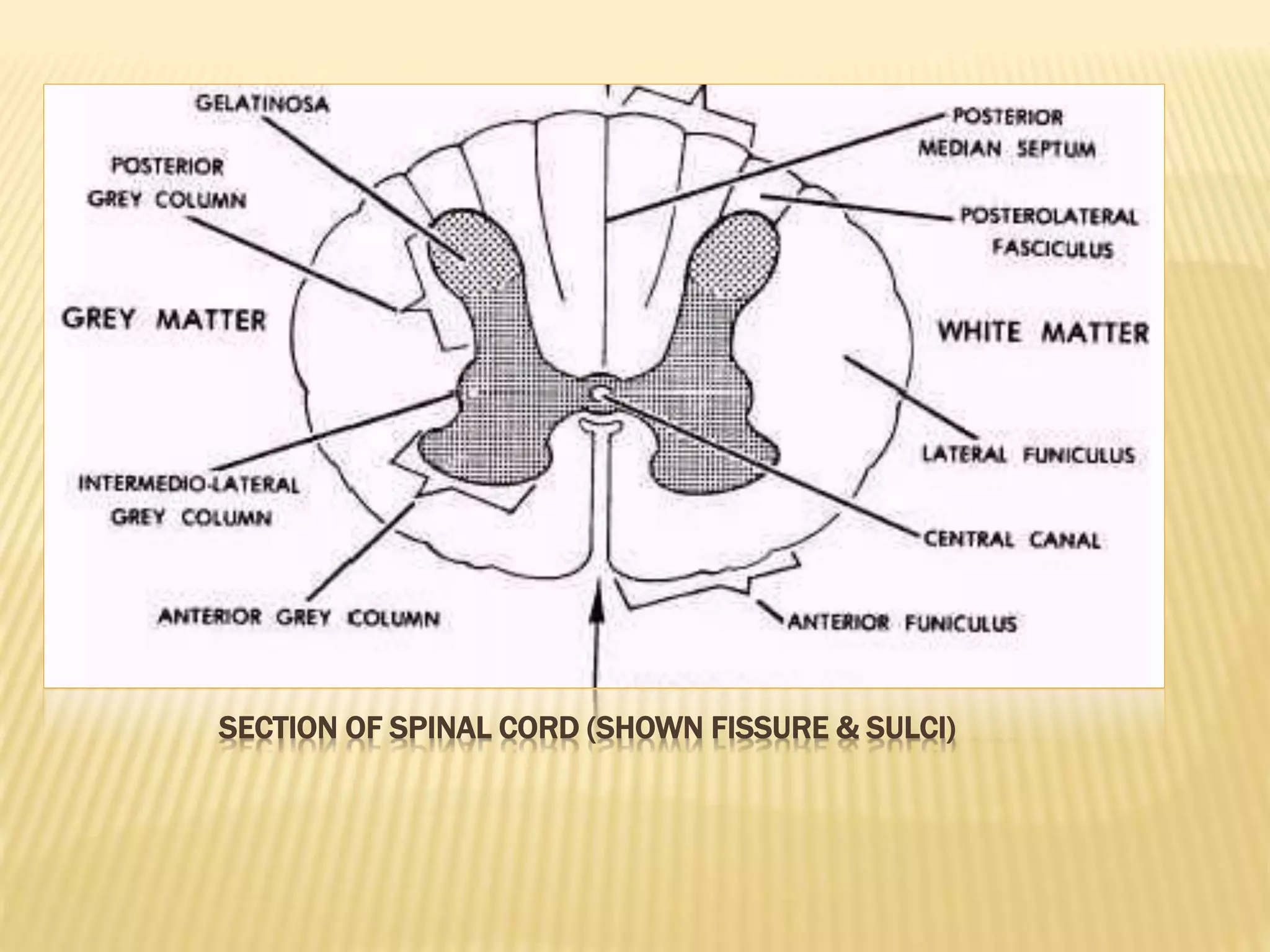
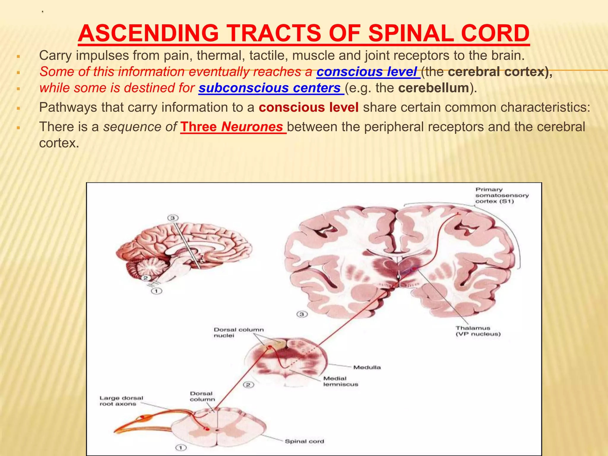
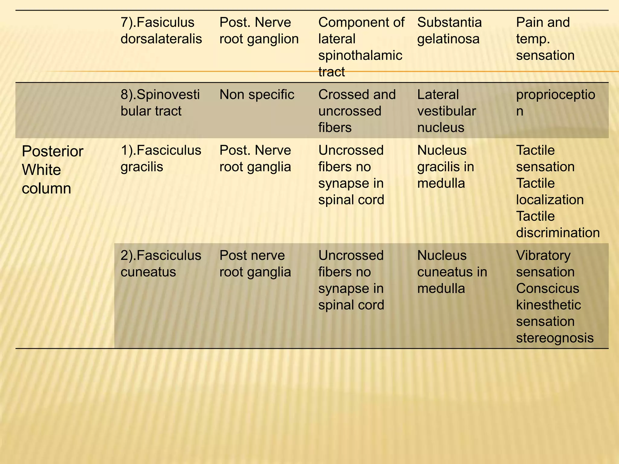
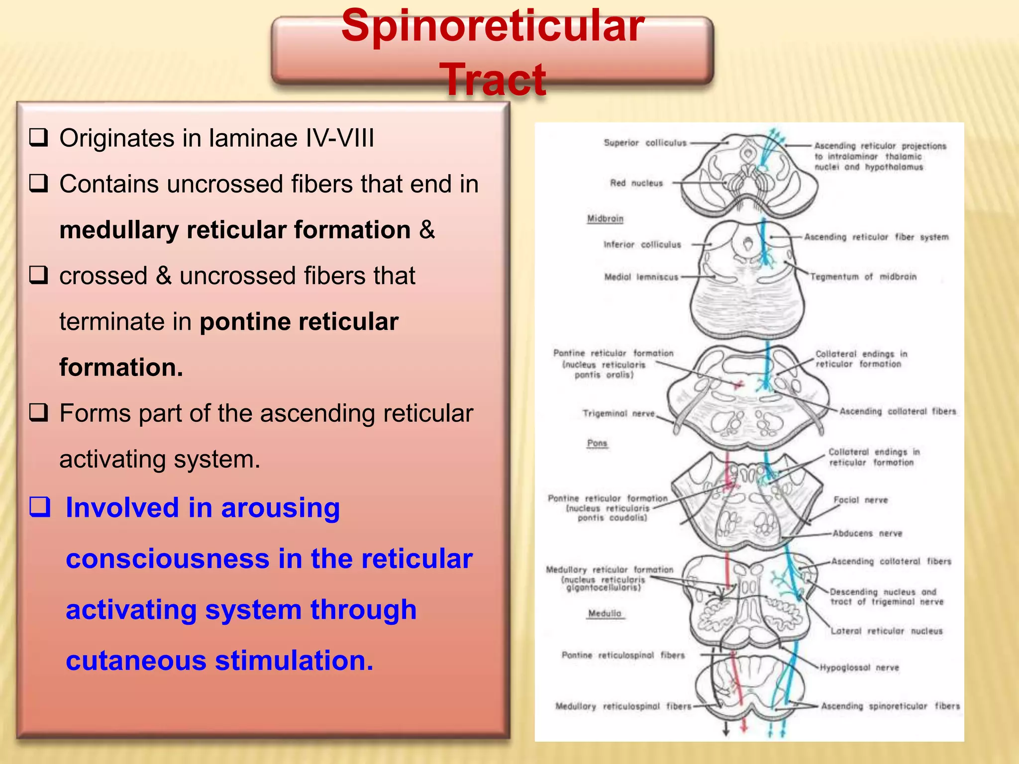
ASCENDING TRACTS
(SENSORY PATHWAY)
AKASH NAYAK
B.P.T 2014
.
CONTENTS:-
 SPINAL CORD IN GENERAL
GRAY MATTER &
WHITE MATTER
 TRACTS IN SPINAL CORD
ASCENDING TRACTS OF SPINAL CORD
(SITUATON, ORIGIN, COURSE, FUNCTION & EFFECTS OF
LESION )
RELATED TERMINOLOGY:-
 White matter
 Grey matter
 Ganglia
 commissures
Anterior(ventral)
Posterior (dorsal)
SPINAL CORD
The spinal cord is a long, thin, tubular
structure made up of neural tissue, that extends
from the medulla oblongata in the brainstem to the
lumbar region of the vertebral column. It carry
information periphery to brain and brain to periphery.
Situati
on &
extent
Coveri
ng
Shape
&
Length
enlarg
ement
s
Segme
nts
Spinal
nerves
Fissure
&
sulci
From
foreman
magnu
m to
lower
border
of Ist
lumbar
vertebra
e
Dura
matter
Pia
matter
Arachno
id
matter
Cylindric
al in
shape
45 cm
(male)
43 cm
(female)
Cervical
Lumbar
enlarge
ments
31
segment
s
Cervical
=8
Thoracic
12
Lumbar
5
Sacral 5
Coccyg
eal 1
Total=
31
31 pairs
spinal
nerves
Cervical
=8
Thoracic
12
Lumbar
5
Sacral 5
Coccyg
eal 1
Total=31
pairs
Ant. Median
fissure
Antrolateral
sulcus
Post. Median
S.
post. Median
septum
Post.
Intermediate
sulcus.
Post.
Intermediate
septum
Posterolateral
sulcus
SECTION OF SPINAL CORD (SHOWN FISSURE & SULCI)
.
INTERNAL STRUCTURE OF SPINAL CORD
 Grey matter (inner)
 White matter (outer)
GREY MATTER OF SPINAL CORD:-
 it is a collection of nerve cell bodies, dendrites and part of axon.
 it resembles as ‘wings of butterfly’ or ‘H’ shape.
WHITE MATTER OF SPINAL CORD:-
 It surrounds the grey matter. It formed by the bundles of both myelinated
and unmyelinated fibers.
 The White matter of the spinal cord consists of
 Ascending and Descending tracts.
 It is divided into Dorsal, Lateral & Ventral Columns or Funiculi.
.
TRACTS OF SPINAL CORD
They are classified into:
1).Short tracts:
a. Association or intrinsic tracts:- These tracts connect adjacent segments of spinal
cord on the same side.
b. commissural tracts:- these tracts also connect opposite halves of same segment of
spinal cord.
2). Long tracts: (projection tracts)
connect the spinal cord with other parts of CNS.
a. Ascending tracts (sensory impulses)
b. Descending tracts (motor impilses)
.
ASCENDING TRACTS OF SPINAL CORD
 Carry impulses from pain, thermal, tactile, muscle and joint receptors to the brain.
 Some of this information eventually reaches a conscious level (the cerebral cortex),
 while some is destined for subconscious centers (e.g. the cerebellum).
 Pathways that carry information to a conscious level share certain common characteristics:
 There is a sequence of Three Neurones between the peripheral receptors and the cerebral
cortex.
.
The first-order neurone or primary
afferent neurone) enters the spinal
cord through the dorsal root of a
spinal nerve and its cell body lies in
the dorsal root ganglion.
The main fiber remains on the
ipsilateral side of the cord and
terminates in synaptic contact
with the second neurone which
lies either in the spinal grey
matter or in the medulla
oblongata of the brain stem.
.
•The axon of the
second order neurone
crosses over
(decussates) to the
opposite side of the CNS
and ascends to the
thalamus, where it
terminates.
•The third-order
neurone has its cell
body in the thalamus.
•Its axon passes to the
somatosensory cortex of the
parietal lobe of the
cerebral hemisphere.
 Three major pathways carry sensory information:
• Posterior white column (Gracile & Cuneate fasciculi)
• Lateral white column (Spinothalamic)
• Anterior white column
situation Tract Origin Course Terminatio
n
Function
Anterior White
column
1).Ant.
Spinothalamic
tract
Chief
sensory
nucleus
Crossing in
spinal cord
forms spinal
lemniscus
Ventral
posaterolater
al nucleus of
thalamus
Crude touch
sensation
Lateral
White
Column
1).Lateral
spinothalamic
tract
Substantia
gelatinosa
“ “ “ “ Pain and
temp.
senstation
2).Ventral
spinocerebell
ar tract
Marginal
nucleus
Crossing in
spinal cord
Ant. Lobe of
cerebellum
Subconscious
kinesthetic
sensations
3).Dorsal
spinocerebell
ar tract
Clarke
neucleus
Uncrossed
fibers
Ant. Lobe of
cerebellum
Subconscious
kinesthetic
sensations
4).Spinotectal
tract
Chief sensory
nucleus
Crossing in
spinal cord
Supior
colliculus
Spinovisual
reflex
5).Spinorectic
ular tract
Intermediolate
ral cells
Crossed and
uncrossed
fibers
Reticular
formation of
brainstem
Cnsciousness
and
awareness
6).Spino
olivary tract
Non specific Crossed and
uncrossed
fibers
Olivary
nucleus
Proprioceptio
n
7).Fasiculus
dorsalateralis
Post. Nerve
root ganglion
Component of
lateral
spinothalamic
tract
Substantia
gelatinosa
Pain and
temp.
sensation
8).Spinovesti
bular tract
Non specific Crossed and
uncrossed
fibers
Lateral
vestibular
nucleus
proprioceptio
n
Posterior
White
column
1).Fasciculus
gracilis
Post. Nerve
root ganglia
Uncrossed
fibers no
synapse in
spinal cord
Nucleus
gracilis in
medulla
Tactile
sensation
Tactile
localization
Tactile
discrimination
2).Fasciculus
cuneatus
Post nerve
root ganglia
Uncrossed
fibers no
synapse in
spinal cord
Nucleus
cuneatus in
medulla
Vibratory
sensation
Conscicus
kinesthetic
sensation
stereognosis
 Posterior Column:
 Contains two tracts;
Fasciculus Gracilis (Tracts of
Goll) & Fasciculus
Cuneatus (Tract of Burdach)
 Carry impulses concerned with
proprioception and
discriminative touch
from ipsilateral side of the body
 Contain the axons of primary
afferent neurons that have entered
cord through dorsal roots of spinal
nerves
 FG contains fibers received at
sacral, lumbar and lower thoracic
levels,
 FC contains fibers received at
upper thoracic and cervical levels
• Fibers ascend without interruption
where they terminate upon 2nd
order neurons in nucleus gracilis
and nucleus cuneatus
• The axons of the 2nd order neurons
decussate in the medulla as internal
arcuate fibers and ascend through
the brain stem as medial
lemniscus.
• The medial lemniscus terminates in
the ventral posterior nucleus of
the thalamus (3rd order neurons),
which project to the somatosensory
cortex (thalamocortical fibers)
 ,
• Located lateral and ventral to
the ventral horn.
• Carry impulses concerned
with; pain and thermal
sensations (Lateral tract)
and non- discriminative
touch and pressure
(Anterior tract).
• In brain stem, constitute the
spinal lemniscus.
• Information is sent to the
primary sensory cortex on
the opposite side of the
body.
SPINOTHALAMIC TRACT
Function:
•Carries pain & Temperature to
thalamus and sensory area of the
cerebral cortex.
Neurones: 3 Neurones
• Neurone I: Small cells in the dorsal
root ganglia.
•Neurone II: Cells of substantia
gelatinosa of Rolandi in the
posterior horn.
•Neurone III: Cells of (VP) nucleus of
the thalamus.
•The spinothalamic tract contains
second-order neurones, the cell
bodies of which lie in the contralateral
dorsal horn
Lateral Spinothalamic Tract
Function:
•Carries crude touch & pressure to
thalamus and sensory cortex.
Neurones: 3 Neurones
•Neurone I:
Medium sized cells in the
dorsal root ganglia.
•Neurone II:
Cells of main sensory nucleus
or
(nucleus proprius).
•Neurone III:
Cells of VP nucleus of
thalamus.
 Effect of lesion:
Loss of crude touch sensation
below the level of the lesion.
Anterior Spinothalamic Tract
Spinocerebellar Tracts
 The spinocerebellar system
consists of a sequence of only
two neurons;
 Neurone I:Large cells of dorsal
root ganglia.
 Neurone II: cells of the nucleus
dorsalis (Clark's nucleus.
 Two tracts: Dorsal &Ventral
 Located near the dorsolateral and
ventrolateral surfaces of the cord
 Contain axons of the second order
neurons
 Carry information derived from
muscle spindles, Golgi tendon
organs and tectile receptors to the
cerebellum
 for the control of posture and
coordination of movements.
Posterior Spinocerebellar
Tract
 Present only above level L3
 The cell bodies of 2nd order neuron
lie in Clark’s column
 Axons of 2nd order neuron terminate
ipsilaterally (uncrossed) in the
cerebellar cortex by entering through
the inferior cerebellar peduncle.
 Posterior spinocerebellar tract
convey sensory information to the
same side of the cerebellum
Ventral (Anterior)Spinocerebellar
Tract
 The cell bodies of 2nd order neuron
lie in base of the dorsal horn of the
lumbosacral segments
 Axons of 2nd order neuron cross to
opposite side, ascend as far as the
midbrain, and then make a sharp turn
caudally and enter the superior
cerebellar peduncle
 The fibers cross the midline for a
second time within the cerebellum
before terminating in the cerebellar
cortex
 Ventral spinocerebellar tract convey
sensory information to the same side
of the cerebellum
Spinotectal Tract
 Ascends in the anterolateral part, in
close association with spinothalamic
system.
 Primary afferents reach dorsal horn
through dorsal roots and terminate on
2nd order neurons
 The cell bodies of 2nd order neuron
lie in base of the dorsal horn.
 Axons of 2nd order neuron cross to
opposite side, and project to the
periaquiductal gray matter and
superior colliculus in the
midbrain.
 Involved in reflexive
turning of the head and
eyes toward a point of
cutaneous stimulation.
Spino - olivary Tract
Indirect spinocerebellar pathway
(spino-olivo-cerebellar)
Impulses from the spinal cord are
relayed to the cerebellum via
inferior olivary nucleus.
Conveys sensory information to
the cerebellum.
Fibers arise at all levels of the
spinal cord.
Contribute to movement
coordination associated
primarily with balance.
Spinoreticular
Tract
 Originates in laminae IV-VIII
 Contains uncrossed fibers that end in
medullary reticular formation &
 crossed & uncrossed fibers that
terminate in pontine reticular
formation.
 Forms part of the ascending reticular
activating system.
 Involved in arousing
consciousness in the reticular
activating system through
cutaneous stimulation.
THANK YOU…

Ascending tracts

  • 1.
  • 2.
    . CONTENTS:-  SPINAL CORDIN GENERAL GRAY MATTER & WHITE MATTER  TRACTS IN SPINAL CORD ASCENDING TRACTS OF SPINAL CORD (SITUATON, ORIGIN, COURSE, FUNCTION & EFFECTS OF LESION )
  • 3.
    RELATED TERMINOLOGY:-  Whitematter  Grey matter  Ganglia  commissures Anterior(ventral) Posterior (dorsal)
  • 4.
    SPINAL CORD The spinalcord is a long, thin, tubular structure made up of neural tissue, that extends from the medulla oblongata in the brainstem to the lumbar region of the vertebral column. It carry information periphery to brain and brain to periphery. Situati on & extent Coveri ng Shape & Length enlarg ement s Segme nts Spinal nerves Fissure & sulci From foreman magnu m to lower border of Ist lumbar vertebra e Dura matter Pia matter Arachno id matter Cylindric al in shape 45 cm (male) 43 cm (female) Cervical Lumbar enlarge ments 31 segment s Cervical =8 Thoracic 12 Lumbar 5 Sacral 5 Coccyg eal 1 Total= 31 31 pairs spinal nerves Cervical =8 Thoracic 12 Lumbar 5 Sacral 5 Coccyg eal 1 Total=31 pairs Ant. Median fissure Antrolateral sulcus Post. Median S. post. Median septum Post. Intermediate sulcus. Post. Intermediate septum Posterolateral sulcus
  • 5.
    SECTION OF SPINALCORD (SHOWN FISSURE & SULCI)
  • 6.
    . INTERNAL STRUCTURE OFSPINAL CORD  Grey matter (inner)  White matter (outer) GREY MATTER OF SPINAL CORD:-  it is a collection of nerve cell bodies, dendrites and part of axon.  it resembles as ‘wings of butterfly’ or ‘H’ shape. WHITE MATTER OF SPINAL CORD:-  It surrounds the grey matter. It formed by the bundles of both myelinated and unmyelinated fibers.  The White matter of the spinal cord consists of  Ascending and Descending tracts.  It is divided into Dorsal, Lateral & Ventral Columns or Funiculi.
  • 7.
    . TRACTS OF SPINALCORD They are classified into: 1).Short tracts: a. Association or intrinsic tracts:- These tracts connect adjacent segments of spinal cord on the same side. b. commissural tracts:- these tracts also connect opposite halves of same segment of spinal cord. 2). Long tracts: (projection tracts) connect the spinal cord with other parts of CNS. a. Ascending tracts (sensory impulses) b. Descending tracts (motor impilses)
  • 8.
    . ASCENDING TRACTS OFSPINAL CORD  Carry impulses from pain, thermal, tactile, muscle and joint receptors to the brain.  Some of this information eventually reaches a conscious level (the cerebral cortex),  while some is destined for subconscious centers (e.g. the cerebellum).  Pathways that carry information to a conscious level share certain common characteristics:  There is a sequence of Three Neurones between the peripheral receptors and the cerebral cortex.
  • 9.
    . The first-order neuroneor primary afferent neurone) enters the spinal cord through the dorsal root of a spinal nerve and its cell body lies in the dorsal root ganglion. The main fiber remains on the ipsilateral side of the cord and terminates in synaptic contact with the second neurone which lies either in the spinal grey matter or in the medulla oblongata of the brain stem.
  • 10.
    . •The axon ofthe second order neurone crosses over (decussates) to the opposite side of the CNS and ascends to the thalamus, where it terminates. •The third-order neurone has its cell body in the thalamus. •Its axon passes to the somatosensory cortex of the parietal lobe of the cerebral hemisphere.
  • 11.
     Three majorpathways carry sensory information: • Posterior white column (Gracile & Cuneate fasciculi) • Lateral white column (Spinothalamic) • Anterior white column
  • 12.
    situation Tract OriginCourse Terminatio n Function Anterior White column 1).Ant. Spinothalamic tract Chief sensory nucleus Crossing in spinal cord forms spinal lemniscus Ventral posaterolater al nucleus of thalamus Crude touch sensation Lateral White Column 1).Lateral spinothalamic tract Substantia gelatinosa “ “ “ “ Pain and temp. senstation 2).Ventral spinocerebell ar tract Marginal nucleus Crossing in spinal cord Ant. Lobe of cerebellum Subconscious kinesthetic sensations 3).Dorsal spinocerebell ar tract Clarke neucleus Uncrossed fibers Ant. Lobe of cerebellum Subconscious kinesthetic sensations 4).Spinotectal tract Chief sensory nucleus Crossing in spinal cord Supior colliculus Spinovisual reflex 5).Spinorectic ular tract Intermediolate ral cells Crossed and uncrossed fibers Reticular formation of brainstem Cnsciousness and awareness 6).Spino olivary tract Non specific Crossed and uncrossed fibers Olivary nucleus Proprioceptio n
  • 13.
    7).Fasiculus dorsalateralis Post. Nerve root ganglion Componentof lateral spinothalamic tract Substantia gelatinosa Pain and temp. sensation 8).Spinovesti bular tract Non specific Crossed and uncrossed fibers Lateral vestibular nucleus proprioceptio n Posterior White column 1).Fasciculus gracilis Post. Nerve root ganglia Uncrossed fibers no synapse in spinal cord Nucleus gracilis in medulla Tactile sensation Tactile localization Tactile discrimination 2).Fasciculus cuneatus Post nerve root ganglia Uncrossed fibers no synapse in spinal cord Nucleus cuneatus in medulla Vibratory sensation Conscicus kinesthetic sensation stereognosis
  • 14.
     Posterior Column: Contains two tracts; Fasciculus Gracilis (Tracts of Goll) & Fasciculus Cuneatus (Tract of Burdach)  Carry impulses concerned with proprioception and discriminative touch from ipsilateral side of the body  Contain the axons of primary afferent neurons that have entered cord through dorsal roots of spinal nerves  FG contains fibers received at sacral, lumbar and lower thoracic levels,  FC contains fibers received at upper thoracic and cervical levels
  • 15.
    • Fibers ascendwithout interruption where they terminate upon 2nd order neurons in nucleus gracilis and nucleus cuneatus • The axons of the 2nd order neurons decussate in the medulla as internal arcuate fibers and ascend through the brain stem as medial lemniscus. • The medial lemniscus terminates in the ventral posterior nucleus of the thalamus (3rd order neurons), which project to the somatosensory cortex (thalamocortical fibers)
  • 16.
     , • Locatedlateral and ventral to the ventral horn. • Carry impulses concerned with; pain and thermal sensations (Lateral tract) and non- discriminative touch and pressure (Anterior tract). • In brain stem, constitute the spinal lemniscus. • Information is sent to the primary sensory cortex on the opposite side of the body. SPINOTHALAMIC TRACT
  • 17.
    Function: •Carries pain &Temperature to thalamus and sensory area of the cerebral cortex. Neurones: 3 Neurones • Neurone I: Small cells in the dorsal root ganglia. •Neurone II: Cells of substantia gelatinosa of Rolandi in the posterior horn. •Neurone III: Cells of (VP) nucleus of the thalamus. •The spinothalamic tract contains second-order neurones, the cell bodies of which lie in the contralateral dorsal horn Lateral Spinothalamic Tract
  • 18.
    Function: •Carries crude touch& pressure to thalamus and sensory cortex. Neurones: 3 Neurones •Neurone I: Medium sized cells in the dorsal root ganglia. •Neurone II: Cells of main sensory nucleus or (nucleus proprius). •Neurone III: Cells of VP nucleus of thalamus.  Effect of lesion: Loss of crude touch sensation below the level of the lesion. Anterior Spinothalamic Tract
  • 19.
    Spinocerebellar Tracts  Thespinocerebellar system consists of a sequence of only two neurons;  Neurone I:Large cells of dorsal root ganglia.  Neurone II: cells of the nucleus dorsalis (Clark's nucleus.  Two tracts: Dorsal &Ventral  Located near the dorsolateral and ventrolateral surfaces of the cord  Contain axons of the second order neurons  Carry information derived from muscle spindles, Golgi tendon organs and tectile receptors to the cerebellum  for the control of posture and coordination of movements.
  • 20.
    Posterior Spinocerebellar Tract  Presentonly above level L3  The cell bodies of 2nd order neuron lie in Clark’s column  Axons of 2nd order neuron terminate ipsilaterally (uncrossed) in the cerebellar cortex by entering through the inferior cerebellar peduncle.  Posterior spinocerebellar tract convey sensory information to the same side of the cerebellum
  • 21.
    Ventral (Anterior)Spinocerebellar Tract  Thecell bodies of 2nd order neuron lie in base of the dorsal horn of the lumbosacral segments  Axons of 2nd order neuron cross to opposite side, ascend as far as the midbrain, and then make a sharp turn caudally and enter the superior cerebellar peduncle  The fibers cross the midline for a second time within the cerebellum before terminating in the cerebellar cortex  Ventral spinocerebellar tract convey sensory information to the same side of the cerebellum
  • 22.
    Spinotectal Tract  Ascendsin the anterolateral part, in close association with spinothalamic system.  Primary afferents reach dorsal horn through dorsal roots and terminate on 2nd order neurons  The cell bodies of 2nd order neuron lie in base of the dorsal horn.  Axons of 2nd order neuron cross to opposite side, and project to the periaquiductal gray matter and superior colliculus in the midbrain.  Involved in reflexive turning of the head and eyes toward a point of cutaneous stimulation.
  • 23.
    Spino - olivaryTract Indirect spinocerebellar pathway (spino-olivo-cerebellar) Impulses from the spinal cord are relayed to the cerebellum via inferior olivary nucleus. Conveys sensory information to the cerebellum. Fibers arise at all levels of the spinal cord. Contribute to movement coordination associated primarily with balance.
  • 24.
    Spinoreticular Tract  Originates inlaminae IV-VIII  Contains uncrossed fibers that end in medullary reticular formation &  crossed & uncrossed fibers that terminate in pontine reticular formation.  Forms part of the ascending reticular activating system.  Involved in arousing consciousness in the reticular activating system through cutaneous stimulation.
  • 25.