Download to read offline

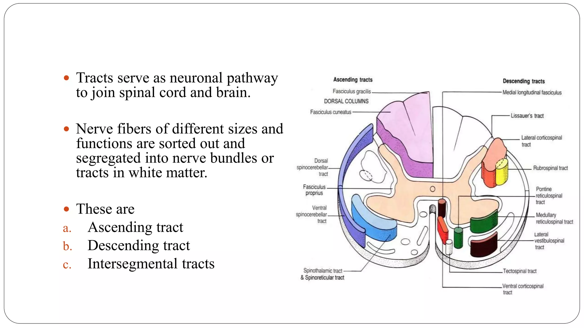
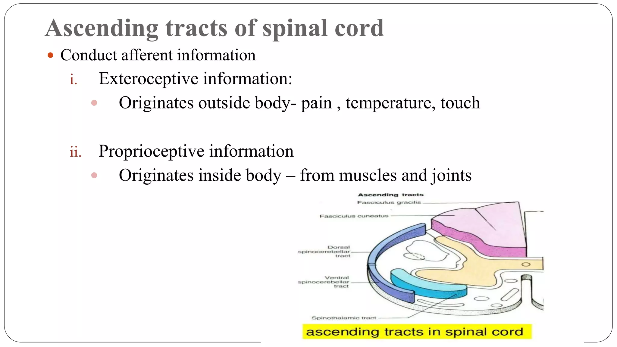


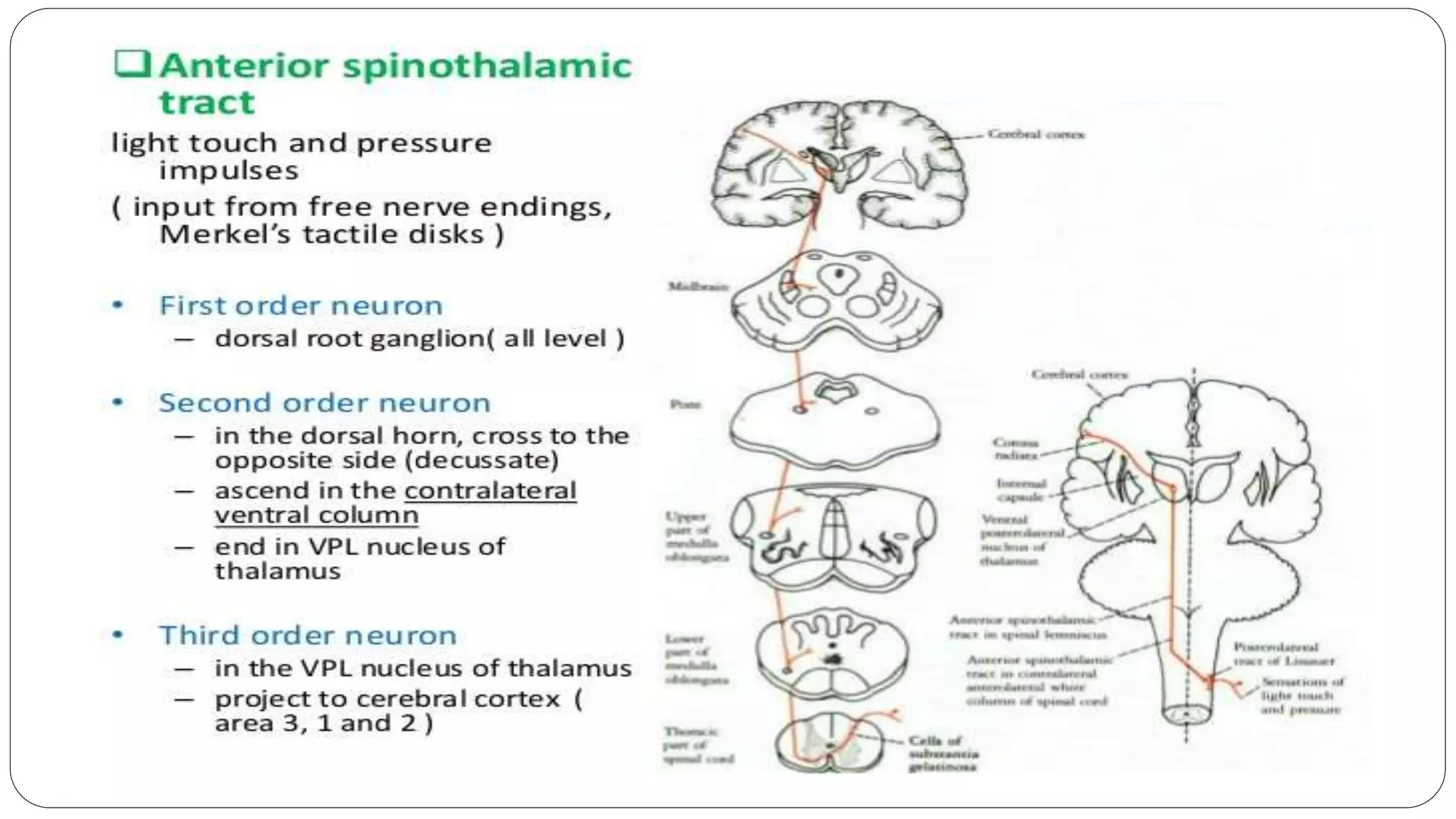


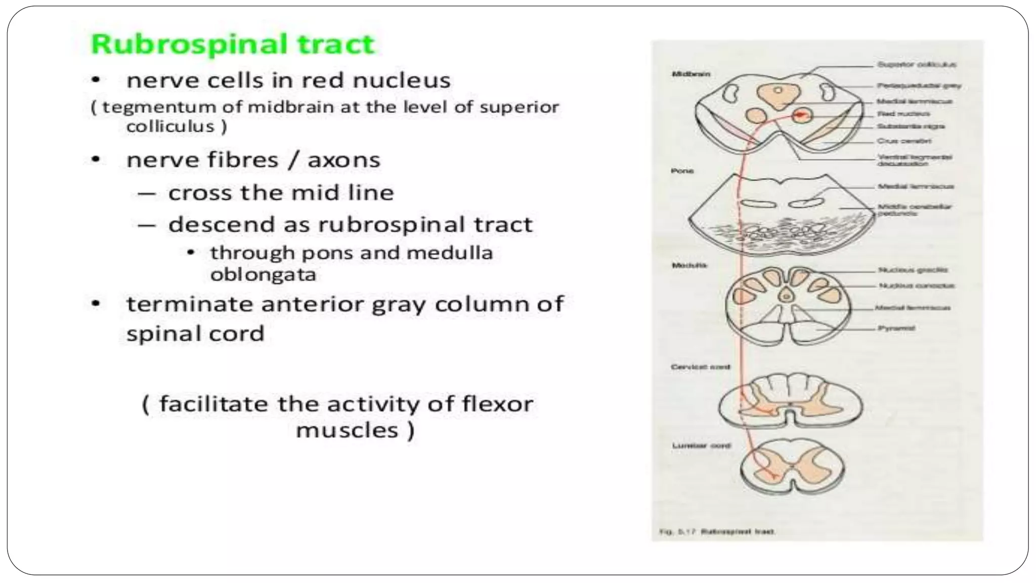


The document summarizes the ascending and descending spinal tracts. There are ascending tracts that conduct sensory information from the body to the brain, including the lateral and anterior spinothalamic tracts for pain and temperature, and the posterior white columns for proprioception. There are also descending tracts that convey motor commands from the brain to the spinal cord to control skeletal muscles, such as the lateral and anterior corticospinal tracts. In addition, there are tracts connecting the spinal cord and cerebellum that are important for motor coordination.


