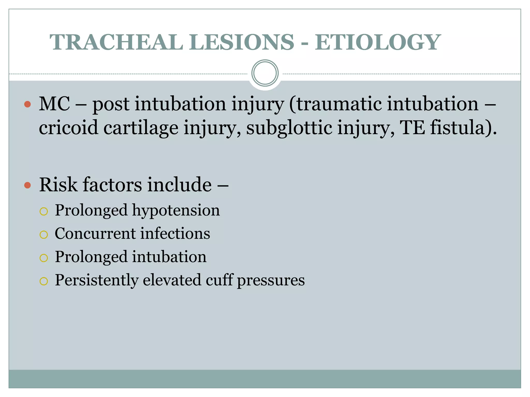
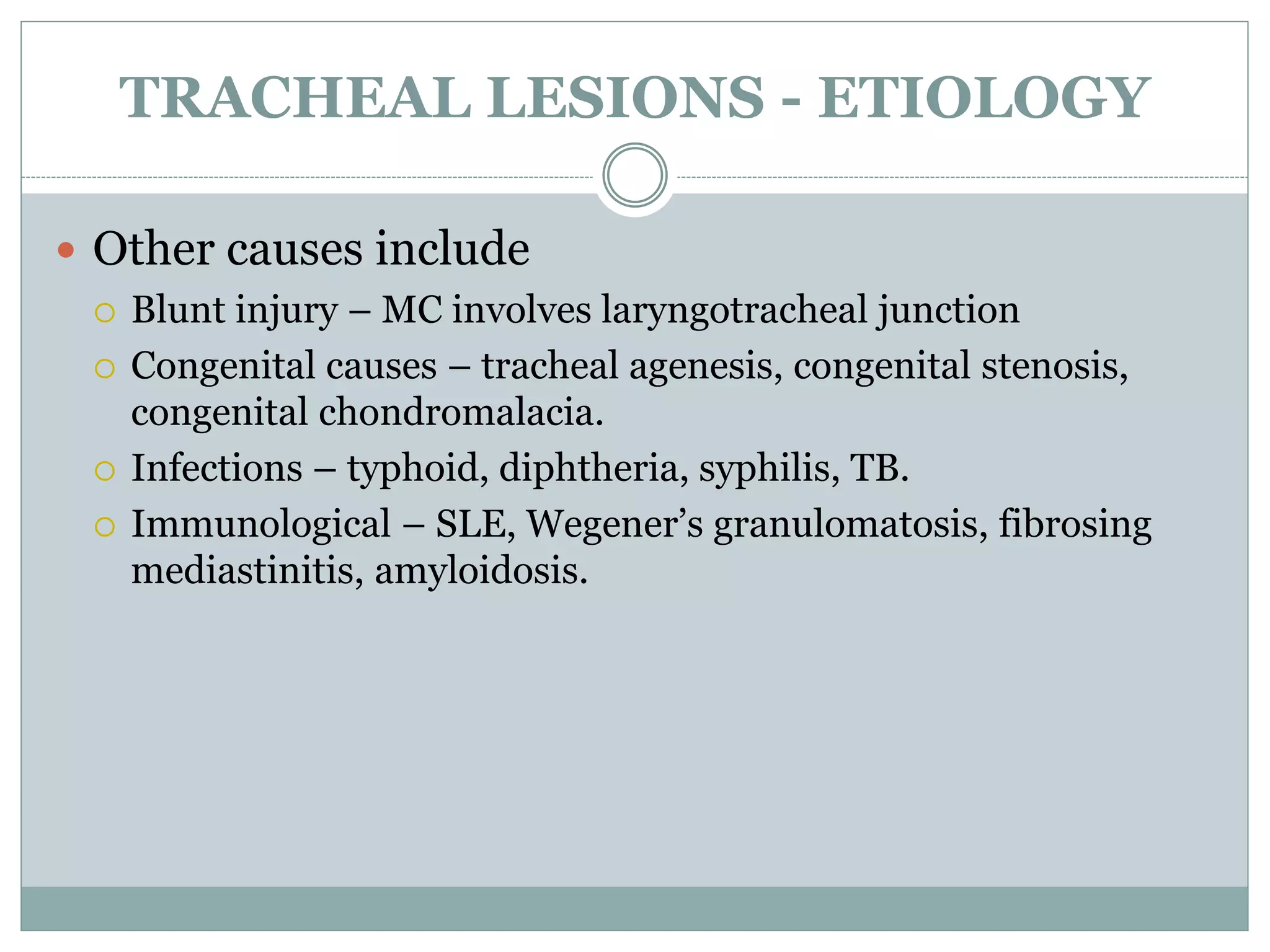
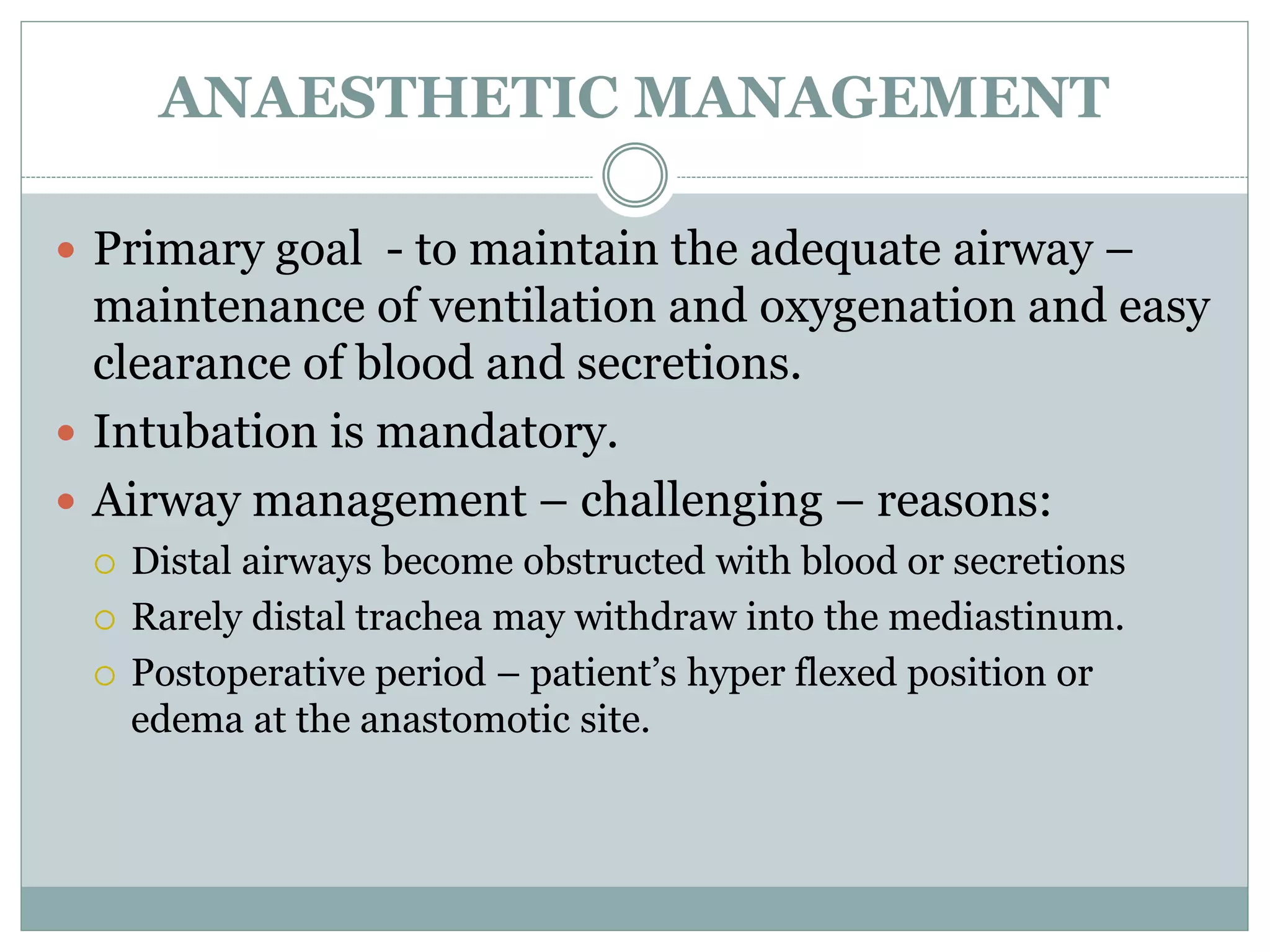
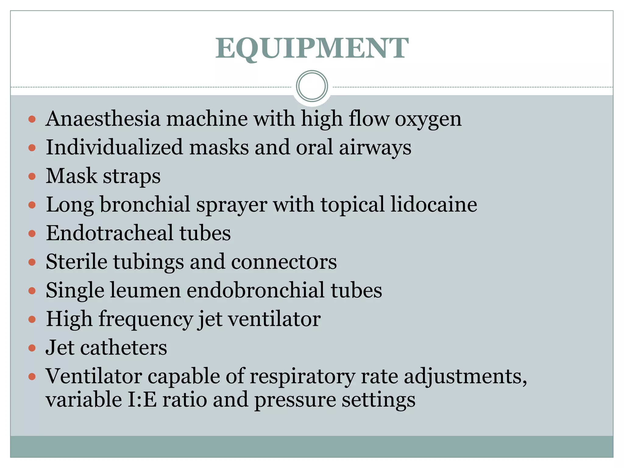
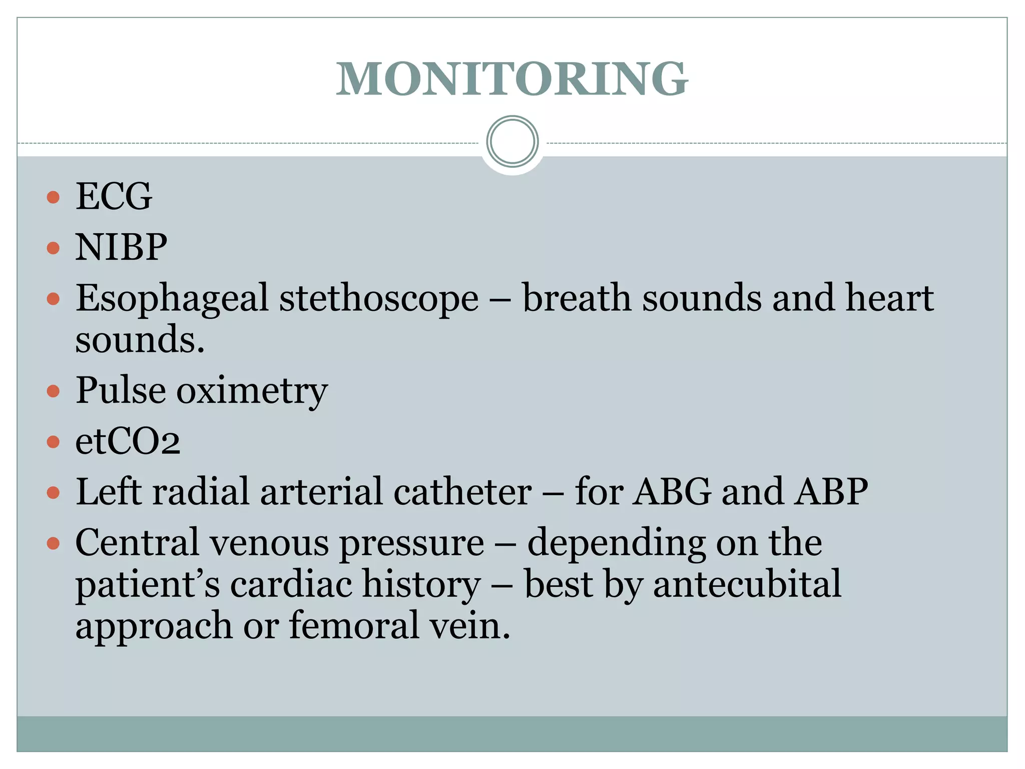
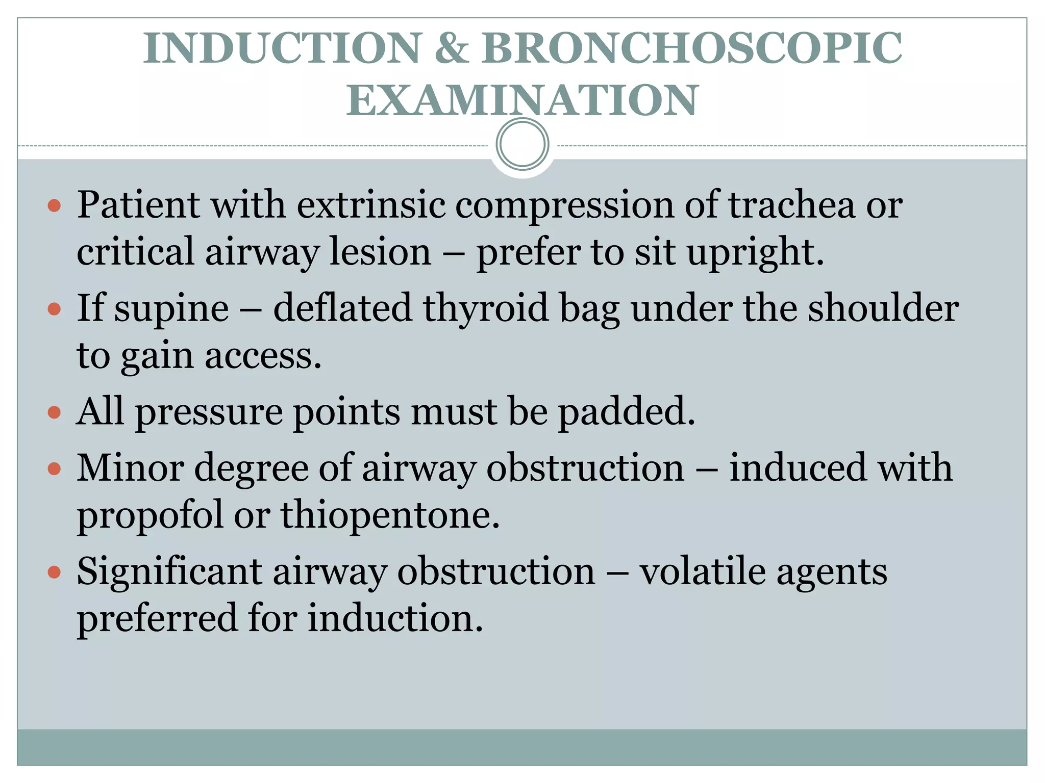
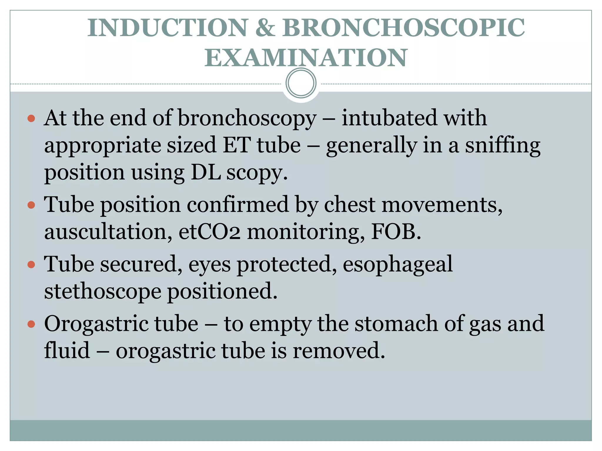
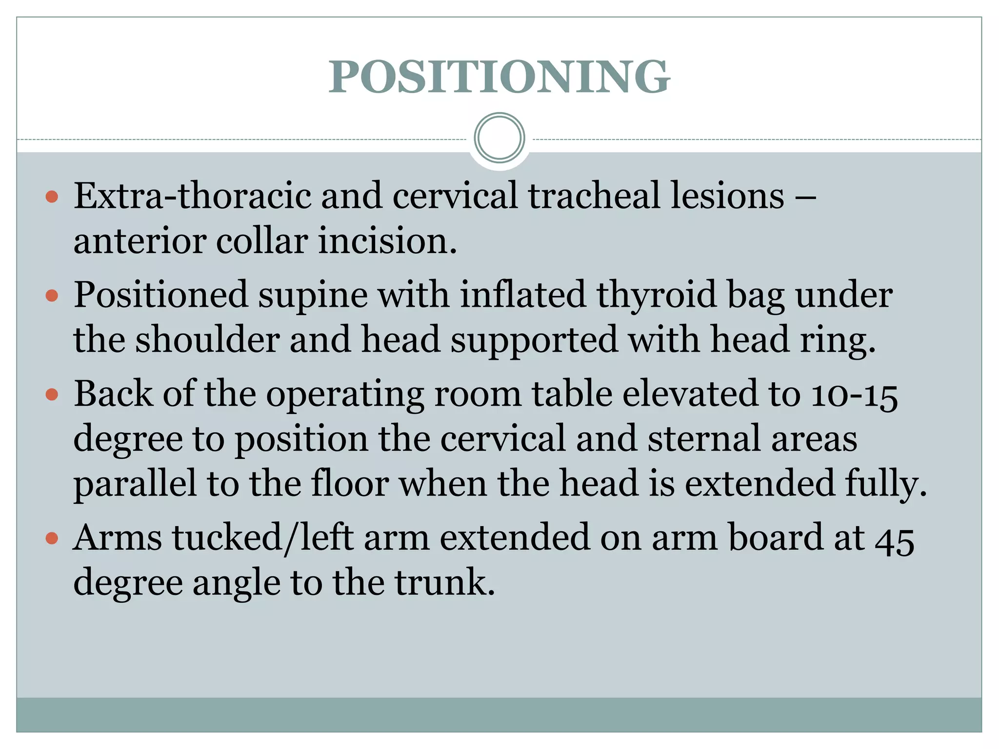
D R . D A V I S K U R I A N
TRACHEAL RESECTION
TRACHEAL LESIONS - ETIOLOGY
 MC – post intubation injury (traumatic intubation –
cricoid cartilage injury, subglottic injury, TE fistula).
 Risk factors include –
 Prolonged hypotension
 Concurrent infections
 Prolonged intubation
 Persistently elevated cuff pressures
TRACHEAL LESIONS - ETIOLOGY
 Second MC cause – tumours – squamous cell and
adenoid cystic carcinomas.
 Sq cell CA – discrete exophytic or ulcerating lesion.
 Adenoid cystic CA – slow growing – spreads either
locally (tracheal submucosa) or directly into the lung
parenchyma or pleura.
 Secondary tumours – thyroid (MC), esophagus, lung,
breast, thymus, head and neck malignancies &
lymphomas.
TRACHEAL LESIONS - ETIOLOGY
 Other causes include
 Blunt injury – MC involves laryngotracheal junction
 Congenital causes – tracheal agenesis, congenital stenosis,
congenital chondromalacia.
 Infections – typhoid, diphtheria, syphilis, TB.
 Immunological – SLE, Wegener’s granulomatosis, fibrosing
mediastinitis, amyloidosis.
PREOP EVALUATION
 Includes
 Detailed history
 Examination of the patient
 All routine blood investigations
 PFT – flow volume loops
 Radiological evaluation
 Diagnostic bronchoscopy
HISTORY AND PHYSICAL EXAMINATION
 History of recent endotracheal intubation or
tracheostomy.
 Presence of coexisiting comorbidities – especially lung,
CVS
 Signs and symptoms of airway obstruction – dyspnea
 Wheeze
 Stridor (4mm or less narrowing)
 Persistent cough
 Hoarseness (involvement of recurrent laryngeal nerve & vocal cords)
 Difficulty in clearing secretions
 Inabilility to tolerate supine position.
HISTORY AND PHYSICAL EXAMINATION
 Mandatory to prove adequate laryngeal airway prior to
tracheal resection and reconstruction.
 Tracheal deviation or extra-thoracic compression – by
palpation.
 Stridor at rest/expiratory effort – elicited.
 Auscultation of the upper airway.
 Range of neck movements (should tolerate hyper flexion
and hyperextension comfortably) & C Spine stability.
 Tracheostomy – should be examined for the patency of
the tube and tracheal stoma.
PULMONARY FUNCTION TESTING
 Ratio of Peak expiratory flow to FEV1 – index of
airway obstruction.
 Flow volume loops
RADIOLOGICAL ASSESSMENT
 Fluoroscopic studies to demonstrate vocal cord
movement, tracheomalacia.
 CT
 MRI
 Other investigations:
 Vocal cord mobility – by Laryngoscopy
ANAESTHETIC MANAGEMENT
 Primary goal - to maintain the adequate airway –
maintenance of ventilation and oxygenation and easy
clearance of blood and secretions.
 Intubation is mandatory.
 Airway management – challenging – reasons:
 Distal airways become obstructed with blood or secretions
 Rarely distal trachea may withdraw into the mediastinum.
 Postoperative period – patient’s hyper flexed position or
edema at the anastomotic site.
PREMEDICATION
 Patients with significant tracheal narrowing –
shouldn't receive premedication unless they are in
the hands of a person skilled in airway management.
 Over sedation and CNS depression should be
avoided.
 BZD – sedative and anxiolytic properties.
 Antisialagogues – atropine etc. can be disastrous in
patients with tracheal stenosis- tenacious secretions.
EQUIPMENT
 Anaesthesia machine with high flow oxygen
 Individualized masks and oral airways
 Mask straps
 Long bronchial sprayer with topical lidocaine
 Endotracheal tubes
 Sterile tubings and connect0rs
 Single leumen endobronchial tubes
 High frequency jet ventilator
 Jet catheters
 Ventilator capable of respiratory rate adjustments,
variable I:E ratio and pressure settings
MONITORING
 ECG
 NIBP
 Esophageal stethoscope – breath sounds and heart
sounds.
 Pulse oximetry
 etCO2
 Left radial arterial catheter – for ABG and ABP
 Central venous pressure – depending on the
patient’s cardiac history – best by antecubital
approach or femoral vein.
INDUCTION & BRONCHOSCOPIC
EXAMINATION
 Patient with extrinsic compression of trachea or
critical airway lesion – prefer to sit upright.
 If supine – deflated thyroid bag under the shoulder
to gain access.
 All pressure points must be padded.
 Minor degree of airway obstruction – induced with
propofol or thiopentone.
 Significant airway obstruction – volatile agents
preferred for induction.
INDUCTION & BRONCHOSCOPIC
EXAMINATION
 Muscle relaxants must be avoided before securing
the airway.
 After adequate anaesthesia – DL scopy – topical
anaesthesia to oropharynx and glottis – face mask
reapplied – volatile agent+O2 continued – deepen
the plane – DL scopy – topical anaesthesia below the
vocal cord – bronchoscopy.
INDUCTION & BRONCHOSCOPIC
EXAMINATION
 Rigid bronchoscopy –
 To visualize the nature and extent of lesion
 Potential difficulty in ET tube placement
 Opportunity for tracheal dilation to provide airway of adequate
size – serial dilation with rigid pediatric bronchoscopes.
 Tumor handling and dilation must be done with care
–
 Dislodgement can cause further airway obstruction
 Bleeding can further compromise the airway.
INDUCTION & BRONCHOSCOPIC
EXAMINATION
 At the end of bronchoscopy – intubated with
appropriate sized ET tube – generally in a sniffing
position using DL scopy.
 Tube position confirmed by chest movements,
auscultation, etCO2 monitoring, FOB.
 Tube secured, eyes protected, esophageal
stethoscope positioned.
 Orogastric tube – to empty the stomach of gas and
fluid – orogastric tube is removed.
POSITIONING
 Extra-thoracic and cervical tracheal lesions –
anterior collar incision.
 Positioned supine with inflated thyroid bag under
the shoulder and head supported with head ring.
 Back of the operating room table elevated to 10-15
degree to position the cervical and sternal areas
parallel to the floor when the head is extended fully.
 Arms tucked/left arm extended on arm board at 45
degree angle to the trunk.
POSITIONING
 Lesions of distal trachea – explored through – right
posterolateral thoracotomy in the fourth interspace
or the bed of 4th rib.
 Patient in left lateral decubitus position.
 Right arm – drapped and prepped into the surgical
field – for the surgeon to manipulate the arm to gain
access to the neck.
CERVICAL TRACHEAL
RECONSTRUCTION
 Low cervical collar incision.
 Anterior dissection of trachea – careful dissection around
innominate artery and structures around the trachea.
 Anaesthesia maintained by oral ET tube – prior to
tracheal division N2O eliminated from the gas mixture –
maintained by O2+inhalational agent – surgical tape
around the trachea below the lesion – ET tube cuff
deflated prior placement of lateral sutures – trachea
resected – distal part intubated across the field using
flexible, sterile, reinforced tube – connected to circuit.
CERVICAL TRACHEAL
RECONSTRUCTION
 After resection – two free ends of trachea are
anastomosed by traction sutures – patient’s neck
flexed from above – once all the sutures have been
put – reinforced tube is removed – the tube in the
proximal trachea is readvanced into the distal
trachea under supervision.
 Prior to the final airway exchange – airway is
suctioned of blood and secretions.
 Anastomotic site is checked for leaks with sustained
positive pressure breath 30 mm Hg.
 Patient’s head is flexed and supported.
TRACHEAL RECONSTRUCTION
CERVICAL TRACHEAL
RECONSTRUCTION
 At the end – patient must be breathing
spontaneously – awake extubation – head should be
supported in a hyperflexed position to avoid undue
traction at anastomotic site.
 Equipments for re-intubation must be kept ready
including a FOB.
 Upper airway edema – MCC of respiratory distress in
the immediate post operative period.
 Patient must be transported with supplemental
oxygen.
DISTAL TRACHEA AND CARINA
RECONSTRUCTION
 Preferred approach is right posterolateral
thoracotomy.
 General principles are the same for cervical tracheal
reconstruction.
 Thoracic epidural before induction of anaesthesia –
then patient in left lateral position.
 Positive pressure ventilation and ms relaxants of
intermediate duration of action – with appropriate
monitoring.
 Ms relaxants must be promptly reversed at the end
of the procedure.
DISTAL TRACHEA AND CARINA
RECONSTRUCTION
 Initial resection – performed with the tube proximal
to the airway lesion – once trachea divided – too
short to hold an ET tube – Left mainstem bronchus
intubated from the operative field and one lung
ventilation is employed.
 Second endobronchial tube introduced into right
mainstem bronchus and CPAP or HFPPV via jet
ventilator.
 SPO2 monitored with a pulse oximeter.
 HFPPV may also be used for left lung ventilation.
DISTAL TRACHEA AND CARINA
RECONSTRUCTION
 End of procedure – ET tube is withdrawn into
proximal trachea – ventilation proceeds through
area of anastomosis – position of tube confirmed by
FOB.
 Routinely right mainstem bronchus is anastomosed
to distal trachea.
 Left mainstem bronchus is re-implanted in an end to
side anastomosis to the bronchus intermedius or
distal trachea.
 Extubated similar to cervical approach and
transported.
DISTAL TRACHEA AND CARINA
RECONSTRUCTION
 Most widely employed method – O2+/-N2O with
volatile anasthetic.
 Inhalational agents – bronchodilators.
 Isoflurane and sevoflurane – pleasant induction, less
arrythmogenic and less hepatotoxicity compared
with halothane (less pungent than both iso and
sevo).
POSTOPERATIVE CARE
 ICU admission
 Chest XRay – to r/o pneumothorax
 O2 via high flow, humidified system via face mask –
adequate oxygenation as well as thinning of secretions.
 Head kept in a flexed position.
 Chest physiotherapy.
 Blind nasotracheal suction done with caution in patient
with inadequate cough reflex – take care not to cause
perforation at the anastomotic site, airway edema,
vomiting and aspiration.
 Flexible FOB guided pulmonary toilet to clear off
secretions.
POSTOPERATIVE CARE
 Equipments for reintubation must be kept ready.
 Laryngeal edema – another important complication
can present as stridor or hoarseness – management
includes placing the patient in a sitting position to
promote venous drainage, controlled fluid
administration and nebulisation with mixture of
adrenaline and saline, IV steroids.
 Pain contol – IV, epidural, intrapleural analgesia,
intercostal blocks or PCA.
Tracheal resection - anesthesia

Tracheal resection - anesthesia

  • 1.
    D R .D A V I S K U R I A N TRACHEAL RESECTION
  • 2.
    TRACHEAL LESIONS -ETIOLOGY  MC – post intubation injury (traumatic intubation – cricoid cartilage injury, subglottic injury, TE fistula).  Risk factors include –  Prolonged hypotension  Concurrent infections  Prolonged intubation  Persistently elevated cuff pressures
  • 3.
    TRACHEAL LESIONS -ETIOLOGY  Second MC cause – tumours – squamous cell and adenoid cystic carcinomas.  Sq cell CA – discrete exophytic or ulcerating lesion.  Adenoid cystic CA – slow growing – spreads either locally (tracheal submucosa) or directly into the lung parenchyma or pleura.  Secondary tumours – thyroid (MC), esophagus, lung, breast, thymus, head and neck malignancies & lymphomas.
  • 4.
    TRACHEAL LESIONS -ETIOLOGY  Other causes include  Blunt injury – MC involves laryngotracheal junction  Congenital causes – tracheal agenesis, congenital stenosis, congenital chondromalacia.  Infections – typhoid, diphtheria, syphilis, TB.  Immunological – SLE, Wegener’s granulomatosis, fibrosing mediastinitis, amyloidosis.
  • 5.
    PREOP EVALUATION  Includes Detailed history  Examination of the patient  All routine blood investigations  PFT – flow volume loops  Radiological evaluation  Diagnostic bronchoscopy
  • 6.
    HISTORY AND PHYSICALEXAMINATION  History of recent endotracheal intubation or tracheostomy.  Presence of coexisiting comorbidities – especially lung, CVS  Signs and symptoms of airway obstruction – dyspnea  Wheeze  Stridor (4mm or less narrowing)  Persistent cough  Hoarseness (involvement of recurrent laryngeal nerve & vocal cords)  Difficulty in clearing secretions  Inabilility to tolerate supine position.
  • 7.
    HISTORY AND PHYSICALEXAMINATION  Mandatory to prove adequate laryngeal airway prior to tracheal resection and reconstruction.  Tracheal deviation or extra-thoracic compression – by palpation.  Stridor at rest/expiratory effort – elicited.  Auscultation of the upper airway.  Range of neck movements (should tolerate hyper flexion and hyperextension comfortably) & C Spine stability.  Tracheostomy – should be examined for the patency of the tube and tracheal stoma.
  • 8.
    PULMONARY FUNCTION TESTING Ratio of Peak expiratory flow to FEV1 – index of airway obstruction.  Flow volume loops
  • 9.
  • 10.
  • 11.
    RADIOLOGICAL ASSESSMENT  Fluoroscopicstudies to demonstrate vocal cord movement, tracheomalacia.  CT  MRI  Other investigations:  Vocal cord mobility – by Laryngoscopy
  • 12.
    ANAESTHETIC MANAGEMENT  Primarygoal - to maintain the adequate airway – maintenance of ventilation and oxygenation and easy clearance of blood and secretions.  Intubation is mandatory.  Airway management – challenging – reasons:  Distal airways become obstructed with blood or secretions  Rarely distal trachea may withdraw into the mediastinum.  Postoperative period – patient’s hyper flexed position or edema at the anastomotic site.
  • 13.
    PREMEDICATION  Patients withsignificant tracheal narrowing – shouldn't receive premedication unless they are in the hands of a person skilled in airway management.  Over sedation and CNS depression should be avoided.  BZD – sedative and anxiolytic properties.  Antisialagogues – atropine etc. can be disastrous in patients with tracheal stenosis- tenacious secretions.
  • 14.
    EQUIPMENT  Anaesthesia machinewith high flow oxygen  Individualized masks and oral airways  Mask straps  Long bronchial sprayer with topical lidocaine  Endotracheal tubes  Sterile tubings and connect0rs  Single leumen endobronchial tubes  High frequency jet ventilator  Jet catheters  Ventilator capable of respiratory rate adjustments, variable I:E ratio and pressure settings
  • 15.
    MONITORING  ECG  NIBP Esophageal stethoscope – breath sounds and heart sounds.  Pulse oximetry  etCO2  Left radial arterial catheter – for ABG and ABP  Central venous pressure – depending on the patient’s cardiac history – best by antecubital approach or femoral vein.
  • 16.
    INDUCTION & BRONCHOSCOPIC EXAMINATION Patient with extrinsic compression of trachea or critical airway lesion – prefer to sit upright.  If supine – deflated thyroid bag under the shoulder to gain access.  All pressure points must be padded.  Minor degree of airway obstruction – induced with propofol or thiopentone.  Significant airway obstruction – volatile agents preferred for induction.
  • 17.
    INDUCTION & BRONCHOSCOPIC EXAMINATION Muscle relaxants must be avoided before securing the airway.  After adequate anaesthesia – DL scopy – topical anaesthesia to oropharynx and glottis – face mask reapplied – volatile agent+O2 continued – deepen the plane – DL scopy – topical anaesthesia below the vocal cord – bronchoscopy.
  • 18.
    INDUCTION & BRONCHOSCOPIC EXAMINATION Rigid bronchoscopy –  To visualize the nature and extent of lesion  Potential difficulty in ET tube placement  Opportunity for tracheal dilation to provide airway of adequate size – serial dilation with rigid pediatric bronchoscopes.  Tumor handling and dilation must be done with care –  Dislodgement can cause further airway obstruction  Bleeding can further compromise the airway.
  • 19.
    INDUCTION & BRONCHOSCOPIC EXAMINATION At the end of bronchoscopy – intubated with appropriate sized ET tube – generally in a sniffing position using DL scopy.  Tube position confirmed by chest movements, auscultation, etCO2 monitoring, FOB.  Tube secured, eyes protected, esophageal stethoscope positioned.  Orogastric tube – to empty the stomach of gas and fluid – orogastric tube is removed.
  • 20.
    POSITIONING  Extra-thoracic andcervical tracheal lesions – anterior collar incision.  Positioned supine with inflated thyroid bag under the shoulder and head supported with head ring.  Back of the operating room table elevated to 10-15 degree to position the cervical and sternal areas parallel to the floor when the head is extended fully.  Arms tucked/left arm extended on arm board at 45 degree angle to the trunk.
  • 21.
    POSITIONING  Lesions ofdistal trachea – explored through – right posterolateral thoracotomy in the fourth interspace or the bed of 4th rib.  Patient in left lateral decubitus position.  Right arm – drapped and prepped into the surgical field – for the surgeon to manipulate the arm to gain access to the neck.
  • 22.
    CERVICAL TRACHEAL RECONSTRUCTION  Lowcervical collar incision.  Anterior dissection of trachea – careful dissection around innominate artery and structures around the trachea.  Anaesthesia maintained by oral ET tube – prior to tracheal division N2O eliminated from the gas mixture – maintained by O2+inhalational agent – surgical tape around the trachea below the lesion – ET tube cuff deflated prior placement of lateral sutures – trachea resected – distal part intubated across the field using flexible, sterile, reinforced tube – connected to circuit.
  • 23.
    CERVICAL TRACHEAL RECONSTRUCTION  Afterresection – two free ends of trachea are anastomosed by traction sutures – patient’s neck flexed from above – once all the sutures have been put – reinforced tube is removed – the tube in the proximal trachea is readvanced into the distal trachea under supervision.  Prior to the final airway exchange – airway is suctioned of blood and secretions.  Anastomotic site is checked for leaks with sustained positive pressure breath 30 mm Hg.  Patient’s head is flexed and supported.
  • 24.
  • 25.
    CERVICAL TRACHEAL RECONSTRUCTION  Atthe end – patient must be breathing spontaneously – awake extubation – head should be supported in a hyperflexed position to avoid undue traction at anastomotic site.  Equipments for re-intubation must be kept ready including a FOB.  Upper airway edema – MCC of respiratory distress in the immediate post operative period.  Patient must be transported with supplemental oxygen.
  • 26.
    DISTAL TRACHEA ANDCARINA RECONSTRUCTION  Preferred approach is right posterolateral thoracotomy.  General principles are the same for cervical tracheal reconstruction.  Thoracic epidural before induction of anaesthesia – then patient in left lateral position.  Positive pressure ventilation and ms relaxants of intermediate duration of action – with appropriate monitoring.  Ms relaxants must be promptly reversed at the end of the procedure.
  • 27.
    DISTAL TRACHEA ANDCARINA RECONSTRUCTION  Initial resection – performed with the tube proximal to the airway lesion – once trachea divided – too short to hold an ET tube – Left mainstem bronchus intubated from the operative field and one lung ventilation is employed.  Second endobronchial tube introduced into right mainstem bronchus and CPAP or HFPPV via jet ventilator.  SPO2 monitored with a pulse oximeter.  HFPPV may also be used for left lung ventilation.
  • 28.
    DISTAL TRACHEA ANDCARINA RECONSTRUCTION  End of procedure – ET tube is withdrawn into proximal trachea – ventilation proceeds through area of anastomosis – position of tube confirmed by FOB.  Routinely right mainstem bronchus is anastomosed to distal trachea.  Left mainstem bronchus is re-implanted in an end to side anastomosis to the bronchus intermedius or distal trachea.  Extubated similar to cervical approach and transported.
  • 29.
    DISTAL TRACHEA ANDCARINA RECONSTRUCTION  Most widely employed method – O2+/-N2O with volatile anasthetic.  Inhalational agents – bronchodilators.  Isoflurane and sevoflurane – pleasant induction, less arrythmogenic and less hepatotoxicity compared with halothane (less pungent than both iso and sevo).
  • 30.
    POSTOPERATIVE CARE  ICUadmission  Chest XRay – to r/o pneumothorax  O2 via high flow, humidified system via face mask – adequate oxygenation as well as thinning of secretions.  Head kept in a flexed position.  Chest physiotherapy.  Blind nasotracheal suction done with caution in patient with inadequate cough reflex – take care not to cause perforation at the anastomotic site, airway edema, vomiting and aspiration.  Flexible FOB guided pulmonary toilet to clear off secretions.
  • 31.
    POSTOPERATIVE CARE  Equipmentsfor reintubation must be kept ready.  Laryngeal edema – another important complication can present as stridor or hoarseness – management includes placing the patient in a sitting position to promote venous drainage, controlled fluid administration and nebulisation with mixture of adrenaline and saline, IV steroids.  Pain contol – IV, epidural, intrapleural analgesia, intercostal blocks or PCA.