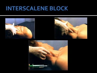
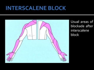
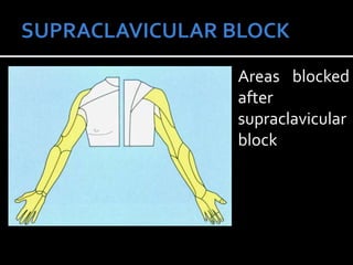
DR. DAVIS KURIAN
 INDICATIONS :
 surgery or manipulation on clavicle,
shoulder or upper arm (except the
medial aspect.
 therapeutic-frozen shoulder, post
herpetic neuralgia, tumour related
pain etc
 Donot reliably block the inferior trunk, hence
requires supplementation at ulnar nerve for
surgeries of hand and forearm.
 CONTRAINDICATIONS:
 SPECIFIC:
 Infection or malignant disease in the neck.
 Infection of the skin in the puncture area.
 Contralateral paresis of the phrenic or recurrent
laryngeal nerves.
 Anticoagulation treatment.
 Distorted anatomy - e.g. due to prior surgical
interventions or trauma to the neck.
 RELATIVE:
 Hemorrhagic diathesis.
 Local nerve injury (as there may be doubt
whether the cause is surgery or anesthesia).
 Severe chronic obstructive pulmonary
disease
 Before any regional block
 Check that the emergency equipment is
present and in working order.
 Sterile precautions.
 Intravenous access, ECG monitoring, pulse
oximetry, intubation kit, emergency
medication, ventilation facilities.
 ANTERIOR APPROACH (WINNIE’S)
 POSTERIOR APPROACH (PIPPA’STECH)
 Blocks can be given using – landmarks alone,
nerve stimulator guided, USG guided or with
both USG and nerve stimulator guided.
WINNIE’S ANTERIOR ROUTE:
SINGLE SHOT:
POSITION – supine, head turned to opposite
side
LANDMARKS
Sternocleidomastoid muscle, interscalene groove
between the scalenus anterior and scalenus medius
muscles transverse process (C6), external jugular
vein.
 Turn head to opposite side + slight lift (200).
 Roll finger posterolaterally from the posterior
border of sternocliedomastoid over the
anterior scalene muscle to reach the
interscalene groove.
 The injection site in the interscalene groove
lies at thelevel of the cricoid, opposite the
transverse process of C6 (Chassaignac's
tubercle).
 Line extended laterally from the cricoid cartilage
to intersect the interscalene groove indicates
the level of the transverse process of C6.
Although the external jugular vein often overlies
this point of intersection, it is not a constant or
reliable landmark.
 When there are anatomical difficulties, it is
helpful for the patient to inhale deeply or to try
and blow out the cheeks. The scalene muscles
then tense up and the interscalene groove
becomes more easily palpable.
 INJECTIONTECHNIQUE:
The traditional technique first described by Winnie
is a classic paresthesia technique.
Prepare the area, infiltrate the skin, isolate the
area with middle and index fingers.
22- to 25-gauge, 4-cm needle is inserted
perpendicular to the skin at 45-degree caudally,
medially and slightly posterior.
 When the needle is positioned superficially,
paresthesias usually occur in the area of the
elbow, index finger and thumb. Paresthesias
in the shoulder region also frequently occur.
 These result from stimulation of the
suprascapular nerve, which is often located in
the connective tissue sheath
 Electrical nerve stimulator can also be used –
where motor response of the arm or shoulder is
also equally efficacious when compared with
classical parasthesia technique.
 If a blunt needle bevel is used, a “click” may be
detected as the needle passes through the
prevertebral fascia. If bone is encountered within
2 cm of the skin, it is likely to be a transverse
process, and the needle may be “walked” across
this structure to locate the nerve.
 Normal anatomy – no parasthesia in 2-2.5cm,
position of needle must be corrected.
 Contraction of the diaphragm indicates
phrenic nerve stimulation and anterior needle
placement; the needle should be redirected
posteriorly to locate the brachial plexus.
 After negative aspiration, 10 to 40 mL of
solution is injected incrementally, depending
on the desired extent of blockade. After each
4-5ml, aspiration is done.
 After successful injection, the entire area is
massaged in order to ensure even distribution
of the local anesthetic. This also provides
hematoma prophylaxis
With a nerve stimuator the goal is stimulation
of the brachial plexus with a current intensity
of 0.2-0.5 mA (0.1 ms).The following motor
responses result in a similar success rate:
 Pectoralis muscle
 Deltoid muscle
 Triceps muscle
 Biceps muscle
 Any twitch of the hand or forearm
CONTINUOUS INTERSCALENE BLOCK:
 identification of the posterior edge of the
sternocleidomastoid muscle at the level of
the superior thyroid notch.
 block needle is introduced at an angle of 30"
caudally and slightly laterally, in the direction
of the transition from the middle to the
lateral third of the clavicle.
 Position of needle confirmed with ms twitch
and local anaesthetic injected.
POSTERIORTECHNIQUE:
 first described by Kappis in 191 2, and was
republished by Pippa in 1990 as a ”loss of
resistance” technique.
 Sitting, with the neck flexed (to relax the
cervical muscles) and supported by an
assistant (the lateral recumbent position can
be used as an alternative)
LANDMARKS
 Spinous processes of the sixth (C6) and
seventh (C7 - vertebra prominens) cervical
vertebrae.
 The mid-point between the spinous
processes of C6 and C7 is marked.The
puncture site is located approximately 3 cm
lateral to this point.
 Level of the cricoid cartilage (target
direction).
 The needle is introduced at the sagittal level and
perpendicular to the skin, aiming approximately
for the level of the ipsilateral cricoid cartilage.
 At a depth of about 3.5-6 cm, contact is made
with the transverse process of C7. The needle is
withdrawn slightly, the injection direction is
corrected slightly cranially, and one advances
past the transverse process a further 1.5-2 cm
deeper.
 Then the normal process follows.
 COMPLICATIONS:
 Risk of dural puncture if performed too
medially.
 Risk of pneumothorax
 SIDE EFFECTS & COMPLICATIONS:
 Ipsilateral phrenic nerve block
diaphragmatic paresis altered pulmonary
function.
 Severe hypotension and bradycardia (i.e., the
Bezold-Jarisch reflex) have been reported in
awake, sitting patients undergoing shoulder
surgery under an interscalene block d/t
stimulation of intracardiac mechanoreceptors
by decreased venous return, which produces
an abrupt withdrawal of sympathetic tone
and enhanced parasympathetic output.
 Nerve damage and neuritis – rarely seen.
 Local anaesthetic toxicity.
 Associated horner’s syndrome can develop
due to stellate ganglion block.
 Rarely pneumothorax
Usual areas of
blockade after
interscalene
block
 Injection of a local anesthetic into the area of
the brachial plexus trunks in the caudal part
of the interscalene groove, in its most
compact part above the clavicle.
 The first percutaneous supraclavicular block
was performed in 1911 by German surgeon
Diedrich Kulenkampff on himself.
 Blockade occurs at the distal trunk–proximal
division level.
 At this point, the brachial plexus is compact and
a small volume of solution produces rapid onset
of reliable blockade of the brachial plexus.
 Additional advantage is that the block can also
be performed with the patient's arm in any
position.
 The supraclavicular block is often called the
"spinal anesthesia of the upper extremity"
because of its ubiquitous application for
upper extremity surgery.
 INDICATIONS
 Surgery on the upper arm, forearm and hand.
 As such no therapeutic indications.
 CONTRAINDICATIONS
 Infections or malignant diseases in the area of the
throat and neck or at the site of injection.
 Bleeding disorders & anticoagulant therapy.
 Contralateral pneumothorax
 Severe COPD
LANDMARKS:
 The clavicular head of sternocleidomastoid.
 Interscalene groove
 The interscalene groove - caudal part in the
supraclavicular fossa – difficult to identify –
covered by omohyoid muscle.
 The midpoint of the clavicle. The injection point is
located about 1.5-2 cm lateral to the clavicular
head of the sternocleidomastoid muscle and 2 cm
above the clavicle.
 Subclavian artery – close proximity to trunks –
pulse is an important landmark for injection.
TECHNIQUE
 Patient in supine position with the head turned
away from the side to be blocked.
 Arm adducted – preferably pulled down to
ipsilateral knee as far as possible.
 Interscalene groove identified and mark should be
made approximately 1.5 to 2.0 cm posterior to the
midpoint of the clavicle. Palpation of the
subclavian artery at this site confirms the
landmark.
 Needle is directed in a caudally, slightly
lateral, and posterior direction until a
paresthesia or motor response is elicited or
the first rib is encountered.
 If the first rib is encountered without
elicitation of a paresthesia, the needle can be
systematically walked anteriorly and
posteriorly along the rib until the plexus or
the subclavian artery is located.
 If artery – slight posteromedial shift – brachial
plexus.
 Avoid too much posteromedial orientation as
the dome of pleura is in close proximity to
first rib – chance of pneumothorax.
 Modified plumb-bob approach uses similar
patient positioning, although the needle
entry site is at the point where the lateral
border of the sternocleidomastoid muscle
inserts into the clavicle.
 Aspirate and inject – massage after injection
– to avoid hematoma and spread of the drug.
SIDE EFFECTS
Concomitant block of the following nerves
and ganglia:
 Vagus nerve
 Recurrent laryngeal nerve
 Phrenic nerve.
 Stellate ganglion
COMPLICATIONS
 Pneumothorax
 Neural injury
 lntravascular injection
 Stellate ganglion block
 CNS intoxication
Areas blocked
after
supraclavicular
block
 MILLER’SANAESTHESIA 7TH EDN.
 ANESTHESIOLOGY – BY
DAVID.E.LONGNECKER
 DANILO JANKOVIC REGIONAL NERVE
BLOCK AND INFILTRATIONTHERAPY
TEXTBOOK 3RD EDN.
 NYSORAWEBSITE – www.nysora.com
Interscalene  & supraclavicular nerve blocks

Interscalene & supraclavicular nerve blocks

  • 1.
  • 2.
     INDICATIONS : surgery or manipulation on clavicle, shoulder or upper arm (except the medial aspect.  therapeutic-frozen shoulder, post herpetic neuralgia, tumour related pain etc
  • 3.
     Donot reliablyblock the inferior trunk, hence requires supplementation at ulnar nerve for surgeries of hand and forearm.
  • 4.
     CONTRAINDICATIONS:  SPECIFIC: Infection or malignant disease in the neck.  Infection of the skin in the puncture area.  Contralateral paresis of the phrenic or recurrent laryngeal nerves.  Anticoagulation treatment.  Distorted anatomy - e.g. due to prior surgical interventions or trauma to the neck.
  • 5.
     RELATIVE:  Hemorrhagicdiathesis.  Local nerve injury (as there may be doubt whether the cause is surgery or anesthesia).  Severe chronic obstructive pulmonary disease
  • 6.
     Before anyregional block  Check that the emergency equipment is present and in working order.  Sterile precautions.  Intravenous access, ECG monitoring, pulse oximetry, intubation kit, emergency medication, ventilation facilities.
  • 7.
     ANTERIOR APPROACH(WINNIE’S)  POSTERIOR APPROACH (PIPPA’STECH)  Blocks can be given using – landmarks alone, nerve stimulator guided, USG guided or with both USG and nerve stimulator guided.
  • 8.
    WINNIE’S ANTERIOR ROUTE: SINGLESHOT: POSITION – supine, head turned to opposite side LANDMARKS Sternocleidomastoid muscle, interscalene groove between the scalenus anterior and scalenus medius muscles transverse process (C6), external jugular vein.
  • 9.
     Turn headto opposite side + slight lift (200).  Roll finger posterolaterally from the posterior border of sternocliedomastoid over the anterior scalene muscle to reach the interscalene groove.  The injection site in the interscalene groove lies at thelevel of the cricoid, opposite the transverse process of C6 (Chassaignac's tubercle).
  • 10.
     Line extendedlaterally from the cricoid cartilage to intersect the interscalene groove indicates the level of the transverse process of C6. Although the external jugular vein often overlies this point of intersection, it is not a constant or reliable landmark.  When there are anatomical difficulties, it is helpful for the patient to inhale deeply or to try and blow out the cheeks. The scalene muscles then tense up and the interscalene groove becomes more easily palpable.
  • 12.
     INJECTIONTECHNIQUE: The traditionaltechnique first described by Winnie is a classic paresthesia technique. Prepare the area, infiltrate the skin, isolate the area with middle and index fingers. 22- to 25-gauge, 4-cm needle is inserted perpendicular to the skin at 45-degree caudally, medially and slightly posterior.
  • 13.
     When theneedle is positioned superficially, paresthesias usually occur in the area of the elbow, index finger and thumb. Paresthesias in the shoulder region also frequently occur.  These result from stimulation of the suprascapular nerve, which is often located in the connective tissue sheath
  • 14.
     Electrical nervestimulator can also be used – where motor response of the arm or shoulder is also equally efficacious when compared with classical parasthesia technique.  If a blunt needle bevel is used, a “click” may be detected as the needle passes through the prevertebral fascia. If bone is encountered within 2 cm of the skin, it is likely to be a transverse process, and the needle may be “walked” across this structure to locate the nerve.
  • 15.
     Normal anatomy– no parasthesia in 2-2.5cm, position of needle must be corrected.  Contraction of the diaphragm indicates phrenic nerve stimulation and anterior needle placement; the needle should be redirected posteriorly to locate the brachial plexus.
  • 16.
     After negativeaspiration, 10 to 40 mL of solution is injected incrementally, depending on the desired extent of blockade. After each 4-5ml, aspiration is done.  After successful injection, the entire area is massaged in order to ensure even distribution of the local anesthetic. This also provides hematoma prophylaxis
  • 17.
    With a nervestimuator the goal is stimulation of the brachial plexus with a current intensity of 0.2-0.5 mA (0.1 ms).The following motor responses result in a similar success rate:  Pectoralis muscle  Deltoid muscle  Triceps muscle  Biceps muscle  Any twitch of the hand or forearm
  • 18.
    CONTINUOUS INTERSCALENE BLOCK: identification of the posterior edge of the sternocleidomastoid muscle at the level of the superior thyroid notch.  block needle is introduced at an angle of 30" caudally and slightly laterally, in the direction of the transition from the middle to the lateral third of the clavicle.  Position of needle confirmed with ms twitch and local anaesthetic injected.
  • 19.
    POSTERIORTECHNIQUE:  first describedby Kappis in 191 2, and was republished by Pippa in 1990 as a ”loss of resistance” technique.  Sitting, with the neck flexed (to relax the cervical muscles) and supported by an assistant (the lateral recumbent position can be used as an alternative)
  • 20.
    LANDMARKS  Spinous processesof the sixth (C6) and seventh (C7 - vertebra prominens) cervical vertebrae.  The mid-point between the spinous processes of C6 and C7 is marked.The puncture site is located approximately 3 cm lateral to this point.  Level of the cricoid cartilage (target direction).
  • 21.
     The needleis introduced at the sagittal level and perpendicular to the skin, aiming approximately for the level of the ipsilateral cricoid cartilage.  At a depth of about 3.5-6 cm, contact is made with the transverse process of C7. The needle is withdrawn slightly, the injection direction is corrected slightly cranially, and one advances past the transverse process a further 1.5-2 cm deeper.  Then the normal process follows.
  • 23.
     COMPLICATIONS:  Riskof dural puncture if performed too medially.  Risk of pneumothorax
  • 24.
     SIDE EFFECTS& COMPLICATIONS:  Ipsilateral phrenic nerve block diaphragmatic paresis altered pulmonary function.
  • 25.
     Severe hypotensionand bradycardia (i.e., the Bezold-Jarisch reflex) have been reported in awake, sitting patients undergoing shoulder surgery under an interscalene block d/t stimulation of intracardiac mechanoreceptors by decreased venous return, which produces an abrupt withdrawal of sympathetic tone and enhanced parasympathetic output.
  • 26.
     Nerve damageand neuritis – rarely seen.  Local anaesthetic toxicity.  Associated horner’s syndrome can develop due to stellate ganglion block.  Rarely pneumothorax
  • 27.
    Usual areas of blockadeafter interscalene block
  • 28.
     Injection ofa local anesthetic into the area of the brachial plexus trunks in the caudal part of the interscalene groove, in its most compact part above the clavicle.  The first percutaneous supraclavicular block was performed in 1911 by German surgeon Diedrich Kulenkampff on himself.
  • 29.
     Blockade occursat the distal trunk–proximal division level.  At this point, the brachial plexus is compact and a small volume of solution produces rapid onset of reliable blockade of the brachial plexus.  Additional advantage is that the block can also be performed with the patient's arm in any position.
  • 30.
     The supraclavicularblock is often called the "spinal anesthesia of the upper extremity" because of its ubiquitous application for upper extremity surgery.
  • 31.
     INDICATIONS  Surgeryon the upper arm, forearm and hand.  As such no therapeutic indications.  CONTRAINDICATIONS  Infections or malignant diseases in the area of the throat and neck or at the site of injection.  Bleeding disorders & anticoagulant therapy.  Contralateral pneumothorax  Severe COPD
  • 32.
    LANDMARKS:  The clavicularhead of sternocleidomastoid.  Interscalene groove
  • 33.
     The interscalenegroove - caudal part in the supraclavicular fossa – difficult to identify – covered by omohyoid muscle.  The midpoint of the clavicle. The injection point is located about 1.5-2 cm lateral to the clavicular head of the sternocleidomastoid muscle and 2 cm above the clavicle.  Subclavian artery – close proximity to trunks – pulse is an important landmark for injection.
  • 34.
    TECHNIQUE  Patient insupine position with the head turned away from the side to be blocked.  Arm adducted – preferably pulled down to ipsilateral knee as far as possible.  Interscalene groove identified and mark should be made approximately 1.5 to 2.0 cm posterior to the midpoint of the clavicle. Palpation of the subclavian artery at this site confirms the landmark.
  • 35.
     Needle isdirected in a caudally, slightly lateral, and posterior direction until a paresthesia or motor response is elicited or the first rib is encountered.  If the first rib is encountered without elicitation of a paresthesia, the needle can be systematically walked anteriorly and posteriorly along the rib until the plexus or the subclavian artery is located.
  • 36.
     If artery– slight posteromedial shift – brachial plexus.  Avoid too much posteromedial orientation as the dome of pleura is in close proximity to first rib – chance of pneumothorax.
  • 37.
     Modified plumb-bobapproach uses similar patient positioning, although the needle entry site is at the point where the lateral border of the sternocleidomastoid muscle inserts into the clavicle.  Aspirate and inject – massage after injection – to avoid hematoma and spread of the drug.
  • 38.
    SIDE EFFECTS Concomitant blockof the following nerves and ganglia:  Vagus nerve  Recurrent laryngeal nerve  Phrenic nerve.  Stellate ganglion
  • 39.
    COMPLICATIONS  Pneumothorax  Neuralinjury  lntravascular injection  Stellate ganglion block  CNS intoxication
  • 40.
  • 41.
     MILLER’SANAESTHESIA 7THEDN.  ANESTHESIOLOGY – BY DAVID.E.LONGNECKER  DANILO JANKOVIC REGIONAL NERVE BLOCK AND INFILTRATIONTHERAPY TEXTBOOK 3RD EDN.  NYSORAWEBSITE – www.nysora.com