Digital Subtraction
Angiography
What’s DSA?
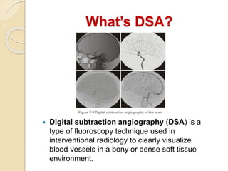
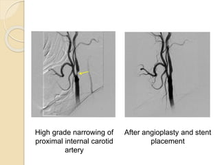
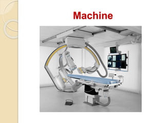
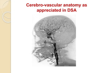
 Digital subtraction angiography (DSA) is a
type of fluoroscopy technique used in
interventional radiology to clearly visualize
blood vessels in a bony or dense soft tissue
environment.
Etymology
ANGIO?
ANGIO means blood vessel
And angiography is the radiological study of blood
vessel in the body after the
introduction of iodinated contrast media.
SUBTRACTION?
It is simply a technique by which bone structures
images are subtracted or canceled out from a
film of bones plus opacified vessels, leaving an
unobscured image of the vessels.
After angioplasty and stent
placement
High grade narrowing of
proximal internal carotid
artery
The syngo iFlow
Machine
Principle
 Blood vessels are not normally seen in
an x-ray image because of low
contrast.
 Dense fluid with high atomic numbers
increase the contrast. e.g. Iodine.
 Iodine absorbs photons more than
blood and tissue.
 The first contrast media used for
intravascular injection were called
high-osmolar contrast media (HOCM).
Block Diagram
Procedure
 Images are acquired by exposing an area of
interest.
 Image includes all overlying structure besides the
blood vessels in this area.
 Procedure:
◦ A mask image is acquired.
◦ Images of the same area at the rate of 1-6 frames/sec is
obtained while injecting the contrast.
 During angiography, patients may be sedated to reduce
anxiety.
 Their heart rate and rhythm, breathing, and oxygen
saturation are monitored throughout the procedure.
 Patient clean draped .
 A local anesthetic is usually used in the area where the
catheter is to be inserted, most commonly the femoral
artery.
 First, a small incision is given, medicut is inserted into the
artery. Fluoroscopy is used to guide the needle to the
proper position .
 The needle is then removed after placing guide wire in the
artery and vascular sheath is inserted over the guide wire .
The catheter is then inserted along the guide wire through
the sheath.
Routine
 When the catheter is in the correct position, the wire is pulled
out and dye is injected through the catheter.
 Images are acquired during contrast injection. Injections can
be made directly into the artery of interest (selective
arteriography)
 Complications from an arteriogram are very rare, but there is
some risk. Most problems that occur can be detected at the
time of the procedure or immediately after the procedure.
The artery may be injured at the puncture site or along the
artery where the catheter is passed.
 Oral fluid is given and analgesics are given if required.
 Special care should be given in case of children and
geriatric patient since additional patience is required.
Materials used:
 Catheters
 Vascular sheath
 Medicut
 Guidewires
 Contrast
 Connector/100
cm.tubing
 Surgical blade
Saline
Disposable syringes
Local anesthesia
Heparin
Surgical gloves
Elastoplast
Picard Pigtail
Sim-1
Catheters
VASCULAR SHEATH
MEDICUT
GUIDE WIRES
ELASTOPLAST STRIPS
CONTRAST
ANESTHESIA ANTICOAGULANT
PVA-PARTICLESGEL FOAM
Cerebro-vascular anatomy as
appreciated in DSA
Therapeutic usage
 Thrombectomy
THANK YOU!!

Digital subtraction angiography

  • 1.
  • 2.
    What’s DSA?  Digitalsubtraction angiography (DSA) is a type of fluoroscopy technique used in interventional radiology to clearly visualize blood vessels in a bony or dense soft tissue environment.
  • 3.
    Etymology ANGIO? ANGIO means bloodvessel And angiography is the radiological study of blood vessel in the body after the introduction of iodinated contrast media. SUBTRACTION? It is simply a technique by which bone structures images are subtracted or canceled out from a film of bones plus opacified vessels, leaving an unobscured image of the vessels.
  • 4.
    After angioplasty andstent placement High grade narrowing of proximal internal carotid artery
  • 5.
  • 6.
  • 7.
    Principle  Blood vesselsare not normally seen in an x-ray image because of low contrast.  Dense fluid with high atomic numbers increase the contrast. e.g. Iodine.  Iodine absorbs photons more than blood and tissue.  The first contrast media used for intravascular injection were called high-osmolar contrast media (HOCM).
  • 8.
  • 9.
    Procedure  Images areacquired by exposing an area of interest.  Image includes all overlying structure besides the blood vessels in this area.  Procedure: ◦ A mask image is acquired. ◦ Images of the same area at the rate of 1-6 frames/sec is obtained while injecting the contrast.
  • 10.
     During angiography,patients may be sedated to reduce anxiety.  Their heart rate and rhythm, breathing, and oxygen saturation are monitored throughout the procedure.  Patient clean draped .  A local anesthetic is usually used in the area where the catheter is to be inserted, most commonly the femoral artery.  First, a small incision is given, medicut is inserted into the artery. Fluoroscopy is used to guide the needle to the proper position .  The needle is then removed after placing guide wire in the artery and vascular sheath is inserted over the guide wire . The catheter is then inserted along the guide wire through the sheath. Routine
  • 11.
     When thecatheter is in the correct position, the wire is pulled out and dye is injected through the catheter.  Images are acquired during contrast injection. Injections can be made directly into the artery of interest (selective arteriography)  Complications from an arteriogram are very rare, but there is some risk. Most problems that occur can be detected at the time of the procedure or immediately after the procedure. The artery may be injured at the puncture site or along the artery where the catheter is passed.  Oral fluid is given and analgesics are given if required.  Special care should be given in case of children and geriatric patient since additional patience is required.
  • 12.
    Materials used:  Catheters Vascular sheath  Medicut  Guidewires  Contrast  Connector/100 cm.tubing  Surgical blade Saline Disposable syringes Local anesthesia Heparin Surgical gloves Elastoplast
  • 13.
  • 14.
  • 15.
  • 16.
  • 17.
  • 18.