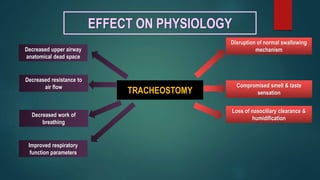
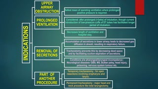
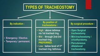
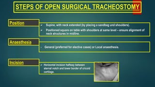
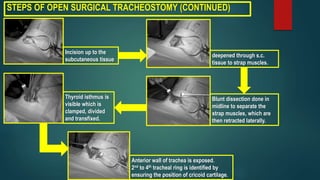
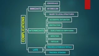
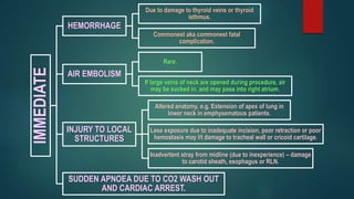
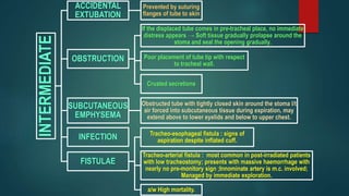
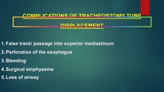
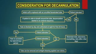
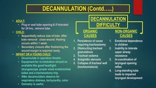
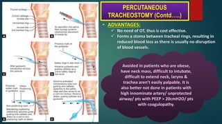
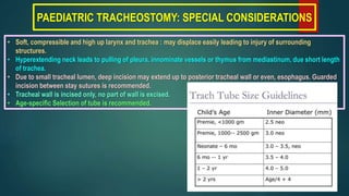
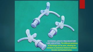
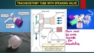
TRACHEOSTOMY
- AN OVERVIEW
-----------DR. SAYAN BANERJEE
DEFINITIONS
 TRACHEOSTOMY : (“stoma” meaning “mouth / mouth-like opening’)
Creation of a stoma at the skin surface that leads to the tracheal lumen.
 TRACHEOTOMY : (“tomo”- meaning “a cut /section”)
Creation of a surgical opening in the trachea.
EFFECT ON PHYSIOLOGY
TRACHEOSTOMY
Disruption of normal swallowing
mechanism
Compromised smell & taste
sensation
Loss of nasociliary clearance &
humidification
Decreased upper airway
anatomical dead space
Decreased resistance to
air flow
Decreased work of
breathing
Improved respiratory
function parameters
INDICATIONS
UPPER AIRWAY
OBSTRUCTION
PROLONGED
VENTILATION
REMOVAL OF
SECRETIONS
PART OF
ANOTHER
PROCEDURE
INDICATIONS
UPPER AIRWAY
OBSTRUCTION
CONGENITAL
Laryngeal web/cysts,
B/L choanal atresia,
Tracheoesophageal
fistula,
Craniofacial
anomalies,
Subglottic/tracheal
stenosis
ACQUIRED
Infective : Acute epiglottitis, Diphtheria, Acute layngo-tracheo-
bronchitis, Ludwig’s angina
Trauma : External injury to larynx/trachea, maxillofacial injury,
corrosive injury, inhalational injury
Neoplasm : Tumours of larynx, pharynx, tongue,
upper trachea
Foreign Body : Foreign body lodged in
larynx
Vocal cords palsy : B/L abductor paralysis,
Bulbar palsy
PROLONGED
VENTILATION
REMOVAL OF
SECRETIONS
PART OF ANOTHER
PROCEDURE
INDICATIONS
UPPER
AIRWAY
OBSTRUCTION
PROLONGED
VENTILATION
Safest mean of assisting ventilation where prolonged
positive pressure is required.
Considered after prolonged (>3wks) of intubation, though current
introduction of low-pressure cuffs of ET tubes has facilitated longer
period of intubation.
Decreases length of ventilation and
hospital stay.
REMOVAL OF
SECRETIONS
Accumulation of secretions in lower airway leads to decreased gas
diffusion in alveoli, resulting in respiratory failure.
Tracheostomy prevents this by decreasing dead space
and by facilitating suction-aspiration of secretions.
Conditions a/w pharyngeal/laryngeal incompetence :
Neurological diseases- GBS, MS, Bulbar palsy, head injury,
poisoning, nervous system tumour etc.
PART OF
ANOTHER
PROCEDURE
Temporary tracheostomy : for open
resections involving oropharynx and
larynx.
Permanent tracheostomy : major head and
neck procedure like total laryngectomy.
TYPES OF TRACHEOSTOMY
By indication :
• Emergency / Elective.
• Temporary / permanent.
By position of
tracheostoma :
• High : above isthmus
via 1st tracheal ring.
• Mid : through 2nd-3rd
tracheal ring,
PREFERRED.
• Low : below level of 3rd
tracheal ring /isthmus.
By surgical procedure :
• Open Surgical
tracheostomy.
• Mini-tracheostomy /
cricothyrotomy.
• Percutaneous
dilatational
tracheostomy.
STEPS OF OPEN SURGICAL TRACHEOSTOMY
 General (preferred for elective cases) or Local anaesthesia.
 Supine, with neck extended (by placing a sandbag und shoulders).
 Positioned square on table with shoulders at same level – ensure alignment of
neck structures in midline.
Anaesthesia
Incision  Horizontal incision halfway between
sternal notch and lower border of cricoid
cartilage.
Position
STEPS OF OPEN SURGICAL TRACHEOSTOMY (CONTINUED)
Incision up to the
subcutaneous tissue
deepened through s.c.
tissue to strap muscles.
Blunt dissection done in
midline to separate the
strap muscles, which are
then retracted laterally.
Thyroid isthmus is
visible which is
clamped, divided
and transfixed.
Anterior wall of trachea is exposed.
2nd to 4th tracheal ring is identified by
ensuring the position of cricoid cartilage.
Checklist before
entering trachea
Appropriately sized
tracheostomy tube with
deflated and patent cuff with
no leak.
Properly working ventilatory
circuit and connecting
equipment to ensure
uninterrupted ventilation
following tracheostomy.
Proper communication
between surgeon and
anaesthetist.
Pre-oxygenation
A vertical slit is made on anterior wall
of trachea between stay sutures.
Once Trachea is opened, surgeon can visualize the E.T. tube
Anaesthetist starts withdrawing E.T. tube under guidance of Surgeon
When the tip of E.T tube is just above the tracheotomy, withdrawal
stopped and tracheostomy tube is inserted.
Cuff inflated and connected to ventilator
Incision closed loosely and tube secured in position with sutures
and/or tape
STEPS OF OPEN SURGICAL TRACHEOSTOMY (CONTINUED)
POST-OPERATIVE CARE
Positioning of tube
• Original tube secured in position for at least 3 days to form a tract.
• Tube should be changed after 7 days and sutures removed.
Cuff deflation
Cuff of Portex tube deflated for 10 minutes every 2 hours to prevent pressure necrosis &
dilatation of trachea; Cuff should be permanently deflated once there is no risk for
aspiration. In most cases no need for inflation after 12 hours.
Suction of
secretions and
humidification • Frequent suctioning and hot water bath humidifiers or nebulizes may be required in
early post operative period.
• In non-ventilated patients covering the tube with gauze soaked in normal saline allows
retention of moisture.
Swallowing
• Due to pressure of cuff and due to limited movement of laryngopharynx.
• Soft diet with maneuvers to assist swallowing and to prevent aspiration.
SUCTION OF
SECRETIONS
COMPLICATIONS
IMMEDIATE
HEMORRHAGE
AIR EMBOLISM
INJURY TO LOCAL STRUCTURES
INTERMEDIATE
ACCIDENTAL EXTUBATION
OBSTRUCTION
SUBCUTANEOUS EMPHYSEMA
INFECTION
FISTULAE
LATE
TRACHEO-CUTANEOUS FISTULA
TRACHEAL STENOSIS
IMMEDIATE
HEMORRHAGE
Due to damage to thyroid veins or thyroid
isthmus.
Commonest aka commonest fatal
complication.
AIR EMBOLISM
Rare.
If large veins of neck are opened during procedure, air
may be sucked in, and may pass into right atrium.
INJURY TO LOCAL
STRUCTURES
Altered anatomy, e.g. Extension of apex of lung in
lower neck in emphysematous patients.
Less exposure due to inadequate incision, poor retraction or poor
hemostasis may l/t damage to tracheal wall or cricoid cartilage.
Inadvertent stray from midline (due to inexperience) – damage
to carotid sheath, esophagus or RLN.
SUDDEN APNOEA DUE TO CO2 WASH OUT
AND CARDIAC ARREST.
INTERMEDIATE
ACCIDENTAL
EXTUBATION
Prevented by suturing
flanges of tube to skin
OBSTRUCTION
If the displaced tube comes in pre-tracheal place, no immediate
distress appears → Soft tissue gradually prolapse around the
stoma and seal the opening gradually.
Poor placement of tube tip with respect
to tracheal wall.
Crusted secretions
SUBCUTANEOUS
EMPHYSEMA
Obstructed tube with tightly closed skin around the stoma l/t
air forced into subcutaneous tissue during expiration, may
extend above to lower eyelids and below to upper chest.
INFECTION
FISTULAE
Tracheo-esophageal fistula : signs of
aspiration despite inflated cuff.
Tracheo-arterial fistula : most common in post-irradiated patients
with low tracheostomy; presents with massive haemorrhage with
nearly no pre-monitory sign ;Innominate artery is m.c. involved;
Managed by immediate exploration.
a/w High mortality.
COMPLICATIONS OF TRACHEOSTOMY TUBE
DISPLACEMENT
1.False track/ passage into superior mediastinum
2.Perforation of the esophagus
3.Bleeding
4.Surgical emphysema
5.Loss of airway
CONSIDERATION FOR DECANNULATION
Initial cuff is replaced with an uncuffed fenestrated tube.
If patient is able to breath around the tube, decannulation
started in an ordered sequence.
Tube is blocked by day and unblocked at night for first 24 hours.
Patient tolerates?
Tube is occluded
for total 24 hour
period.
Tube can be removed and airtight dressing applied over stoma.
Yes No
Patient tolerates?
Yes
No
Patient tolerates?
Yes
Downsize tube, assurance,
counselling, retry after a few
days
DECANNULATION (Contd…..)
PERCUTANEOUS TRACHEOSTOMY
 Commonest procedure used now for provision of alternative airway in ITU patients.
 Most commonly used technique – Dilatation technique by Ciaglia.
PREOPERATIVE CRITERIA
•The ability to hyperextend the neck
•Presence of at least 1 cm distance
between cricoid cartilage and
suprasternal notch ensuring that the
patient will be able to be reintubated in
case of accidental extubation.
CONTRAINDICATIONS
•Children (younger than 12 years of age)
•Obese
•Patients with severe coagulopathies
STEPS • Position as surgical tracheostomy.
• Trachea punctured using cannula around needle. Position ensured by air aspired in water
filled syringe attached to needle.
• Needle removed.
• Guide wire inserted through cannula.
• Cannula withdrawn to allow series of dilators with increased diameter to pass over guide
wire.
• When passage is wide open, appropriately sized tracheostomy tube inserted and secured
in position.
(1) Cannula,
(2) Scalpel,
(3) Syringe,
(4) Cloth tie,
(5) Guide wire,
(6) Dilator,
(7) Tracheostomy tube,
(8) Obturator
PERCUTANEOUS
TRACHEOSTOM
Y KIT
• ADVANTAGES:
 No need of OT, thus is cost effective.
 Forms a stoma between tracheal rings, resulting in
reduced blood loss as there is usually no disruption
of blood vessels.
Avoided in patients who are obese,
have neck mass, difficult to intubate,
difficult to extend neck, larynx &
trachea aren’t easily palpable. It is
also better not done in patients with
high innominate artery/ unprotected
airway/ pts with PEEP > 20cmH2O/ pts
with coagulopathy.
PERCUTANEOUS
TRACHEOSTOMY (Contd…..)
PAEDIATRIC TRACHEOSTOMY: SPECIAL CONSIDERATIONS
• Soft, compressible and high up larynx and trachea : may displace easily leading to injury of surrounding
structures.
• Hyperextending neck leads to pulling of pleura, innominate vessels or thymus from mediastinum, due short length
of trachea.
• Due to small tracheal lumen, deep incision may extend up to posterior tracheal wall or even, esophagus. Guarded
incision between stay sutures is recommended.
• Tracheal wall is incised only, no part of wall is excised.
• Age-specific Selection of tube is recommended.
TRACHEOSTOMY
TUBES
FULLER’S BIVALVE METALLIC TRACHEOSTOMY TUBE
• Outer tube bi-valved. The 2 blades when pressed together,
help in smooth entry of tube.
• Inner tube is longer & has a vent for phonation.
• Pt phonates by closing main tube opening.
• Vent also helps in decannulation of tube.
JACKSON’S METALLIC TRACHEOSTOMY TUBE
• Made of German silver (alloy of Ag + Cu + P).
• Has obturator (pilot), inner tube & outer tube.
• Inner tube is longer than outer tube for its removal &
cleaning. Outer tube maintains patency. Pilot is inserted
into outer tube for smooth & non-traumatic insertion of
tube.
• Lock prevents expulsion of tube during cough.
TRACHEOSTOMY TUBE WITH SPEAKING VALVE
Tracheostomy

Tracheostomy

  • 1.
  • 2.
    DEFINITIONS  TRACHEOSTOMY :(“stoma” meaning “mouth / mouth-like opening’) Creation of a stoma at the skin surface that leads to the tracheal lumen.  TRACHEOTOMY : (“tomo”- meaning “a cut /section”) Creation of a surgical opening in the trachea.
  • 3.
    EFFECT ON PHYSIOLOGY TRACHEOSTOMY Disruptionof normal swallowing mechanism Compromised smell & taste sensation Loss of nasociliary clearance & humidification Decreased upper airway anatomical dead space Decreased resistance to air flow Decreased work of breathing Improved respiratory function parameters
  • 4.
  • 5.
    INDICATIONS UPPER AIRWAY OBSTRUCTION CONGENITAL Laryngeal web/cysts, B/Lchoanal atresia, Tracheoesophageal fistula, Craniofacial anomalies, Subglottic/tracheal stenosis ACQUIRED Infective : Acute epiglottitis, Diphtheria, Acute layngo-tracheo- bronchitis, Ludwig’s angina Trauma : External injury to larynx/trachea, maxillofacial injury, corrosive injury, inhalational injury Neoplasm : Tumours of larynx, pharynx, tongue, upper trachea Foreign Body : Foreign body lodged in larynx Vocal cords palsy : B/L abductor paralysis, Bulbar palsy PROLONGED VENTILATION REMOVAL OF SECRETIONS PART OF ANOTHER PROCEDURE
  • 6.
    INDICATIONS UPPER AIRWAY OBSTRUCTION PROLONGED VENTILATION Safest mean ofassisting ventilation where prolonged positive pressure is required. Considered after prolonged (>3wks) of intubation, though current introduction of low-pressure cuffs of ET tubes has facilitated longer period of intubation. Decreases length of ventilation and hospital stay. REMOVAL OF SECRETIONS Accumulation of secretions in lower airway leads to decreased gas diffusion in alveoli, resulting in respiratory failure. Tracheostomy prevents this by decreasing dead space and by facilitating suction-aspiration of secretions. Conditions a/w pharyngeal/laryngeal incompetence : Neurological diseases- GBS, MS, Bulbar palsy, head injury, poisoning, nervous system tumour etc. PART OF ANOTHER PROCEDURE Temporary tracheostomy : for open resections involving oropharynx and larynx. Permanent tracheostomy : major head and neck procedure like total laryngectomy.
  • 8.
    TYPES OF TRACHEOSTOMY Byindication : • Emergency / Elective. • Temporary / permanent. By position of tracheostoma : • High : above isthmus via 1st tracheal ring. • Mid : through 2nd-3rd tracheal ring, PREFERRED. • Low : below level of 3rd tracheal ring /isthmus. By surgical procedure : • Open Surgical tracheostomy. • Mini-tracheostomy / cricothyrotomy. • Percutaneous dilatational tracheostomy.
  • 10.
    STEPS OF OPENSURGICAL TRACHEOSTOMY  General (preferred for elective cases) or Local anaesthesia.  Supine, with neck extended (by placing a sandbag und shoulders).  Positioned square on table with shoulders at same level – ensure alignment of neck structures in midline. Anaesthesia Incision  Horizontal incision halfway between sternal notch and lower border of cricoid cartilage. Position
  • 11.
    STEPS OF OPENSURGICAL TRACHEOSTOMY (CONTINUED) Incision up to the subcutaneous tissue deepened through s.c. tissue to strap muscles. Blunt dissection done in midline to separate the strap muscles, which are then retracted laterally. Thyroid isthmus is visible which is clamped, divided and transfixed. Anterior wall of trachea is exposed. 2nd to 4th tracheal ring is identified by ensuring the position of cricoid cartilage.
  • 12.
    Checklist before entering trachea Appropriatelysized tracheostomy tube with deflated and patent cuff with no leak. Properly working ventilatory circuit and connecting equipment to ensure uninterrupted ventilation following tracheostomy. Proper communication between surgeon and anaesthetist. Pre-oxygenation A vertical slit is made on anterior wall of trachea between stay sutures. Once Trachea is opened, surgeon can visualize the E.T. tube Anaesthetist starts withdrawing E.T. tube under guidance of Surgeon When the tip of E.T tube is just above the tracheotomy, withdrawal stopped and tracheostomy tube is inserted. Cuff inflated and connected to ventilator Incision closed loosely and tube secured in position with sutures and/or tape STEPS OF OPEN SURGICAL TRACHEOSTOMY (CONTINUED)
  • 13.
    POST-OPERATIVE CARE Positioning oftube • Original tube secured in position for at least 3 days to form a tract. • Tube should be changed after 7 days and sutures removed. Cuff deflation Cuff of Portex tube deflated for 10 minutes every 2 hours to prevent pressure necrosis & dilatation of trachea; Cuff should be permanently deflated once there is no risk for aspiration. In most cases no need for inflation after 12 hours. Suction of secretions and humidification • Frequent suctioning and hot water bath humidifiers or nebulizes may be required in early post operative period. • In non-ventilated patients covering the tube with gauze soaked in normal saline allows retention of moisture. Swallowing • Due to pressure of cuff and due to limited movement of laryngopharynx. • Soft diet with maneuvers to assist swallowing and to prevent aspiration.
  • 14.
  • 15.
    COMPLICATIONS IMMEDIATE HEMORRHAGE AIR EMBOLISM INJURY TOLOCAL STRUCTURES INTERMEDIATE ACCIDENTAL EXTUBATION OBSTRUCTION SUBCUTANEOUS EMPHYSEMA INFECTION FISTULAE LATE TRACHEO-CUTANEOUS FISTULA TRACHEAL STENOSIS
  • 16.
    IMMEDIATE HEMORRHAGE Due to damageto thyroid veins or thyroid isthmus. Commonest aka commonest fatal complication. AIR EMBOLISM Rare. If large veins of neck are opened during procedure, air may be sucked in, and may pass into right atrium. INJURY TO LOCAL STRUCTURES Altered anatomy, e.g. Extension of apex of lung in lower neck in emphysematous patients. Less exposure due to inadequate incision, poor retraction or poor hemostasis may l/t damage to tracheal wall or cricoid cartilage. Inadvertent stray from midline (due to inexperience) – damage to carotid sheath, esophagus or RLN. SUDDEN APNOEA DUE TO CO2 WASH OUT AND CARDIAC ARREST.
  • 17.
    INTERMEDIATE ACCIDENTAL EXTUBATION Prevented by suturing flangesof tube to skin OBSTRUCTION If the displaced tube comes in pre-tracheal place, no immediate distress appears → Soft tissue gradually prolapse around the stoma and seal the opening gradually. Poor placement of tube tip with respect to tracheal wall. Crusted secretions SUBCUTANEOUS EMPHYSEMA Obstructed tube with tightly closed skin around the stoma l/t air forced into subcutaneous tissue during expiration, may extend above to lower eyelids and below to upper chest. INFECTION FISTULAE Tracheo-esophageal fistula : signs of aspiration despite inflated cuff. Tracheo-arterial fistula : most common in post-irradiated patients with low tracheostomy; presents with massive haemorrhage with nearly no pre-monitory sign ;Innominate artery is m.c. involved; Managed by immediate exploration. a/w High mortality.
  • 19.
    COMPLICATIONS OF TRACHEOSTOMYTUBE DISPLACEMENT 1.False track/ passage into superior mediastinum 2.Perforation of the esophagus 3.Bleeding 4.Surgical emphysema 5.Loss of airway
  • 20.
    CONSIDERATION FOR DECANNULATION Initialcuff is replaced with an uncuffed fenestrated tube. If patient is able to breath around the tube, decannulation started in an ordered sequence. Tube is blocked by day and unblocked at night for first 24 hours. Patient tolerates? Tube is occluded for total 24 hour period. Tube can be removed and airtight dressing applied over stoma. Yes No Patient tolerates? Yes No Patient tolerates? Yes Downsize tube, assurance, counselling, retry after a few days
  • 21.
  • 22.
    PERCUTANEOUS TRACHEOSTOMY  Commonestprocedure used now for provision of alternative airway in ITU patients.  Most commonly used technique – Dilatation technique by Ciaglia. PREOPERATIVE CRITERIA •The ability to hyperextend the neck •Presence of at least 1 cm distance between cricoid cartilage and suprasternal notch ensuring that the patient will be able to be reintubated in case of accidental extubation. CONTRAINDICATIONS •Children (younger than 12 years of age) •Obese •Patients with severe coagulopathies STEPS • Position as surgical tracheostomy. • Trachea punctured using cannula around needle. Position ensured by air aspired in water filled syringe attached to needle. • Needle removed. • Guide wire inserted through cannula. • Cannula withdrawn to allow series of dilators with increased diameter to pass over guide wire. • When passage is wide open, appropriately sized tracheostomy tube inserted and secured in position. (1) Cannula, (2) Scalpel, (3) Syringe, (4) Cloth tie, (5) Guide wire, (6) Dilator, (7) Tracheostomy tube, (8) Obturator PERCUTANEOUS TRACHEOSTOM Y KIT
  • 23.
    • ADVANTAGES:  Noneed of OT, thus is cost effective.  Forms a stoma between tracheal rings, resulting in reduced blood loss as there is usually no disruption of blood vessels. Avoided in patients who are obese, have neck mass, difficult to intubate, difficult to extend neck, larynx & trachea aren’t easily palpable. It is also better not done in patients with high innominate artery/ unprotected airway/ pts with PEEP > 20cmH2O/ pts with coagulopathy. PERCUTANEOUS TRACHEOSTOMY (Contd…..)
  • 24.
    PAEDIATRIC TRACHEOSTOMY: SPECIALCONSIDERATIONS • Soft, compressible and high up larynx and trachea : may displace easily leading to injury of surrounding structures. • Hyperextending neck leads to pulling of pleura, innominate vessels or thymus from mediastinum, due short length of trachea. • Due to small tracheal lumen, deep incision may extend up to posterior tracheal wall or even, esophagus. Guarded incision between stay sutures is recommended. • Tracheal wall is incised only, no part of wall is excised. • Age-specific Selection of tube is recommended.
  • 25.
  • 29.
    FULLER’S BIVALVE METALLICTRACHEOSTOMY TUBE • Outer tube bi-valved. The 2 blades when pressed together, help in smooth entry of tube. • Inner tube is longer & has a vent for phonation. • Pt phonates by closing main tube opening. • Vent also helps in decannulation of tube. JACKSON’S METALLIC TRACHEOSTOMY TUBE • Made of German silver (alloy of Ag + Cu + P). • Has obturator (pilot), inner tube & outer tube. • Inner tube is longer than outer tube for its removal & cleaning. Outer tube maintains patency. Pilot is inserted into outer tube for smooth & non-traumatic insertion of tube. • Lock prevents expulsion of tube during cough.
  • 31.
    TRACHEOSTOMY TUBE WITHSPEAKING VALVE