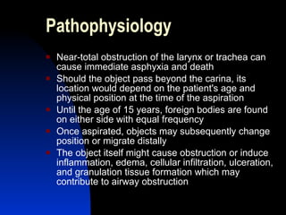
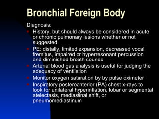
FOREIGN BODY ASPIRATION  (CHOKING) Myrna D.C. San Pedro, MD, FPPS
How common is choking? About 3,000 people die/year from choking Figure remained unchanged last 20 years There has been a progressive decline in the childhood deaths from choking: 1968, 650 children died 1990, 261 children died http://www.clevelandclinic.org/health/
Choking was the 4th leading cause of unintentional injury death in 2000.
Epidemiology  Ages affected  Age <3 years old: 50%  Age <10 years old: 95%  Preschoolers: 75-90% <1 year 10-15% 1-2 years 40-50% 2-3 years 15-25% >3 years 15-20%
Epidemiology Sex: boys more often than girls, 2:1 Increased incidence at holiday time  Known object aspiration in <40% Large case series indicate that nuts and peanuts account for an estimated half of all foreign body aspirations in children  Choking deaths related to toy use 68% of the time Latex Balloons 29% of choking deaths Marbles, Balls 19% of choking deaths
Choke Hazards A 1995 study found round objects to be most dangerous in choking deaths in children  According to Centers for Disease Control and Prevention, of the  > 17,000 cases of pediatric choking in 2001:  60% related to food 31% related to non-food substances 9% related to undetermined objects Of the food-related choking incidents,19% from candy or gum Of the choking incidents from non-food objects, 13% were related to coins
Location of Impacted Foreign Bodies Larynx   1-5% Trachea   5-15% L Main Bronchus   30-35% R Main Bronchus   30-40% L Lobar Bronchus  5-15% R Lobar Bronchus  5-15%
Pathophysiology Near-total obstruction of the larynx or trachea can cause immediate asphyxia and death  Should the object pass beyond the carina, its location would depend on the patient's age and physical position at the time of the aspiration Until the age of 15 years, foreign bodies are found on either side with equal frequency Once aspirated, objects may subsequently change position or migrate distally The object itself might cause obstruction or induce inflammation, edema, cellular infiltration, ulceration, and granulation tissue formation which may contribute to airway obstruction
Pathophysiology Distal to the obstruction, air trapping leading to local emphysema, atelectasis, hypoxic vasoconstriction, post-obstructive pneumonia and possible volume loss, necrotizing pneumonia or abscess, suppurative pneumonia, or bronchiectasis may occur The likelihood of complications increases after 24-48 hours, making quick removal of the foreign body urgent
Laryngeal Foreign Body Clinical Manifestations: Hoarseness  Croupy cough Aphonia Hemoptysis Dyspnea with wheezing  Cyanosis
Laryngeal Foreign Body Diagnosis: Lateral and anteroposterior x-rays of the neck will show indirect evidence if radiolucent or exact location if opaque foreign bodies Lodged anteriorly, in the larynx Behind soft-tissue shadows, hypopharynx or cervical esophagus Sagittal plane, larynx Coronal plane, esophagus Direct laryngoscopy confirms diagnosis and provides access for removal For severe dyspnea, do tracheotomy first before laryngoscopy
Tracheal Foreign Body Characteristic signs: Asthmatoid wheeze Audible slap and palpable thud produced by temporary expiratory impaction at the subglottic level Diagnosis: Occasionally by a chest x-ray In most cases, definitive only by a bronchoscopy
Bronchial Foreign Body Clinical Manifestations: Initial: cough, blood-streaked sputum & metallic taste Depends on degree of obstruction and stage patient seen: Nonobstructive – asymptomatic Near-total obstruction – signs of asphyxia Slight obstruction –wheezing Greater degree of obstruction – emphysema or atelectasis If persistent – chronic bronchopulmonary disease
Bronchial Foreign Body Clinical Manifestations: Most often, aspirated into right lung: Immediate: choking, gagging and paroxysmal coughing Latent period – occasional cough or slight wheezing Recurrent lobar pneumonia or intractable asthma Rarely, hemoptysis Vegetal –  arachidic bronchitis  – cough septic fever and dyspnea
Bronchial Foreign Body Diagnosis: History, but should always be considered in acute or chronic pulmonary lesions whether or not suggested PE: distally, limited expansion, decreased vocal fremitus, impaired or hyperresonant percussion and diminished breath sounds Arterial blood gas analysis is useful for judging the adequacy of ventilation Monitor oxygen saturation by by pulse oximeter Inspiratory posteroanterior (PA) chest x-rays to look for unilateral hyperinflation, lobar or segmental atelectasis, mediastinal shift, or pneumomediastinum
Bronchial Foreign Body Diagnosis: Expiratory chest radiographs are more sensitive for air trapping than inspiratory chest radiographs, also lateral decubitus views Fluoroscopy useful in check valve obstruction when little or no air escapes during expiration leading to obstructive overinflation Even extensive x-rays will not completely rule out presence of a foreign body; most foreign bodies are radiolucent but <20% of aspirated foreign bodies are radiopaque Only bronchoscopy definitive
Emergency Management <1 year:  5 Back slaps and 5 chest thrusts Blind finger sweeps should  not  be done
Heimlich Maneuver
Treatment Almost all aspirated foreign bodies can be extracted bronchoscopically If rigid bronchoscopy is unsuccessful, surgical bronchotomy or segmental resection may be necessary  Chronic bronchial obstruction with bronchiectasis and destruction of lung parenchyma may require segmental or lobar resection Antibiotics for secondary infections Steroids for inflammation Treat complications
Be master of your habits, Or they will master you. Thank You!

Foreign body aspiration

  • 1.
    FOREIGN BODY ASPIRATION (CHOKING) Myrna D.C. San Pedro, MD, FPPS
  • 2.
    How common ischoking? About 3,000 people die/year from choking Figure remained unchanged last 20 years There has been a progressive decline in the childhood deaths from choking: 1968, 650 children died 1990, 261 children died http://www.clevelandclinic.org/health/
  • 3.
    Choking was the4th leading cause of unintentional injury death in 2000.
  • 4.
    Epidemiology Agesaffected Age <3 years old: 50% Age <10 years old: 95% Preschoolers: 75-90% <1 year 10-15% 1-2 years 40-50% 2-3 years 15-25% >3 years 15-20%
  • 5.
    Epidemiology Sex: boysmore often than girls, 2:1 Increased incidence at holiday time Known object aspiration in <40% Large case series indicate that nuts and peanuts account for an estimated half of all foreign body aspirations in children Choking deaths related to toy use 68% of the time Latex Balloons 29% of choking deaths Marbles, Balls 19% of choking deaths
  • 6.
    Choke Hazards A1995 study found round objects to be most dangerous in choking deaths in children According to Centers for Disease Control and Prevention, of the > 17,000 cases of pediatric choking in 2001: 60% related to food 31% related to non-food substances 9% related to undetermined objects Of the food-related choking incidents,19% from candy or gum Of the choking incidents from non-food objects, 13% were related to coins
  • 7.
    Location of ImpactedForeign Bodies Larynx 1-5% Trachea 5-15% L Main Bronchus 30-35% R Main Bronchus 30-40% L Lobar Bronchus 5-15% R Lobar Bronchus 5-15%
  • 8.
    Pathophysiology Near-total obstructionof the larynx or trachea can cause immediate asphyxia and death Should the object pass beyond the carina, its location would depend on the patient's age and physical position at the time of the aspiration Until the age of 15 years, foreign bodies are found on either side with equal frequency Once aspirated, objects may subsequently change position or migrate distally The object itself might cause obstruction or induce inflammation, edema, cellular infiltration, ulceration, and granulation tissue formation which may contribute to airway obstruction
  • 9.
    Pathophysiology Distal tothe obstruction, air trapping leading to local emphysema, atelectasis, hypoxic vasoconstriction, post-obstructive pneumonia and possible volume loss, necrotizing pneumonia or abscess, suppurative pneumonia, or bronchiectasis may occur The likelihood of complications increases after 24-48 hours, making quick removal of the foreign body urgent
  • 10.
    Laryngeal Foreign BodyClinical Manifestations: Hoarseness Croupy cough Aphonia Hemoptysis Dyspnea with wheezing Cyanosis
  • 11.
    Laryngeal Foreign BodyDiagnosis: Lateral and anteroposterior x-rays of the neck will show indirect evidence if radiolucent or exact location if opaque foreign bodies Lodged anteriorly, in the larynx Behind soft-tissue shadows, hypopharynx or cervical esophagus Sagittal plane, larynx Coronal plane, esophagus Direct laryngoscopy confirms diagnosis and provides access for removal For severe dyspnea, do tracheotomy first before laryngoscopy
  • 12.
    Tracheal Foreign BodyCharacteristic signs: Asthmatoid wheeze Audible slap and palpable thud produced by temporary expiratory impaction at the subglottic level Diagnosis: Occasionally by a chest x-ray In most cases, definitive only by a bronchoscopy
  • 13.
    Bronchial Foreign BodyClinical Manifestations: Initial: cough, blood-streaked sputum & metallic taste Depends on degree of obstruction and stage patient seen: Nonobstructive – asymptomatic Near-total obstruction – signs of asphyxia Slight obstruction –wheezing Greater degree of obstruction – emphysema or atelectasis If persistent – chronic bronchopulmonary disease
  • 14.
    Bronchial Foreign BodyClinical Manifestations: Most often, aspirated into right lung: Immediate: choking, gagging and paroxysmal coughing Latent period – occasional cough or slight wheezing Recurrent lobar pneumonia or intractable asthma Rarely, hemoptysis Vegetal – arachidic bronchitis – cough septic fever and dyspnea
  • 15.
    Bronchial Foreign BodyDiagnosis: History, but should always be considered in acute or chronic pulmonary lesions whether or not suggested PE: distally, limited expansion, decreased vocal fremitus, impaired or hyperresonant percussion and diminished breath sounds Arterial blood gas analysis is useful for judging the adequacy of ventilation Monitor oxygen saturation by by pulse oximeter Inspiratory posteroanterior (PA) chest x-rays to look for unilateral hyperinflation, lobar or segmental atelectasis, mediastinal shift, or pneumomediastinum
  • 16.
    Bronchial Foreign BodyDiagnosis: Expiratory chest radiographs are more sensitive for air trapping than inspiratory chest radiographs, also lateral decubitus views Fluoroscopy useful in check valve obstruction when little or no air escapes during expiration leading to obstructive overinflation Even extensive x-rays will not completely rule out presence of a foreign body; most foreign bodies are radiolucent but <20% of aspirated foreign bodies are radiopaque Only bronchoscopy definitive
  • 17.
    Emergency Management <1year: 5 Back slaps and 5 chest thrusts Blind finger sweeps should not be done
  • 18.
  • 19.
    Treatment Almost allaspirated foreign bodies can be extracted bronchoscopically If rigid bronchoscopy is unsuccessful, surgical bronchotomy or segmental resection may be necessary Chronic bronchial obstruction with bronchiectasis and destruction of lung parenchyma may require segmental or lobar resection Antibiotics for secondary infections Steroids for inflammation Treat complications
  • 20.
    Be master ofyour habits, Or they will master you. Thank You!

Editor's Notes

  • #5 Factors: Orally focused development, No molar teeth, unable to chew and play when eating or distracted