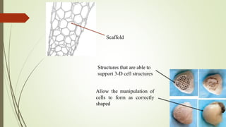
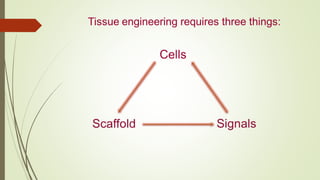
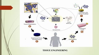
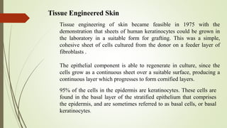
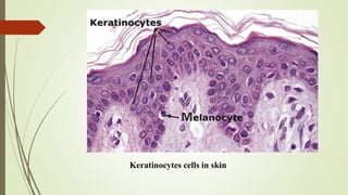
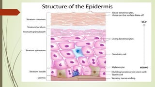
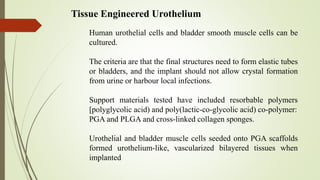
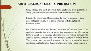
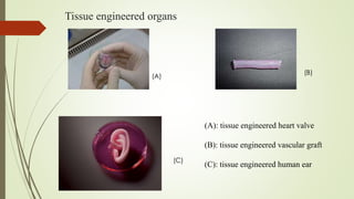
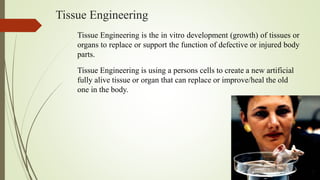
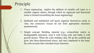
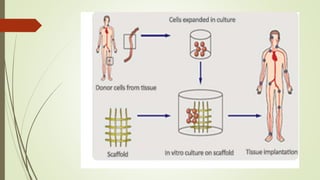
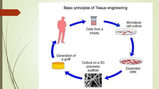
1. Tissue engineering involves growing tissues or organs in vitro to replace damaged body parts. Cells are seeded onto a scaffold and bathed in growth factors to grow new tissue.
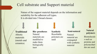
2. Common scaffolds include collagen, polymers like PLLA, and ceramics. Cells used include stem cells, keratinocytes for skin, and bladder cells.
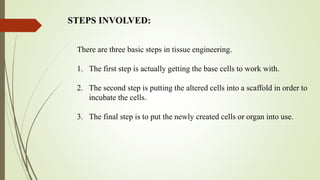
3. The process involves obtaining cells, seeding them onto a scaffold, and incubating the construct to grow new tissue which can then be implanted.
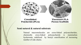
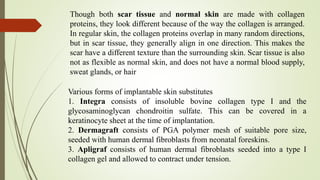
Introduction
Definition
History
Principle
Cell sources
What cells can be used?
Scaffolds
Biomaterials
Bioreactor
How tissue engineering is done?
How does tissue engineering differ from cloning?
Tissue engineering of specific structures
Application of tissue engineering
Limitations
Conclusion
References
Biomaterials for tissue engineering slideshareBukar Abdullahi
An overview of Tissue Engineering with some basics in Biomaterials and Synthetic Polymers. Further references should be considered as I presented this a specific target audience.
A presentation on Tissue Engineering made by Deepak Rajput. It was presented as a seminar requirement at the University of Tennessee Space Institute in Spring 2009.
TISSUE DEVELOPMENT WITH TISSUE ENGINEERING APPROACHFelix Obi
Tissue Engineering is the development and practice of combining scaffolds, cells, and suitable biochemical factors (regulatory factors or Signals) into functional tissues. The goal of tissue engineering is to assemble functional constructs that restore, maintain, or improve damaged tissues or whole organs.
Cells are the building blocks of tissue, and tissues are the basic unit of function in the body. Generally, groups of cells make and secrete their own support structures, called extracellular matrix. This matrix, or scaffold, does more than just support the cells; it also acts as a relay station for various signaling molecules. Thus, cells receive messages from many sources that become available from the local environment. Each signal can start a chain of responses that determine what happens to the cell. By understanding how individual cells respond to signals, interact with their environment, and organize into tissues and organisms, Tissue Engineers are now able to manipulate these processes to amend damaged tissues or even create new ones.
Introduction
Artificial skin
Invention
Structure of human skin
Importance of skin
Key development
Biomaterials
Methods to produce artificial skin
Application
Problems
Future development
Conclusions
references
Engineering bone tissue using human Embryonic Stem CellsBalaganesh Kuruba
Bone defects lead by traumatic injuries, congenital malformations and other surgical rescissions rises the immediate need for a more evolved and safer approaches in tissue repair at alarming rates for the downgrading issues with existing strategies which needs to be addressed. Currently practiced treatment methods addressing the issue with bone defects are invasive, traumatic and are not cost effective. Yet, issues of immune rejection either immediately or in the later stages have been reported claiming its ineffectiveness in some selective case studies.
Previous work by researchers carried out the "Biomimetic" approach to provide the cells with the microenvironment and in situ conditions for the cells seeded into the 3D Osteogenic scaffolds enriched with growth supplments. Here, we address the issue of non-availability of therapeutic cells - a major problem with current translational medicine by proposing the use of Human Embryonic Stem Cells in generating strong and structurally rigid bone tissue. Inducing the production of Mesenchymal Progenitor cells from Human Embryonic Stem cells in Serum supplemented expansion medium and elimination of bone Fibroblast growth factor produced high quality MPCs which were induced in osteogenic medium to result in bone differentiating cells. Culturing these MPCs produced from three different protocols into 3D Scaffold and 3D-Endoret Osteogenic Scaffold produced tissue constructs which are analysed both biochemically and Histologically to check for the Bone tissue differentiation parameters such as Bone sialoprotein deposition, Osteopontin accumulation and Collagen deposition. Matrix mineralization in these constructs were studied by uCT imaging and safety studies were conducted by studying Orthotopic implantation models in SCID mouse. And the results are expected to be optimistically affirmative which shall lay a new foundation and pioneer a whole new approach in the field of Tissue Engineering.
CAR-T (Cell Therapy) Nomenclature Review & Brand Equity Study. April 15, 2015Bill Smith
CAR-T (Cell Therapy) Nomenclature Review & Brand Equity Study. April 15, 2015.
Brand Acumen. The Global Leader in Pharmaceutical Name Development and Submission Strategy.
Final presentation for BIOL405, NSC, Spring 2014. Presented by Kevin Hugins and Duy-Khiem Chanh Pham. This presentation addressed the use of Chimeric Antigen Receptors for gene therapy for cancer. Gene therapy was first conceptualized to alter debilitating fates of genetic diseases. Gene therapy technology can help introduce new functional DNA to replace mutated genes. The idea first arose in 1972 when Friedmann and Roblin authored a paper, “Gene therapy for human genetic disease?”, demonstrating that exogenous DNA can be taken up by mammalian cells (1). They proposed that the same procedure could be done on humans to correct genetic defects by introducing therapeutic DNA. Currently, genetic modification of T lymphocytes has been the major area of research for treating malignant tumors. This technique seeks to create chimeric antigen receptor (CAR) in T cells by genetically modifying them in vitro and reintroduce them back into blood circulation. The T cells are unique to every patient and the chimeric antigen receptors are unique to the tumor that it is targeting.
Indian Dental Academy: will be one of the most relevant and exciting training center with best faculty and flexible training programs for dental professionals who wish to advance in their dental practice,Offers certified courses in Dental implants,Orthodontics,Endodontics,Cosmetic Dentistry, Prosthetic Dentistry, Periodontics and General Dentistry.
what is tissue engineering
Sources of tissue grafting
Strategies for tissue engineering
Stem cells
Several strategies are now available for developing new organs and tissues
What is the scaffold?
Ideal properties of scaffold
Scaffolding procedures
BIOMATERIALS AND SCAFFOLDS
CAD-CAM technique for scaffolding design
CELL CULTURE METHODS
TISSUE-ENGINEERED DENTAL TISSUES
Biomaterials were defined as “any substance, other than a drug, or a combination of substances, synthetic or natural in origin, which can be used for any period of time, as a whole or as a part of a system, which treats, augments or replaces any tissue, organ or function of the body”
Stem Cells and Tissue Engineering: past, present and futureAna Rita Ramos
Tissue engineering brings together the principles of the life sciences and medicine with engineering. New biomaterials; advances in genomics and proteomics and increased understanding of healing processes contributed to the increase of this area over the past decade.
Stem cell biology is paving the way for the generation of unlimited cells of specific phenotypes for incorporation
into engineered tissue constructs.
Multi-source connectivity as the driver of solar wind variability in the heli...Sérgio Sacani
The ambient solar wind that flls the heliosphere originates from multiple
sources in the solar corona and is highly structured. It is often described
as high-speed, relatively homogeneous, plasma streams from coronal
holes and slow-speed, highly variable, streams whose source regions are
under debate. A key goal of ESA/NASA’s Solar Orbiter mission is to identify
solar wind sources and understand what drives the complexity seen in the
heliosphere. By combining magnetic feld modelling and spectroscopic
techniques with high-resolution observations and measurements, we show
that the solar wind variability detected in situ by Solar Orbiter in March
2022 is driven by spatio-temporal changes in the magnetic connectivity to
multiple sources in the solar atmosphere. The magnetic feld footpoints
connected to the spacecraft moved from the boundaries of a coronal hole
to one active region (12961) and then across to another region (12957). This
is refected in the in situ measurements, which show the transition from fast
to highly Alfvénic then to slow solar wind that is disrupted by the arrival of
a coronal mass ejection. Our results describe solar wind variability at 0.5 au
but are applicable to near-Earth observatories.
(May 29th, 2024) Advancements in Intravital Microscopy- Insights for Preclini...Scintica Instrumentation
Intravital microscopy (IVM) is a powerful tool utilized to study cellular behavior over time and space in vivo. Much of our understanding of cell biology has been accomplished using various in vitro and ex vivo methods; however, these studies do not necessarily reflect the natural dynamics of biological processes. Unlike traditional cell culture or fixed tissue imaging, IVM allows for the ultra-fast high-resolution imaging of cellular processes over time and space and were studied in its natural environment. Real-time visualization of biological processes in the context of an intact organism helps maintain physiological relevance and provide insights into the progression of disease, response to treatments or developmental processes.
In this webinar we give an overview of advanced applications of the IVM system in preclinical research. IVIM technology is a provider of all-in-one intravital microscopy systems and solutions optimized for in vivo imaging of live animal models at sub-micron resolution. The system’s unique features and user-friendly software enables researchers to probe fast dynamic biological processes such as immune cell tracking, cell-cell interaction as well as vascularization and tumor metastasis with exceptional detail. This webinar will also give an overview of IVM being utilized in drug development, offering a view into the intricate interaction between drugs/nanoparticles and tissues in vivo and allows for the evaluation of therapeutic intervention in a variety of tissues and organs. This interdisciplinary collaboration continues to drive the advancements of novel therapeutic strategies.
Seminar of U.V. Spectroscopy by SAMIR PANDASAMIR PANDA
Spectroscopy is a branch of science dealing the study of interaction of electromagnetic radiation with matter.
Ultraviolet-visible spectroscopy refers to absorption spectroscopy or reflect spectroscopy in the UV-VIS spectral region.
Ultraviolet-visible spectroscopy is an analytical method that can measure the amount of light received by the analyte.
A brief information about the SCOP protein database used in bioinformatics.
The Structural Classification of Proteins (SCOP) database is a comprehensive and authoritative resource for the structural and evolutionary relationships of proteins. It provides a detailed and curated classification of protein structures, grouping them into families, superfamilies, and folds based on their structural and sequence similarities.
Deep Behavioral Phenotyping in Systems Neuroscience for Functional Atlasing a...Ana Luísa Pinho
Functional Magnetic Resonance Imaging (fMRI) provides means to characterize brain activations in response to behavior. However, cognitive neuroscience has been limited to group-level effects referring to the performance of specific tasks. To obtain the functional profile of elementary cognitive mechanisms, the combination of brain responses to many tasks is required. Yet, to date, both structural atlases and parcellation-based activations do not fully account for cognitive function and still present several limitations. Further, they do not adapt overall to individual characteristics. In this talk, I will give an account of deep-behavioral phenotyping strategies, namely data-driven methods in large task-fMRI datasets, to optimize functional brain-data collection and improve inference of effects-of-interest related to mental processes. Key to this approach is the employment of fast multi-functional paradigms rich on features that can be well parametrized and, consequently, facilitate the creation of psycho-physiological constructs to be modelled with imaging data. Particular emphasis will be given to music stimuli when studying high-order cognitive mechanisms, due to their ecological nature and quality to enable complex behavior compounded by discrete entities. I will also discuss how deep-behavioral phenotyping and individualized models applied to neuroimaging data can better account for the subject-specific organization of domain-general cognitive systems in the human brain. Finally, the accumulation of functional brain signatures brings the possibility to clarify relationships among tasks and create a univocal link between brain systems and mental functions through: (1) the development of ontologies proposing an organization of cognitive processes; and (2) brain-network taxonomies describing functional specialization. To this end, tools to improve commensurability in cognitive science are necessary, such as public repositories, ontology-based platforms and automated meta-analysis tools. I will thus discuss some brain-atlasing resources currently under development, and their applicability in cognitive as well as clinical neuroscience.
Richard's aventures in two entangled wonderlandsRichard Gill
Since the loophole-free Bell experiments of 2020 and the Nobel prizes in physics of 2022, critics of Bell's work have retreated to the fortress of super-determinism. Now, super-determinism is a derogatory word - it just means "determinism". Palmer, Hance and Hossenfelder argue that quantum mechanics and determinism are not incompatible, using a sophisticated mathematical construction based on a subtle thinning of allowed states and measurements in quantum mechanics, such that what is left appears to make Bell's argument fail, without altering the empirical predictions of quantum mechanics. I think however that it is a smoke screen, and the slogan "lost in math" comes to my mind. I will discuss some other recent disproofs of Bell's theorem using the language of causality based on causal graphs. Causal thinking is also central to law and justice. I will mention surprising connections to my work on serial killer nurse cases, in particular the Dutch case of Lucia de Berk and the current UK case of Lucy Letby.
Professional air quality monitoring systems provide immediate, on-site data for analysis, compliance, and decision-making.
Monitor common gases, weather parameters, particulates.
2. Tissue Engineering
Tissue Engineering is the in vitro development (growth) of tissues or
organs to replace or support the function of defective or injured body
parts.
Tissue Engineering is using a persons cells to create a new artificial
fully alive tissue or organ that can replace or improve/heal the old
one in the body.
3. Principle
1. Tissue engineering implies the addition of suitable cell types to a
suitable support matrix, through which an organised and functional
tissue is formed (resembling the tissue engineering)
2. Epithelial and endothelial cell layers organize themselves easily in
vitro, but connective tissues do not form appropriate structures
spontaneously.
3. Simple concept: Building material (e.g., extracellular matrix or
biodegradable polymer), seed it with living cells and bathe it with
growth factors. When the cells multiply, they fill up the scaffold and
grow into three-dimensional tissue, and once implanted in the body,
the cells recreate their intended tissue functions.
4.
5.
6. Initially approved by FDA for treatment of venous leg ulcers, later on in 2000 for
treatment of diabetic foot ulcers.
Apligraf contains two types of cells – an outer layer of protective skin cells, and
an inner layer of cells contained within collagen. Both types of cells contain
substances similar to those found in human skin. Apligraf does not contain
certain things in skin such as hair follicles, sweat glands or blood vessels.
Pro-osteon is a coralline hydroxyapatite, bone like graft used to fill defects in
bone.
In 1960’s artificial skin was being used to treat burn victims, later on modified
into synthetic fibres for artificial skin grafts.
1n 1990’s pro-osteon coral derived bone graft material was introduced and 1996
integra’s was approved for as an tissue regeneration product.
In 1998 “Apligraf ” approved for treatment of ulcers,
8. Cell substrate and Support material
Nature of the support material depends on the information and
suitability for the adherent cell types.
It is divided into 5 broad classes:
Traditional
Abiotic
materials
(metals and
ceramics)
Bio- prostheses
Natural
materials are
modified to
make them
biologically
inert
Synthetic
Resorbable
polymers are
used
Semi-natural Natural
polymers
Natural
material are
conjugated
with synthetic
material
Biomolecule
s such as
proteins and
polysacchari
des are used
9. BIOINERT RESORBABLE BIOACTIVE
Support material
Bio-Compatibility
1. No material can be totally inert when implanted but the group known as “bioinert”
only provoke the formation of scar tissues (eg: stainless steel in artificial hips)
2. Resorbable materials dissolve when implanted with generation of harmless
dissolution products (eg: polymers like PLLA using suturing)
Poly(L-lactic) acid is a biodegradable thermoplastic a aliphatic polyester derived
from renewable resources, such as corn starch (in the United States), tapioca roots, chips
or starch (mostly in Asia), or sugarcane.
3. Bioactive material stimulate a biological response from the body (eg: synthetic
hydroxyapatite ceramics and bioactive glasses.
10. Bioprostheses
1. It is formed by the extensive cross-linking of natural tissue eg: porcin heart
valves and tendons
2. These are designed and fabricated primarily to function as long as possible
independently and without modification by surrounding tissue
eg: collagen based connective tissue stabilized by glutaraldehyde, can survive
unchanged for many years.
Traditional support materials are not used because they do not
integrate within reasonable period.
12. 1. Resorbable polymers are used which are hydrolysed and then
phagocytosed, the greatest advantage of such material is their easy
and cheap production in a controllable & reproducible manner at
large scale.
2. Less compatibility than natural polymers
3. Synthetic polymers used are PGA [poly(glycolic acid)],
PLA[Poly(l-lactic acid)], polycarbonate, polycaprolactone.
4. PLA, PGA and PLGA[poly(lactic-co-glycolic acid) are most widely
used, PLA is amorphous and hydrophobic degrading to release lactic
acid.
13. Semi natural & natural substrate
Natural macromolecules are cross-linked polysaccharides,
chemically cross-linked polysaccharide is mammalian
hyaluronan, stabilized by benzyl esterification of increasing
number of side chains.
14. Collagen sponges are also used, prepared from various insoluble and
aggregated collagen eg: collagen scaffold in tubular shape, with smooth
muscles and endothelial cells
Tissue engineered
heart valve
Tissue engineered
vascular graft
15. Types of cells
1. Autologous cells are obtained from the same individual to which
they will be re-implanted. Autologous cells have the fewest
problems with rejection and pathogen transmission, however in
some cases might not be available
2. Allogeneic cells come from the body of a donor of the same
species.
3. Xenogenic cells are these isolated from individuals of another
species. In particular animal cells have been used quite extensively in
experiments aimed at the construction of cardiovascular implants.
4. Syngenic or isogenic cells are isolated from genetically identical
organisms, such as twins, clones, or highly inbred research animal
models
16. Stem cells are undifferentiated cells with the ability to divide in culture and
give rise to different forms of specialized cells. According to their source
stem cells are divided into "adult" and "embryonic" stem cells, the first class
being multipotent and the latter mostly pluripotent; some cells are
totipotent, in the earliest stages of the embryo.
Scaffolds
Cells are often implanted or 'seeded' into an artificial structure capable of
supporting three-dimensional tissue formation. Scaffolds usually serve at
least one of the following purposes:
1. Allow cell attachment and migration
2. Deliver and retain cells and biochemical factors
3. Enable diffusion of vital cell nutrients and expressed products
17. Allow the manipulation of
cells to form as correctly
shaped
Structures that are able to
support 3-D cell structures
Scaffold
18. Stem cells
Undifferentiated cells with ability to divide in culture & give rise to
different forms of specialized cells.
Characteristic Features:
1. They are capable of dividing & renewing themselves for long
periods
2. They are unspecialized
3. They can give rise to specialized cell types.
Pluripotent
cells
Totipotent
cells
Multipotent
cells
20. Embryonic stem cell lines are cultures of cells derived from epiblast
tissue of inner cell mass of a blastocyst or earlier morula stage
embryos — approximately 4 to 5 days old in humans & consisting of
50–150 cells. ES cells are pluripotent & give rise during
development to all derivatives of 3 primary germ layers:
1. ectoderm,
2. endoderm &
3. mesoderm.
Embryonic stem cells
21.
22. Totipotent stem cells can differentiate into embryonic & extra embryonic
cell types. Such cells can construct a complete, viable organism. These cells
are produced from fusion of an egg & sperm cell. Eg: Fertilized egg
Multipotent stem cells give rise to a limited range of cells within a tissue
type. Eg: Hematopoietic stem cells
Pluripotent stem cells are descendants of totipotent cells & can
differentiate into nearly all cells, but cannot give rise to an entire
organism. i.e. cells derived from any of three germ layers
23.
24. Unipotent cells can produce only one cell type, their own, but have the property
of self-renewal, which distinguishes them from non-stem cells. E.g. muscle stem
cells.
26. Mesenchymal
Mesenchymal stem cells, or MSCs, are multipotent stromal cells
that can differentiate into a variety of cell types, including:
osteoblasts (bone cells), chondrocytes (cartilage cells), and
adipocytes (fat cells).
27. There are three basic steps in tissue engineering.
1. The first step is actually getting the base cells to work with.
2. The second step is putting the altered cells into a scaffold in order to
incubate the cells.
3. The final step is to put the newly created cells or organ into use.
STEPS INVOLVED:
30. 95% of the cells in the epidermis are keratinocytes. These cells are
found in the basal layer of the stratified epithelium that comprises
the epidermis, and are sometimes referred to as basal cells, or basal
keratinocytes.
Tissue Engineered Skin
Tissue engineering of skin became feasible in 1975 with the
demonstration that sheets of human keratinocytes could be grown in
the laboratory in a suitable form for grafting. This was a simple,
cohesive sheet of cells cultured from the donor on a feeder layer of
fibroblasts .
The epithelial component is able to regenerate in culture, since the
cells grow as a continuous sheet over a suitable surface, producing a
continuous layer which progresses to form cornified layers.
33. Though both scar tissue and normal skin are made with collagen
proteins, they look different because of the way the collagen is arranged.
In regular skin, the collagen proteins overlap in many random directions,
but in scar tissue, they generally align in one direction. This makes the
scar have a different texture than the surrounding skin. Scar tissue is also
not as flexible as normal skin, and does not have a normal blood supply,
sweat glands, or hair
Various forms of implantable skin substitutes
1. Integra consists of insoluble bovine collagen type I and the
glycosaminoglycan chondroitin sulfate. This can be covered in a
keratinocyte sheet at the time of implantation.
2. Dermagraft consists of PGA polymer mesh of suitable pore size,
seeded with human dermal fibroblasts from neonatal foreskins.
3. Apligraf consists of human dermal fibroblasts seeded into a type I
collagen gel and allowed to contract under tension.
36. Human urothelial cells and bladder smooth muscle cells can be
cultured.
The criteria are that the final structures need to form elastic tubes
or bladders, and the implant should not allow crystal formation
from urine or harbour local infections.
Support materials tested have included resorbable polymers
[polyglycolic acid) and poly(lactic-co-glycolic acid) co-polymer:
PGA and PLGA and cross-linked collagen sponges.
Urothelial and bladder muscle cells seeded onto PGA scaffolds
formed urothelium-like, vascularized bilayered tissues when
implanted
Tissue Engineered Urothelium
38. ARTIFICIAL BONE GRAFTS: PRO OSTEON
Safe, strong, and cost effective bone grafts are now performed
using synthetic material known as Pro Osteon Implant 500.
It is sterile, biocompatible (meaning the body’s immune system
does not reject it), and it is easily sculpted to fill a defect in
fractured bones.
Pro Osteon mimics the internal structure of human bone. This
synthetic material is made by subjecting a common, non-decorative
form to coral to a patented chemical process which converts the
coral to hydroxyapatite, the same mineral content of human bone.
The porous, interconnected structure of the coral remains intact,
providing an ideal matrix through which new bone tissue can grow.
39. Using Pro Osteon on a long bone, the surgeon determines the amount of bone
graft he needs and shapes the Pro Osteon block to fit into the damaged area.
The graft area is stabilized with a metal plate, screws, or some other form of
internal fixation.
pro-osteon
complex
40. Bioengineered Tissue Implants Regenerate Damaged Knee Cartilage
ScienceDaily(July 5, 2006)
Cartilage was removed from 23 patients with an average age of 36 years.
After growing the cells in culture for 14 days, the researchers seeded them
onto scaffolds made of esterified hyaluronic acid, grew them for another 14
days on the scaffolds, and then implanted them into the injured knees of the
study patients.
Cartilage regeneration was seen in ten of 23 patients, including in some
patients with pre-existing early osteoarthritis of the knee secondary to
traumatic injury. Maturation of the implanted, tissue-engineered cartilage was
evident as early as 11 months after implantation