
Recommended
More Related Content
What's hot
What's hot (20)
Viewers also liked
Viewers also liked (17)
Similar to Chirag
Similar to Chirag (20)
Recently uploaded
Recently uploaded (20)
Chirag
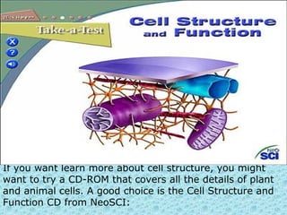
- 1. If you want learn more about cell structure, you might want to try a CD-ROM that covers all the details of plant and animal cells. A good choice is the Cell Structure and Function CD from NeoSCI:
- 2. Cell structure and function A cell has many different parts starting with the outer shell the plasma membrane which protects and regulates molecules from entering and exiting the cell. Within the cell is cytoplasm a semi fluid that contains organelles and where cell functions are completed. There are two kinds of cells prokaryotic and eukaryotic cells. Prokaryotic cells lack a nucleus and are smaller and less complicated than eukaryotic cells. Bacteria and archaea are prokaryotic cells and are used in biotechnology products. Eukaryotic cells are plant and animal cells and have a nucleus. Eukaryotic cells each have their own function in an organism. For example gamete cells also known as egg or sperm cells are for reproduction. Hormone secreting cells like somatotropes produce growth hormones, and white blood cells fight off infections in our bodies.
- 3. Prokaryotic cell Cell Organelles and Metabolism The cell is composed of many organelles each having there own function. The nucleus is comprised of the nuclear envelope (encloses the nucleus), chromatin (DNA and protein) and nucleolus (produces subunits of ribosomes). Important organelles in the cell are lysosomes that digest cell parts, a vesicle that transports and stores substances, and golgi apparatus that modifies and distributes secretory products. Mitochondrion an organelle in the cytoplasm performs cellular respiration which coverts energy of glucose into chemical energy of ATP molecules. During this process mitochondria uses up oxygen and gives off carbon dioxide. This process of cellular respiration of mitochondrion is very important to cellular metabolism.
- 4. Eukaryotic cell Tissue Types Tissue is cells of the same type organized together to perform a common function. There are four tissue type connective, muscular, nervous, and epithelial tissues.
- 5. Connective tissue binds and supports other tissues together. There are four kinds of connective tissue fibrous, supportive, and fluid. Loose and dense fibrous tissue is important and present in lungs, arteries and the urinary bladder. Cartilage and bone are types of supportive connective tissue and blood and lymph are types of fluid connective tissue. Connective tissue is made up of ground substance , stem cell, fibroblast and many other characteristic shown in the diagram below.
- 6. Muscular tissue is composed of muscle fibers which contain protein filaments and myosin filaments. Skeletal, smooth, and cardiac muscle are the three types of vertebrate muscular tissues. Skeletal muscle is voluntary when it contracts body parts move. Skeletal muscle is attached to bones by tendons. They are cylindrical and long and with multiple nuclei in the striated cells. Smooth or visceral muscle is involuntary with a single nuclei for each spindle-shaped cells. Smooth muscle lines blood vessels and the digestive tract. Cardiac muscle is only found in the heart and has characteristics from both smooth and skeletal. Its has branching striated cells like skeletal but with only a single nuclei and it too in involuntary like smooth.
- 7. Epithelial tissues are cells of uniform type and are joined by a basement membrane composed of carbohydrates and proteins. These tissues are to protect, absorb molecules or nutrients, and help sweep impurities away depending on the location in the body. Epithelial tissues protect the lining of the lungs, blood vessels, nose, mouth, and esophagus. Epithelial tissues lines kidney tubules, small intestines, and digestive tract.
- 10. An animal cell can be comprised of anything from a single cell organism to a heart muscle cell in a human. While cells vary greatly in their abilities and functions, they all contain certain specific parts, known as organelles
- 12. This is a diagram of an animal cell. I based my information on this diagrams: [1][2][3] [4] [5]. there are some structures I didn't add because it was not clear to me how they should look like. For all have doubts between cytoskeleton, microfibers and microtubules. If you would like to make another language version of it, message me and can make an image without text for you or message me the names in the language you want them to appear. [6]
- 14. The CD also contains interactive lab investigations that allow students to perform many different cytology techniques:
- 15. KU BIOL 150 - Principles of Cellular and Mollecular Biology Laboratory An integrated lecture and laboratory course for biology majors and students planning to take additional courses in biology. This course covers basic biochemistry, cell structure and function, molecular biology, genetics, physiology, and development of plants and animals. Two hours of laboratory per week.
- 16. •Structure - 2 primary building blocks include protein (about 60% of the membrane) and lipid, or fat (about 40% of the membrane). The primary lipid is called phospholipid, and molecules of phospholipid form a 'phospholipid bilayer' (two layers of phospholipid molecules). This bilayer forms because the two 'ends' of phospholipid molecules have very different characteristics: one end is polar (or hydrophilic) and one (the hydrocarbon tails below) is non-polar (or hydrophobic):
- 17. • At the heart of the immune response is the ability to distinguish between self and nonself. Every body cell carries distinctive molecules that distinguish it as "self." Normally the body's defenses do not attack tissues that carry a self marker; rather, immune cells coexist peaceably with other body cells in a state known as self-tolerance (Source: National Cancer Institute).
- 18. The cyotoskeleton represents the cell's skeleton. Like the bony skeletons that give us stability, the cytoskeleton gives our cells shape, strength, and the ability to move, but it does much more than that. The cytoskeleton is made up of three types of fibers that constantly shrink and grow to meet the needs of the cell: microtubules, microfilaments, and actin filaments. Each type of fiber looks, feels, and functions differently. Microtubules consists of a strong protein called tubulin and they are the 'heavy lifters' of the cytoskeleton. They do the tough physical labor of separating duplicate chromosomes when cells copy themselves and serve as sturdy railway tracks on which countless molecules and materials shuttle to and fro. They also hold the ER and Golgi neatly in stacks and form the main component of flagella and cilia.
- 19. Microfilaments are unusual because they vary greatly according to their location and function in the body. For example, some microfilaments form tough coverings, such as in nails, hair, and the outer layer of skin (not to mention animal claws and scales). Others are found in nerve cells, muscle cells, the heart, and internal organs. In each of these tissues, the filaments are made of different proteins. Actin filament are made up of two chains of the protein actin twisted together. Although actin filaments are the most brittle of the cytoskeletal fibers, they are also the most versatile in terms of the shapes they can take. They can gather together into bundles, weblike networks, or even three-dimensional gels. They shorten or lengthen to allow cells to move and change shape. Together with a protein partner called myosin, actin filaments make possible the muscle contractions necessary for everything from your action on a sports field to the automatic beating of your heart. (Source: NIGM).
- 20. Mitochondria are found exclusively in eukaryotic cells. These organelles are often called the "power plants" of the cell because their main job is to make energy (ATP). Mitochondria are highly unusual-- they contain their own genetic material and protein-making machinery enwrapped in a double membrane. Many scientists believe mitochondria were once free-living bacteria that colonized complex cells sometime during evolution. (Source: NSF).