
Recommended
More Related Content
Similar to Unit 1 Humans and Animals Cells. 4TH OF ESO
Similar to Unit 1 Humans and Animals Cells. 4TH OF ESO (20)
Recently uploaded
Recently uploaded (20)
Unit 1 Humans and Animals Cells. 4TH OF ESO
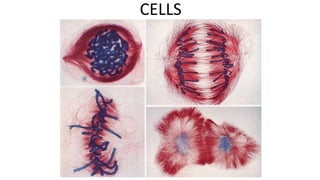
- 1. CELLS
- 2. BRAINSTORMING…. • What is the basic unit of all living things? • What are the two broad categories of cells? • What do you remember about the basic structure of a cell? • List the cell organelles
- 3. CELLULAR THEORY ✔ Cells were discovered in 1677 by Robert Hooke, from a layer of cork. ✔ In 1838, Mathias Schleiden (botanist) and Theodor Schwann (zoologist) proposed part of the Cellular Theory (*). ✔ Since then, this theory has gradually been completed thanks to new scientific and technological discoveries. The theory can be summarized in the following way 1.All living beings are made up of cells. 2. Cells are the smallest unit of beings which have a life of their own. 3. All cells come from another cell * (proposed by Virchow). 4.Every single cell can work independently, although in a coordinated way.
- 4. ✔Why was the microscope so important for the development of biology? ✔Are microorganisms visible to the naked eye? ✔What is a microscope used for? ✔What’s needed to look at a sample under the microscope?
- 5. 1.1. Cell structure All cells exhibit a common basic structure: ⮚A cell membrane, a semipermeable membrane that controls what enters and leaves the cell’s interior ⮚The cytoplasm, the area where all the chemical reactions related to the cell’s activity take place (cell organelles) ⮚Genetic material: Organic biomolecules (DNA, RNA) that directs all the cell functions = control system
- 7. 1.2. Cell’s vital functions Cells are the building blocks of life and perform the three vital functions VITAL FUNCTIONS NUTRITION To transform nutrients into energy for the cell’s activities INTERACTION To detect internal/external changes and adapt to them REPRODUCTION 1.Unicellular organisms= the formation of new individuals. 2.Multicellular organisms=the growth of the body structures
- 8. A. Nutrition This key function allows cells to obtain nutrients and transform them into energy (cell respiration). This energy will be used by cells to renew their cellular structures, as well as, for carrying the rest of cell’s functions. These transformations happen in the cytoplasm through a process called metabolism. This constitute the basis of cell life As a result of the cell metabolism, waste products are produced and they must to be expelled to the external environment. Types of nutrition: 1.Autotrophic nutrition 2.Heterotrophic nutrition What is metabolism? This term refers to the set of chemical processes that happens in the cell’s interior.
- 9. B. Interaction This function allows cell to communicate with the external environment. Meaning, cells adapt to changes that occur around them. Cell would not be able to survive if this function did not exist. They would be unable to activate the essential mechanisms needed to maintain their vital activity
- 10. C. Reproduction Reproduction refers to the formation of new cells from existing ones. 1.In unicellular organisms= production of new individuals. 2.Multicellular organisms= a)replacement of dead cells b) the growth of the individual
- 11. 2. Types of cells Cells are extremely small structures that can only be observed using a microscope Their size can be measured in micrometres, which is equivalent to a thousandth part or a millimetre
- 12. Cell differentiation is the process by which cells change in structure and become capable of carrying out special functions, in other words, cell become different cells so they can do different jobs. Cells then form specialized groups of cells, which join forming tissues and then organs. There are needed changes at three different levels 1.Changes in shape and size 2.Changes in function 3.Changes at a cytoplasmic level ( cell organelles)
- 13. 2.1. Cell organization: From an evolutionary point of view all cells come from a common ancestor There are two types of cells: Type of cells Prokaryotic They are simpler and more primitive Eukaryotic These cells evolved from prokaryotic cells Anaerobic and photosynthetic bacteria combined with prokaryotic cells through endosimbiosis and turned into mitochondria and chloroplasts respectively
- 14. Lynn Margulis – Endosymbiotic theory: - Lynn Margulis (1960’s) - Ancestors of mitochondria and chloroplasts were engulfed by eukaryotic cells. - Evidences: Mitochondria and chloroplasts …. 1. Have double membranes. 2. Reproduce on their own 3. Have their own ribosomes
- 18. DOMAINS RELATED TO PROKARYOTIC CELLS
- 19. REVIEW: Taxonomic classification of living things
- 20. 4. EUKARYOTIC CELLS They are more complex and have a series of advantages over prokaryotic cells. Their cytoplasm contains a wide range of structures that are specialized in performing different functions = Cell organelles Genetic material is found in the nucleus. This way, it is protected and provides better cell stability Eukaryotic cells have a cytoskeleton, a microscopic network of protein filaments and microtubules. Its function is to maintain the cell’s shape and internal organization. It also plays a role in the cell movements
- 21. 4.1. ORGANELLES What are the cell organelles? They are membranous structures found in the cytoplasm. There are different organelles with different jobs, so each organelle is made up of the biomolecules needed to perform its functions 1. Cell membrane 2. Cytoplasm 3. Ribosomes 4. Lysosomes 5. Endoplasmic reticulum ( RER/ SER) 6. Vacuoles 7. Golgi apparatus 8. Mitochondria 9. Chloroplasts 10. Cilia 11. Flagella 12. Nucleus
- 22. NON- MEMBRANOUS CELL ORGANELLES RIBOSOMES: (They are the exception , as they are not membranous structures) They are spherical shaped organelles with no membrane. They can be found scattered throughout the cytoplasm or attached to the rough endoplasmic reticulum. They are in charge of protein synthesis MEMBRANOUS CELL ORGANELLES CATEGORIES: 1. CELL ORGANELLES THAT PROCESS NUTRIENTS Lysosomes, vacuoles, endoplasmic reticulum, Golgi apparatus 2. CELL ORGANELLES THAT PRODUCE ENERGY Mitochondria / chloroplasts 3. CELL ORGANELLES INVOLVE IN MOVEMENT Cilia , flagella, centrioles
- 23. 4.1.1. Organelles that process nutrients
- 24. 4.1.1. Organelles that process nutrients LYSOSOMES They are small vesicles that contain subtances capable of digesting molecules captured by cell. They contain digestive enzymes ENDOPLASMIC RETICULUM It’s a very complex set of tubules and vesicles responsible for manufacturing and transporting different substances, such as lipids and proteins. There are two types: -Rough endoplasmic reticulum: to synthetyse proteins -Smooth endoplasmic reticulum: to produce lipids VACUOLES Structures in charge of storing different substances (mainly water). In animal cells they are more numerous but smaller. In plant cells there is a central and big vacuole. GOLGI APPARATUS This cell organelle is formed by grouped vesicles and flattened sacs; it take substances from the endoplasmic reticulum, modifies them and introduces them into the vesicles for secretion
- 25. 4.1.1. Organelles that produces energy Mitochondria Chloroplasts They are cylindrical organelles made up of a double membrane. The external membrane is smooth and the internal one has folds (cristae). Inside is the matrix, which is mainly made up of genetic material, ribosomes and enzymes Mitochondria are considered the power plants of the eukaryotic cells, as they are in charge of obtaining energy ( cell respiration). They are egg-shaped organelles with a double membrane. They have a series of disc- shaped sacs name thylakoids, which contain the pigment that gives them their green colour. Chloroplasts synthesize organic molecules from inorganic ones using chemical energy obtained from the Sun = photosynthesis. These organelles are only found in the cells of photosynthetic organisms ( plants and algae)
- 27. 4.2. Organelles involved in cell mobility Cilia and flagella They are mobile cell organelles. They are formed by protein fibres from the cytoskeleton Their movement is coordinated by a structure known as centriole. The centriole is formed by protein tubules arranged like cilia and flagella. This is also involved in cellular division Certain eukaryotic cells are able to move in two different ways: 1.Using their appendixes ( cilia and flagella) 2.By changing the viscosity of their cytoplasm
- 28. 4.2.2.Changes in the viscosity of the cytoplasm Proteins found in the cytoskeleton are responsible for changes in the viscosity of cytoplasm. They do this by grouping together or separating themselves. They produce pseudopodia, an extension of cytoplasm, and modify the shape of the cell. Pseudopodia are also used to surround and capture certain materials from the environment ( microbes, food particles…). This process is known as phagocytosis
- 29. 4.3. Nucleus Nucleus It is the control centre of the cell. This organelle contains the genetic material. It is normally located in the centre of the cell. In some cases, such as secreting cells and many plant cells, it can be found in peripheral areas. Its structures varies depending on the moment in life of the cell. It has two different structures: 1.INTERPHASE NUCLEUS (when the cell is not dividing) 2.NUCLEAR DIVISION (when the cell is diving)
- 30. Interphase nucleus Nuclear division It has a porous double membrane that surrounds the nucleoplasm, which is similar to cytoplasm. Inside the nucleoplasm there is chromatin, a substance formed by the double helix of DNA joined to histones (proteins) and nucleolus, a spherical organelle involved in the synthesis of ribosomes Once cell division begins, the nucleus changes deeply: Chromatin condenses into chromosomes. Chromosomes are X-shaped structures. Each chromosome is made up of two chromatin filaments called chromatids, which are joined by a centromere. Both chromatids are identical, so the genetic information is duplicated. The centromere separates two regions in each chromatid, names arms
- 31. The number of chromosomes in gametes ( sex cells) varies from somatic cells. 1.The haploid number (n) is the number or chromosomes in a gamete. 2.The diploid number (2n) refers to the number or chromosomes in somatic cells. There are two sets of haploid cells in a somatic cell, one from each parent
- 32. Animal cell vs plant cell Tour inside the cell
- 33. The cell cycle is the series of events that take place in a cell that cause the division of a parent cell into two daughter cells. These events include the duplication of its DNA (DNA replication) and some of its organelles, and subsequently the partitioning of its cytoplasm and other components into two daughter cells in a process called cell division. Phases: a)Interphase - G1: growth -S: duplication of DNA -G2: cell functions/ preparation for mitosis a)Mitosis (M) The cell cycle
- 35. Mitosis. Prophase 1. Chromatin fibres thicken and shorten to form chromosomes. 2. The nucleolus disappears 3. Protein fibres appear between both poles of the cell to form the mitotic spindle 1. The nuclear membrane disappears, so chromosomes can move freely around the cytoplasm.
- 36. Mitosis. Metaphase 1. Chromosome bond to the mitotic spindle fibres using their centromeres. 2. This union takes place in the equator of the cell. 3. Sister chromatids belonging to each chromosome point to the opposite poles of the cell
- 37. Mitosis. Anaphase 1. The mitotic spindle fibres break into equal halves. This causes the chromosome’s centromere to break. As a result, the two chromatids that made up each chromosome separate into two unconnected half fibres. 2. The mitotic spindle fibres contract, pulling the chromatids towards the opposite poles. 3. From this point onwards, chromatids are considered to be independent chromosomes
- 38. Mitosis. Telophase 1. Once chromatids move to the ends of the poles, the remains of the mitotic spindle fibres disappear. 2. A new nuclear membrane surrounds each group of chromatids, forming two new nuclei. 3. Chromatids expand and turn into chromatin 4. The nucleolus reappears
- 39. Once mitosis ends, the division of the cytoplasm takes place and cell organelles are shared. This process is referred to as cytokinesis and it can take place in two ways: 1.In animal cells, the cytoplasm stretches, thins out and eventually separates. 1.In plant cells, a wall forms which divides the cytoplasm in half Cytokinesis
- 43. Basics about meiosis: • Meiosis is the process in eukaryotic, sexually-reproducing animals that reduces the number of chromosomes in a cell before reproduction • Function of meiosis: • Meiosis is necessary for many sexually-reproducing animals to ensure the same number of chromosomes in the offspring as in the parents. The act of fertilization includes two cells fusing together to become a new zygote. • Meiosis consists of two divisions, both of which follow the same stages as mitosis (prophase, metaphase, anaphase, telophase). • Meiosis is preceded by interphase, in which DNA is replicated to produce chromosomes consisting of two sister chromatids. • A second growth phase called interkinesis may occur between meiosis I and II, however no DNA replication occurs in this stage.
- 46. What is crossing over? The exchange of chromosomal segments between two non-sister chromatids Why does crossing over occur? To provide genetic variation during meiosis as crossing over ensures a combination of the maternal and paternal genes we inherited
- 47. Meiosis. Prophase I ✔ Chromosomes condense ✔ Nuclear membrane dissolves ✔ Homologous chromosomes form bivalents ✔ Crossing over occurs
- 48. Meiosis. Metaphase I Spindle fibres from opposing centrosomes connect to bivalents (at centromeres) and align them along the middle of the cell
- 49. Meiosis. Anaphase I ✔Spindle fibres contract and split the bivalent. ✔Homologous chromosomes move to opposite poles of the cell
- 50. Meiosis. Telophase I ✔ Chromosomes decondense ✔ Nuclear membrane may reform ✔ Cell divides (cytokinesis) to form two haploid daughter cells
- 51. Meiosis II. Prophase II ✔ Chromosomes condense ✔ Nuclear membrane dissolves ✔ Centrosomes move to opposite poles (perpendicular to before) The second division separates sister chromatids (these chromatids may not be identical due to crossing over in prophase I)
- 52. Meiosis. Metaphase II ✔ Spindle fibres from opposing centrosomes attach to chromosomes (at centromere) and align them along the cell equator
- 53. Meiosis. Anaphase II ✔ Spindle fibres contract and separate the sister chromatids. ✔ Chromatids (now called chromosomes) move to opposite poles
- 54. Meiosis. Telophase II ✔ Chromosomes decondense ✔ Nuclear membrane reforms ✔ Cells divide (cytokinesis) to form four haploid daughter cells The final outcome of meiosis is the production of four haploid daughter cells. These cells may all be genetically distinct if crossing over occurs in prophase I (causes recombination of sister chromatids)