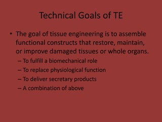
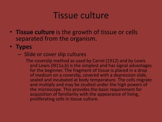
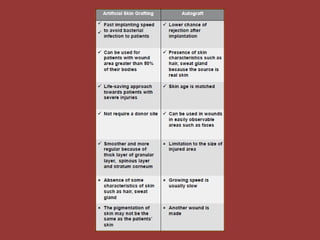
Tissue Engineering
By- Sanju Sah
St. Xavier’s college, Maitighar
Department of Microbiology
Unit 5. Tissue Engineering
5.1 Introduction to Tissue Engineering
5.2 Technical goals of Tissue
Engineering
5.3 Tissue culture Preparation
Slide or cover slip cultures
Flask cultures
Test tube cultures
5.4 Artificial Skin
5.5 Artificial Cartilage
Types of cells
• Autologous
– Harvested from the patient
• Allogeneic cells
– Come from the body of a donor of the same species
• Xenogeneic cells
– Isolated from individuals of another species
• Isogenic cells
– Isolated from genetically identical organisms such as twins,
clones
• Stem cells
– Undifferentiated cells with the ability to divide in cult
Introduction to Tissue Engineering
• Tissue Engineering: A branch of Biomedical
Engineering
• Unification of Molecular Biology, Engineering
and Medicine
• Applied to restore or replace defected tissues
and organs
• As defined by Dr. Langer and Dr.
Vacanti, tissue engineering is
"an interdisciplinary field that applies the principles
of engineering and life sciences toward the
development of biological substitutes that restore,
maintain, or improve tissue function or a whole
organ."
Technical Goals of TE
• The goal of tissue engineering is to assemble
functional constructs that restore, maintain,
or improve damaged tissues or whole organs.
– To fulfill a biomechanical role
– To replace physiological function
– To deliver secretary products
– A combination of above
Tissue culture
• Tissue culture is the growth of tissue or cells
separated from the organism.
• Types
– Slide or cover slip cultures
The coverslip method as used by Carrel (1912) and by Lewis
and Lewis (l911a,b) is the simplest and has signal advantages
for the beginner. The fragment of tissue is placed in a drop
of medium on a coverslip, covered with a depression slide,
sealed and incubated at body temperature. The cells migrate
and multiply and may be studied under the high powers of
the microscope. This provides the basic requirement for
acquisition of familiarity with the appearance of living,
proliferating cells in tissue culture.
– Flask cultures
This methods is used for maintenance of cultures for
longer periods without transfer, developed several
types of culture flasks. In -these ample nutriment and
oxygen are provided, growth of the numerous tissue
fragments is more nearly uniform under identical
conditions
– Test tube cultures
Ordinary Pyrex test tubes are used as the culture
container which is mechanically rotated to expose the
tissues alternately to a fluid nutrient and to oxygen.
Artificial Skin
• When and Why is Skin/ Skin Substitute
Needed?
– When skin is damaged or lost due to severe injury
or burns, bacteria and other microorganisms have
easy access to body.
– To treat a severe burn
– Patients with diabetes will have difficulties in
wound healing, Inflammation will occur, Chronic
Wound will be formed
Skin Autograft
• Transplantation of skin from a donor site to a
receiver site in the same individual
Artificial skin
• Artificial skin is a synthetic equivalent to human skin
which can increase the chance of survival of severely
burned patients by protecting the underlying tissues.
Aims
1. Protection from bacteria
2. Wound healing texture
History of Burns
• Before artificial skin was
developed, burns covering 50%
of the body were considered to
be fatal.
• In 2000, artificial skin cut death
rates in people with over 70%
burns from 100% to 40%.
• Skin is the body’s largest
organs and protects us from
physical damage, disease, and
controls our body’s
temperature.
How Artificial Skin is Made
• Skin is usually donated by other
donors.
• Fibroblasts are removed from
the donated skin and are frozen
until they are needed.
• The fibroblasts are placed on a
polymeric mesh scaffolding,
gather oxygen, and grow new
cells.
• The cells are then transferred to
a culture system.
Artificial Skin cont.
• After 4 weeks the polymer mesh dissolves and leaves behind a new
layer of dermal skin.
• When the growth cycle is completed, they add more nutrients.
• Keratinocytes are added to the collagen and are exposed to air to
form epidermal layers.
• The skin is now completed and is stored in sterile contains until
ready to use.
How Artificial Skin is Used
• Artificial skin is already being
used for burn victims and soon
will be available for other skin
disorders.
• The skin is not used for a
permanent replacement, but to
temporary cover the skin until
your skin can grow back
naturally.
There are two methods to produce
artificial skin. Both of them use fibroblasts
to make it
• Integra Dermal Regeneration Template®
– Semi -synthetic approach to skin regeneration
– Researchers develop a bi-layer membrane system called the
Dermal Regeneration Template
– The first and only FDA approved tissue engineered
product for burn and reconstructive surgery
– Dermal replacement layer is constructed of a porous,
biodegradable matrix of cross-linked bovine tendon collagen
and the glycos-aminoglycan chondroitin 6-sulfate.
• Allows a the wound to establish a new tissue base
– Second layer acts as a temporary replacement
(Epidermal ) – made from silicone polymer
 Following completion of the dermal layer physicians replace
the temporary epidermal with an epidermal auto-graft.
Artificial Cartilage
• Current strategies in human medicine for
treatment of diffuse joint degeneration rely
on replacement of the whole degenerated
joint with inert implants.
• Excellent treatment outcome has been
achieved for up to 15 years or more, but
approximately 20% of treated patients
require revision procedures after this time
(Steadman et al., 2001).
• For younger patients this current state-of-
the-art may translate to two or more
revision surgeries during their lifetime.
• A biological solution to repair damaged
cartilage that would provide life-long pain
relief would be a major medical
achievement.
• Hyaline articular cartilage is a complex
structure, developed and progressively refined
over hundreds of millions of years.
• Cartilage tissue engineered using this triad of
components often exhibit hyaline cartilage
morphology, but the tissue has inferior
mechanical properties when compared to
native joint cartilage
Cells of cartilage tissue engineering
• Articular hyaline cartilage is a very specialized
tissue characterized by low cellularity
extensive extracellular matrix, lack of vascular,
lymphatic, and nervous supply.
• Obvious choice of cell for cartilage repair is
the chondrocyte from hyaline cartilage.
BUT
• Chondrocytes unfortunately tend to dedifferentiation
towards the fibroblast cell lineage when expanded in
culture making them less suitable for transplantation
• the use of autologous chondrocytes is the need for two
surgical procedures, weeks apart to harvest and later
implant the cells, which adds time, cost, anesthetic risk
and risks of donor site morbidity
SO
• Stem cells or cells with chondrogenic potential from
various tissue sources have been investigated for
cartilage repair
Manufacturing process
• Unique building block of articular cartilage matrix is made of
Type II collagen.
• Middle architectural zone called “the netting” is made of
aggregates of proteoglycans called glycosamino- glycans (GAG’s):
This netting holds water i.e.: gives this zone its hydrophilic
character that yields the low friction, fluid wave enabling smooth
joint motion
• Scaffolds are region specific and includes chitosan/ fibrinoger for
bioreplacement.
Advantage and disadvantage
• Advantage
– Can prevent total knee replacement
– Can correct birth defects
– Bring hope and confidence in patient
• Disadvantage
– The risk of complication and infection
– It could be rejected by patient’s body
– expensive
Thank You

Tissue engineering

  • 1.
    Tissue Engineering By- SanjuSah St. Xavier’s college, Maitighar Department of Microbiology
  • 4.
    Unit 5. TissueEngineering 5.1 Introduction to Tissue Engineering 5.2 Technical goals of Tissue Engineering 5.3 Tissue culture Preparation Slide or cover slip cultures Flask cultures Test tube cultures 5.4 Artificial Skin 5.5 Artificial Cartilage
  • 5.
    Types of cells •Autologous – Harvested from the patient • Allogeneic cells – Come from the body of a donor of the same species • Xenogeneic cells – Isolated from individuals of another species • Isogenic cells – Isolated from genetically identical organisms such as twins, clones • Stem cells – Undifferentiated cells with the ability to divide in cult
  • 6.
    Introduction to TissueEngineering • Tissue Engineering: A branch of Biomedical Engineering • Unification of Molecular Biology, Engineering and Medicine • Applied to restore or replace defected tissues and organs
  • 7.
    • As definedby Dr. Langer and Dr. Vacanti, tissue engineering is "an interdisciplinary field that applies the principles of engineering and life sciences toward the development of biological substitutes that restore, maintain, or improve tissue function or a whole organ."
  • 10.
    Technical Goals ofTE • The goal of tissue engineering is to assemble functional constructs that restore, maintain, or improve damaged tissues or whole organs. – To fulfill a biomechanical role – To replace physiological function – To deliver secretary products – A combination of above
  • 11.
    Tissue culture • Tissueculture is the growth of tissue or cells separated from the organism. • Types – Slide or cover slip cultures The coverslip method as used by Carrel (1912) and by Lewis and Lewis (l911a,b) is the simplest and has signal advantages for the beginner. The fragment of tissue is placed in a drop of medium on a coverslip, covered with a depression slide, sealed and incubated at body temperature. The cells migrate and multiply and may be studied under the high powers of the microscope. This provides the basic requirement for acquisition of familiarity with the appearance of living, proliferating cells in tissue culture.
  • 12.
    – Flask cultures Thismethods is used for maintenance of cultures for longer periods without transfer, developed several types of culture flasks. In -these ample nutriment and oxygen are provided, growth of the numerous tissue fragments is more nearly uniform under identical conditions – Test tube cultures Ordinary Pyrex test tubes are used as the culture container which is mechanically rotated to expose the tissues alternately to a fluid nutrient and to oxygen.
  • 13.
  • 14.
    • When andWhy is Skin/ Skin Substitute Needed? – When skin is damaged or lost due to severe injury or burns, bacteria and other microorganisms have easy access to body. – To treat a severe burn – Patients with diabetes will have difficulties in wound healing, Inflammation will occur, Chronic Wound will be formed
  • 15.
    Skin Autograft • Transplantationof skin from a donor site to a receiver site in the same individual
  • 16.
    Artificial skin • Artificialskin is a synthetic equivalent to human skin which can increase the chance of survival of severely burned patients by protecting the underlying tissues. Aims 1. Protection from bacteria 2. Wound healing texture
  • 17.
    History of Burns •Before artificial skin was developed, burns covering 50% of the body were considered to be fatal. • In 2000, artificial skin cut death rates in people with over 70% burns from 100% to 40%. • Skin is the body’s largest organs and protects us from physical damage, disease, and controls our body’s temperature.
  • 18.
    How Artificial Skinis Made • Skin is usually donated by other donors. • Fibroblasts are removed from the donated skin and are frozen until they are needed. • The fibroblasts are placed on a polymeric mesh scaffolding, gather oxygen, and grow new cells. • The cells are then transferred to a culture system.
  • 19.
    Artificial Skin cont. •After 4 weeks the polymer mesh dissolves and leaves behind a new layer of dermal skin. • When the growth cycle is completed, they add more nutrients. • Keratinocytes are added to the collagen and are exposed to air to form epidermal layers. • The skin is now completed and is stored in sterile contains until ready to use.
  • 20.
    How Artificial Skinis Used • Artificial skin is already being used for burn victims and soon will be available for other skin disorders. • The skin is not used for a permanent replacement, but to temporary cover the skin until your skin can grow back naturally.
  • 22.
    There are twomethods to produce artificial skin. Both of them use fibroblasts to make it
  • 23.
    • Integra DermalRegeneration Template® – Semi -synthetic approach to skin regeneration – Researchers develop a bi-layer membrane system called the Dermal Regeneration Template – The first and only FDA approved tissue engineered product for burn and reconstructive surgery – Dermal replacement layer is constructed of a porous, biodegradable matrix of cross-linked bovine tendon collagen and the glycos-aminoglycan chondroitin 6-sulfate. • Allows a the wound to establish a new tissue base – Second layer acts as a temporary replacement (Epidermal ) – made from silicone polymer  Following completion of the dermal layer physicians replace the temporary epidermal with an epidermal auto-graft.
  • 25.
    Artificial Cartilage • Currentstrategies in human medicine for treatment of diffuse joint degeneration rely on replacement of the whole degenerated joint with inert implants. • Excellent treatment outcome has been achieved for up to 15 years or more, but approximately 20% of treated patients require revision procedures after this time (Steadman et al., 2001). • For younger patients this current state-of- the-art may translate to two or more revision surgeries during their lifetime. • A biological solution to repair damaged cartilage that would provide life-long pain relief would be a major medical achievement.
  • 26.
    • Hyaline articularcartilage is a complex structure, developed and progressively refined over hundreds of millions of years. • Cartilage tissue engineered using this triad of components often exhibit hyaline cartilage morphology, but the tissue has inferior mechanical properties when compared to native joint cartilage
  • 27.
    Cells of cartilagetissue engineering • Articular hyaline cartilage is a very specialized tissue characterized by low cellularity extensive extracellular matrix, lack of vascular, lymphatic, and nervous supply. • Obvious choice of cell for cartilage repair is the chondrocyte from hyaline cartilage.
  • 28.
    BUT • Chondrocytes unfortunatelytend to dedifferentiation towards the fibroblast cell lineage when expanded in culture making them less suitable for transplantation • the use of autologous chondrocytes is the need for two surgical procedures, weeks apart to harvest and later implant the cells, which adds time, cost, anesthetic risk and risks of donor site morbidity SO • Stem cells or cells with chondrogenic potential from various tissue sources have been investigated for cartilage repair
  • 29.
    Manufacturing process • Uniquebuilding block of articular cartilage matrix is made of Type II collagen. • Middle architectural zone called “the netting” is made of aggregates of proteoglycans called glycosamino- glycans (GAG’s): This netting holds water i.e.: gives this zone its hydrophilic character that yields the low friction, fluid wave enabling smooth joint motion • Scaffolds are region specific and includes chitosan/ fibrinoger for bioreplacement.
  • 30.
    Advantage and disadvantage •Advantage – Can prevent total knee replacement – Can correct birth defects – Bring hope and confidence in patient • Disadvantage – The risk of complication and infection – It could be rejected by patient’s body – expensive
  • 32.