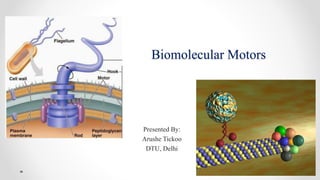
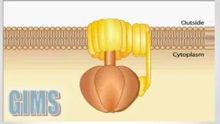
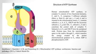
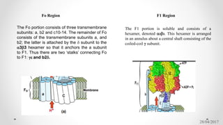
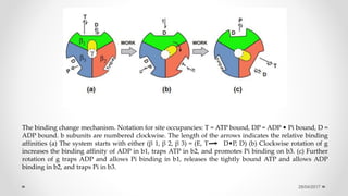
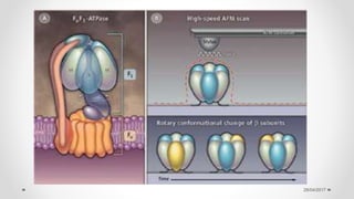
SlideShare a Scribd company logo
Biomolecular Motors
Presented By:
Arushe Tickoo
DTU, Delhi
28/04/2017
The typical biomolecular motor is a protein that uses free energy from ATP (Adenosine Tri-
phosphate) or other nucleotide triphosphate (NTP) hydrolysis as its fuel to produce mechanical
force.
ATP is the universal currency of energy in biological systems and, is generated from glucose by
a sequence of reactions called glycolysis.
The hydrolysis of ATP to ADP and inorganic phosphate is energetically favorable and the pool of
ATP in a cell is far higher than that of its hydrolysis products, hence serving as a remarkable
store of chemical energy that is used to perform the various cellular functions.
Motor proteins are involved in several critical cellular processes and have functions ranging
from ATP synthesis and muscle contraction.
Biomolecular motor
28/04/2017
Flagellated bacteria, such as Escherichia coli, swim by rotating thin helical filaments, each driven at its
base by a reversible rotary motor, powered by an ion flux. A motor is about 45 nm in diameter and is
assembled from about 20 different kinds of parts. It develops maximum torque at stall but can spin several
hundred Hz.
Its direction of rotation is controlled by a sensory system that enables cells to accumulate in regions
deemed more favorable.
No more than 50 nm in Diameter It spins clockwise (CW) orcounterclockwise (CCW) at speeds on the order of
100 Hz, driving long thin helical filaments that enable cells to swim Receptors near the surface of the cell count
molecules of interest (sugars, amino acids, dipeptides) and control the direction of flagellar rotation. If a leg of
the search is deemed favorable, it is extended,
Flagellar motor
28/04/2017
Fig; A schematic diagram
of the flagellar motor
CheY-P
It is the chemotaxis
signaling molecule that
binds to FliM, and FlgM
is the anti-sigma factor
pumped out of the cell by
the transport apparatus;
28/04/2017
A cell swims steadily in a direction roughly parallel to its long axis for about a second—it is said to
“run”—and then moves erratically in place for a small fraction of a second—it is said to “tumble”—
and then swims steadily again in a new direction. When a cell runs at top speed, all of its flagellar
filaments spin CCW, the filaments form a bundle that pushes the cell steadily forward. When a cell
tumbles, one or more filaments spin CW; these filaments leave the bundle, and the cell changes
course
Motors switch from CCW to CW and back again approximately at random. The likelihood of
spinning CW is enhanced by a chemotactic signaling protein, CheY.
When phosphorylated, CheY binds to the cytoplasmic face of the flagellar motor. The
phosphorylation of CheY is catalyzed by a kinase, the activity of which is controlled by
chemoreceptors.
The different components of the motor are named after the genes that encode them. genes for which
mutant cells lacked flagellar filaments were called fla (for flagellum), but after more than 26 had been
found.
fla genes are now called flg, flh, fli, or flj, depending upon their location on the genetic map. Genes for
which mutant cells produce paralyzed flagella are called mot (for motility). Four of the all gene
products that are involved in gene regulation (FlgM, FlhC, FlhD, FliA);
28/04/2017
The M-ring (for membrane) has affinity for inner-
membrane fractions,
The S-ring is seen just above the inner membrane,
The P-ring (for peptidoglycan) is at the right place to be
embedded in the peptidoglycan,
and the L-ring (for lipopolysaccharide) has affinity for
outer-membrane fractions.
It was found that both the M- and S-rings (now called the
MS-ring) comprise different domains of the same protein,
FliF. Therefore, they function as a unit.
28/04/2017
FliG, FliM, and FliN are also referred to as the “switch complex,” since many mutations of fliG, fliM,
and fliN lead to defects in switching (in control of the direction of rotation)
Operons encoding the proteins of the chemotaxis system of E.
colia
Class 1 Class 2 Class 3
flhDC flgAMN fliC
flgBCDEFGHIJKL motABcheAW
flhBAE tar tap cheRBYZ
fliAZY aer
fliDST trg
Class 1 contains the master operon, flhDC, the expression of which is required for transcription of class 2 and class 3
operons.
Class 2 contains eight operons that encode components required for construction of the hook-basal body complex,
and class 3 contains six more that encode components required for filament assembly and motor function
28/04/2017
ATP Synthase
ATP synthase is one of the wonders of the molecular world. ATP synthase is an enzyme, a molecular motor, an
ion pump, and another molecular motor all wrapped together in one amazing nanoscale machine. It plays an
indispensable role in our cells, building most of the ATP that powers our cellular processes.
Human mitochondrial (mt) ATP synthase, or complex V consists of two functional domains: F1, situated in the
mitochondrial matrix, and Fo, located in the inner mitochondrial membrane. Complex V uses the energy created
by the proton electrochemical gradient to phosphorylate ADP to ATP.
ATP synthase consists of two well defined protein entities: the F1 sector, a soluble portion situated in the
mitochondrial matrix, and the Fo sector, bound to the inner mitochondrial membrane. F1 is composed of three
copies of each of subunits α and β, and one each of subunits γ, δ and ε. F1 subunits γ, δ and ε constitute the
central stalk of complex V. Fo consists of a subunit c-ring (probably comprising eight copies, as shown in
bovine mitochondria and one copy each of subunits a, b, d, F6 and the oligomycin sensitivity-conferring
protein (OSCP). Subunits b, d, F6 and OSCP form the peripheral stalk which lies to one side of the complex.
28/04/2017
28/04/2017
Human mitochondrial ATP synthase, or
complex V, consists of two functional domains,
F1 and Fo. F1 comprises 5 different subunits
(three α, three β, and one γ, δ and ε) and is
situated in the mitochondrial matrix. Fo contains
subunits c, a, b, d, F6, OSCP and the accessory
subunits e, f, g and A6L. F1 subunits γ, δ and ε
constitute the central stalk of complex V.
Subunits b, d, F6 and OSCP form the peripheral
stalk. Protons pass from the intermembrane
space to the matrix through Fo, which transfers
the energy created by the proton
electrochemical gradient to F1, where ADP is
phosphorylated to ATP.
Structure of ATP Synthase
Jonckheere I, Smeitink J A M, and Rodenburg R J; Mitochondrial ATP synthase: architecture, function and
pathology (2011), J Inherit Metab Dis
28/04/2017
Fo Region F1 Region
The F1 portion is soluble and consists of a
hexamer, denoted a3b3. This hexamer is arranged
in an annulus about a central shaft consisting of the
coiled-coil γ subunit.
The Fo portion consists of three transmembrane
subunits: a, b2 and c10-14. The remainder of Fo
consists of the transmembrane subunits a, and
b2; the latter is attached by the d subunit to the
a3b3 hexamer so that it anchors the a subunit
to F1. Thus there are two ‘stalks’ connecting Fo
to F1: ge and b2d.
28/04/2017
28/04/2017
28/04/2017
The binding change mechanism. Notation for site occupancies: T = ATP bound, DP = ADP • Pi bound, D =
ADP bound. b subunits are numbered clockwise. The length of the arrows indicates the relative binding
affinities (a) The system starts with either (β 1, β 2, β 3) = (E, T D•P, D) (b) Clockwise rotation of g
increases the binding affinity of ADP in b1, traps ATP in b2, and promotes Pi binding on b3. (c) Further
rotation of g traps ADP and allows Pi binding in b1, releases the tightly bound ATP and allows ADP
binding in b2, and traps Pi in b3.
28/04/2017
In steps 1 and 2 a site binds ADP and phosphate (not necessarily in that order). While trapped in the catalytic
site in step 3, reactants (ADP and Pi ) and product (ATP) are in chemical equilibrium. Step 4 requires the
input of mechanical torque from Fo on g to trap the reactants in the ATP state and to pry open the site
releasing the tightly bound ATP. The way in which this works is found in the shape of the a3b3 hexamer and
the g shaft
At the top of the a3b3 hexamer is a hydrophobic ‘sleeve’ in which the g shaft rotates. Further down, however,
the annulus is offset from the center, so that as g rotates clockwise, it sequentially pushes outwards on each
catalytic site. In addition, the e subunit is located eccentrically and attached to the g and c subunits so that, as
g rotates, it comes into contact sequentially with each b subunit in a conserved region called the DELSEED
sequence (named for the single letter abbreviation of its constituent amino acids). Together, this asymmetric
rotation exerts stress on the catalytic site loosening its grip on ATP so that thermal fluctuations can free it into
solution.
28/04/2017
28/04/2017
A single molecule of F1-ATPase acts as a rotary motor, the smallest known, by direct observation
of its motion. A central rotor of radius approximately 1 nm, formed by its gamma-subunit, turns
in a stator barrel of radius approximately 5nm formed by three alpha- and three beta-subunits. F1-
ATPase, together with the membrane-embedded proton-conducting unit F0, forms the H+-ATP
synthase that reversibly couples transmembrane proton flow to ATP synthesis/hydrolysis in
respiring and photosynthetic cells. In the presence of ATP, the filament rotated for more than 100
revolutions in an anticlockwise direction when viewed from the 'membrane' side. The rotary
torque produced reached more than 40 pN nm(-1) under high load.
Direct observation of the rotation of F1-ATPase.
Noji H, Yasuda R, Yoshida M, Kinosita K Jr.
28/04/2017
In the realm of ATP-driven linear biomotors, the most prominent examples are dynein, kinesin and
myosin, which is involved in muscle contraction. Muscle contraction is a sophisticated process involving
actin and myosin and is a result of actin filaments sliding on myosin heads. Actin filaments are formed of
375 amino acid long subunits that are associated with ATP. Myosin transport along actin is not limited to
muscle contraction, and is involved in several cellular transport processes.
It is thought that the two heads of the myosin move along in a hand-over-hand mechanism based on the
~74nm displacements observed by fluorescent labeling methods.
Interaction of an actin filament
with myosin-coated surface
28/04/2017
1. Itoh, H., et al., (2004) Mechanically driven ATP synthesis by F1-ATPase. Nature,. 427(6973): p. 465-8.
2. Noji, H., et al., (1997) Direct observation of the rotation of F1-ATPase. Nature,. 386(6622): p. 299-302.
3. Oster G, Wang H; ATP Synthase: Two rotary molecular motors working together; University of
California, Berkeley
4. Antoniel M etal., (2014 May) The Oligomycin-Sensitivity Conferring Protein of Mitochondrial ATP
Synthase ; Int J Mol Sci.; 15(5): 7513–7536.
5. Oster G and Wang H; (April 1999) ATP synthase: two motors, two fuels, Elsevier Science
6. Manuela Antoniel, (2014 May) The Oligomycin-Sensitivity Conferring Protein of Mitochondrial ATP
Synthase: Emerging New Roles in Mitochondrial Pathophysiology, International Journal of Molecular
Sciences
References

More Related Content

What's hot

Linker, Adaptor, Homopolymeric Tailing & Terminal Transferase
Linker, Adaptor, Homopolymeric Tailing & Terminal TransferaseLinker, Adaptor, Homopolymeric Tailing & Terminal Transferase
Linker, Adaptor, Homopolymeric Tailing & Terminal Transferase
Utsa Roy
 
Use of Nanotechnology in Diagnosis and Treatment of Cancer
Use of Nanotechnology in Diagnosis and Treatment of CancerUse of Nanotechnology in Diagnosis and Treatment of Cancer
Use of Nanotechnology in Diagnosis and Treatment of Cancer
Anas Indabawa
 
Organ culture- animal tissue culture
Organ culture- animal tissue cultureOrgan culture- animal tissue culture
Organ culture- animal tissue culture
kathantrivedi3
 
Dna nanotechnology
Dna nanotechnologyDna nanotechnology
Dna nanotechnology
shrishaila cd
 
Fluorescence(Forster) Resonance Energy Transfer
Fluorescence(Forster) Resonance Energy TransferFluorescence(Forster) Resonance Energy Transfer
Fluorescence(Forster) Resonance Energy Transfersavvysahana
 
Metabolic flux analysis
Metabolic flux analysisMetabolic flux analysis
Metabolic flux analysis
andysmith007
 
Labelling of dna
Labelling of dnaLabelling of dna
Labelling of dna
christanantony
 
Synthesis of nanoparticles- physical,chemical and biological
Synthesis of nanoparticles- physical,chemical and biologicalSynthesis of nanoparticles- physical,chemical and biological
Synthesis of nanoparticles- physical,chemical and biologicalPriya Nanda
 
Gene expression in bacteria and bacteriophages
Gene expression in bacteria and bacteriophagesGene expression in bacteria and bacteriophages
Gene expression in bacteria and bacteriophages
Vinod Pawar
 
Magnetotactic bacteria.
Magnetotactic bacteria.Magnetotactic bacteria.
Magnetotactic bacteria.
qamrunnisashaikh1997
 
Stem cells niche and therapeutic applications
Stem cells niche and therapeutic applications Stem cells niche and therapeutic applications
Stem cells niche and therapeutic applications Sourabh Harankhedkar
 
immobilized Enzyme reactors- batch and continuous types.
immobilized Enzyme reactors- batch and continuous types. immobilized Enzyme reactors- batch and continuous types.
immobilized Enzyme reactors- batch and continuous types.
University of Mumbai
 
Protein Folding Mechanism
Protein Folding MechanismProtein Folding Mechanism
Protein Folding Mechanism
Sabahat Ali
 
Cell biology Lecture 5
Cell biology Lecture 5Cell biology Lecture 5
Cell biology Lecture 5Suk Namgoong
 
Recombinant protein
Recombinant proteinRecombinant protein
Recombinant protein
Pranitha Prani
 
Plant expression vectors
Plant expression vectorsPlant expression vectors
Plant expression vectors
Abhishek Indurkar
 
liposome mediated gene delivery
liposome mediated gene deliveryliposome mediated gene delivery
liposome mediated gene delivery
Pratibha Kumari Sharma
 
Abzymes
AbzymesAbzymes
Abzymes
jeeva raj
 
Dna microarray (dna chips)
Dna microarray (dna chips)Dna microarray (dna chips)
Dna microarray (dna chips)Rachana Tiwari
 
Tumor formtion , ti ri plasmid , dna trnsfr.
Tumor formtion , ti ri plasmid , dna trnsfr.Tumor formtion , ti ri plasmid , dna trnsfr.
Tumor formtion , ti ri plasmid , dna trnsfr.
Sukirti Vedula
 

What's hot (20)

Linker, Adaptor, Homopolymeric Tailing & Terminal Transferase
Linker, Adaptor, Homopolymeric Tailing & Terminal TransferaseLinker, Adaptor, Homopolymeric Tailing & Terminal Transferase
Linker, Adaptor, Homopolymeric Tailing & Terminal Transferase
 
Use of Nanotechnology in Diagnosis and Treatment of Cancer
Use of Nanotechnology in Diagnosis and Treatment of CancerUse of Nanotechnology in Diagnosis and Treatment of Cancer
Use of Nanotechnology in Diagnosis and Treatment of Cancer
 
Organ culture- animal tissue culture
Organ culture- animal tissue cultureOrgan culture- animal tissue culture
Organ culture- animal tissue culture
 
Dna nanotechnology
Dna nanotechnologyDna nanotechnology
Dna nanotechnology
 
Fluorescence(Forster) Resonance Energy Transfer
Fluorescence(Forster) Resonance Energy TransferFluorescence(Forster) Resonance Energy Transfer
Fluorescence(Forster) Resonance Energy Transfer
 
Metabolic flux analysis
Metabolic flux analysisMetabolic flux analysis
Metabolic flux analysis
 
Labelling of dna
Labelling of dnaLabelling of dna
Labelling of dna
 
Synthesis of nanoparticles- physical,chemical and biological
Synthesis of nanoparticles- physical,chemical and biologicalSynthesis of nanoparticles- physical,chemical and biological
Synthesis of nanoparticles- physical,chemical and biological
 
Gene expression in bacteria and bacteriophages
Gene expression in bacteria and bacteriophagesGene expression in bacteria and bacteriophages
Gene expression in bacteria and bacteriophages
 
Magnetotactic bacteria.
Magnetotactic bacteria.Magnetotactic bacteria.
Magnetotactic bacteria.
 
Stem cells niche and therapeutic applications
Stem cells niche and therapeutic applications Stem cells niche and therapeutic applications
Stem cells niche and therapeutic applications
 
immobilized Enzyme reactors- batch and continuous types.
immobilized Enzyme reactors- batch and continuous types. immobilized Enzyme reactors- batch and continuous types.
immobilized Enzyme reactors- batch and continuous types.
 
Protein Folding Mechanism
Protein Folding MechanismProtein Folding Mechanism
Protein Folding Mechanism
 
Cell biology Lecture 5
Cell biology Lecture 5Cell biology Lecture 5
Cell biology Lecture 5
 
Recombinant protein
Recombinant proteinRecombinant protein
Recombinant protein
 
Plant expression vectors
Plant expression vectorsPlant expression vectors
Plant expression vectors
 
liposome mediated gene delivery
liposome mediated gene deliveryliposome mediated gene delivery
liposome mediated gene delivery
 
Abzymes
AbzymesAbzymes
Abzymes
 
Dna microarray (dna chips)
Dna microarray (dna chips)Dna microarray (dna chips)
Dna microarray (dna chips)
 
Tumor formtion , ti ri plasmid , dna trnsfr.
Tumor formtion , ti ri plasmid , dna trnsfr.Tumor formtion , ti ri plasmid , dna trnsfr.
Tumor formtion , ti ri plasmid , dna trnsfr.
 

Similar to Bio-Molecular motors

Atp synthase , Atp synthase complex 1 to 4.
Atp synthase , Atp synthase complex 1 to 4.Atp synthase , Atp synthase complex 1 to 4.
Atp synthase , Atp synthase complex 1 to 4.
Cherry
 
Ge0102 unit 4
Ge0102 unit 4Ge0102 unit 4
Ge0102 unit 4
Arvind Ramesh
 
BT631-27-Membrane_proteins
BT631-27-Membrane_proteinsBT631-27-Membrane_proteins
BT631-27-Membrane_proteinsRajesh G
 
Oxidative phosphorylation
Oxidative phosphorylationOxidative phosphorylation
Oxidative phosphorylation
devadevi666
 
ATP-Synthase Presentation
ATP-Synthase PresentationATP-Synthase Presentation
ATP-Synthase Presentation
ahmedicine
 
Atp synthase
Atp synthaseAtp synthase
Atp synthase
Lovnish Thakur
 
ATP motors
ATP motorsATP motors
ATP motorsstudent
 
14 atpase
14 atpase14 atpase
14 atpase
Sidal hospital
 
Microtubules and molecular motors
Microtubules and molecular motorsMicrotubules and molecular motors
Microtubules and molecular motorsaljeirou
 
Microtubules and molecular motors
Microtubules and molecular motorsMicrotubules and molecular motors
Microtubules and molecular motorsaljeirou
 
Biological oxidation L5 (oxidative phosphorylation) pdf
Biological oxidation L5 (oxidative phosphorylation) pdfBiological oxidation L5 (oxidative phosphorylation) pdf
Biological oxidation L5 (oxidative phosphorylation) pdf
DIPAK KUMAR SINGHA
 
Oxidative Phosphorylation
Oxidative PhosphorylationOxidative Phosphorylation
ATP SYNTHASE STRUCTURE ATP SYNTHESIS.pptx
ATP SYNTHASE STRUCTURE ATP SYNTHESIS.pptxATP SYNTHASE STRUCTURE ATP SYNTHESIS.pptx
ATP SYNTHASE STRUCTURE ATP SYNTHESIS.pptx
karans002001
 
Protein degradation.pptx how do proteins degrade
Protein degradation.pptx how do proteins degradeProtein degradation.pptx how do proteins degrade
Protein degradation.pptx how do proteins degrade
salmanulislam2
 
Oxidative phosphorylation and photophosphorylation
Oxidative phosphorylation and photophosphorylationOxidative phosphorylation and photophosphorylation
Oxidative phosphorylation and photophosphorylation
Bahauddin Zakariya University lahore
 
Chemiosmotic regeneration of ATP during respiration, fo f1 model of ATpase & ...
Chemiosmotic regeneration of ATP during respiration, fo f1 model of ATpase & ...Chemiosmotic regeneration of ATP during respiration, fo f1 model of ATpase & ...
Chemiosmotic regeneration of ATP during respiration, fo f1 model of ATpase & ...
gohil sanjay bhagvanji
 
Energy & Respiration.pdf
Energy & Respiration.pdfEnergy & Respiration.pdf
Energy & Respiration.pdf
MashyINeedYourHelp
 
Protein 3
Protein 3Protein 3
Protein structure, targeting and sorting
Protein structure, targeting and sortingProtein structure, targeting and sorting
Protein structure, targeting and sortingaljeirou
 

Similar to Bio-Molecular motors (20)

Atp synthase , Atp synthase complex 1 to 4.
Atp synthase , Atp synthase complex 1 to 4.Atp synthase , Atp synthase complex 1 to 4.
Atp synthase , Atp synthase complex 1 to 4.
 
Ge0102 unit 4
Ge0102 unit 4Ge0102 unit 4
Ge0102 unit 4
 
BT631-27-Membrane_proteins
BT631-27-Membrane_proteinsBT631-27-Membrane_proteins
BT631-27-Membrane_proteins
 
Oxidative phosphorylation
Oxidative phosphorylationOxidative phosphorylation
Oxidative phosphorylation
 
ATP-Synthase Presentation
ATP-Synthase PresentationATP-Synthase Presentation
ATP-Synthase Presentation
 
Atp synthase
Atp synthaseAtp synthase
Atp synthase
 
ATP motors
ATP motorsATP motors
ATP motors
 
14 atpase
14 atpase14 atpase
14 atpase
 
Microtubules and molecular motors
Microtubules and molecular motorsMicrotubules and molecular motors
Microtubules and molecular motors
 
Microtubules and molecular motors
Microtubules and molecular motorsMicrotubules and molecular motors
Microtubules and molecular motors
 
Biological oxidation L5 (oxidative phosphorylation) pdf
Biological oxidation L5 (oxidative phosphorylation) pdfBiological oxidation L5 (oxidative phosphorylation) pdf
Biological oxidation L5 (oxidative phosphorylation) pdf
 
Msc thesis
Msc thesisMsc thesis
Msc thesis
 
Oxidative Phosphorylation
Oxidative PhosphorylationOxidative Phosphorylation
Oxidative Phosphorylation
 
ATP SYNTHASE STRUCTURE ATP SYNTHESIS.pptx
ATP SYNTHASE STRUCTURE ATP SYNTHESIS.pptxATP SYNTHASE STRUCTURE ATP SYNTHESIS.pptx
ATP SYNTHASE STRUCTURE ATP SYNTHESIS.pptx
 
Protein degradation.pptx how do proteins degrade
Protein degradation.pptx how do proteins degradeProtein degradation.pptx how do proteins degrade
Protein degradation.pptx how do proteins degrade
 
Oxidative phosphorylation and photophosphorylation
Oxidative phosphorylation and photophosphorylationOxidative phosphorylation and photophosphorylation
Oxidative phosphorylation and photophosphorylation
 
Chemiosmotic regeneration of ATP during respiration, fo f1 model of ATpase & ...
Chemiosmotic regeneration of ATP during respiration, fo f1 model of ATpase & ...Chemiosmotic regeneration of ATP during respiration, fo f1 model of ATpase & ...
Chemiosmotic regeneration of ATP during respiration, fo f1 model of ATpase & ...
 
Energy & Respiration.pdf
Energy & Respiration.pdfEnergy & Respiration.pdf
Energy & Respiration.pdf
 
Protein 3
Protein 3Protein 3
Protein 3
 
Protein structure, targeting and sorting
Protein structure, targeting and sortingProtein structure, targeting and sorting
Protein structure, targeting and sorting
 

More from arushe143

Landfill design and operation
Landfill design and operationLandfill design and operation
Landfill design and operation
arushe143
 
Memberane used in Biosensor
Memberane used in BiosensorMemberane used in Biosensor
Memberane used in Biosensor
arushe143
 
biopharmaceuticals
biopharmaceuticalsbiopharmaceuticals
biopharmaceuticals
arushe143
 
Environment biotechnology
Environment biotechnologyEnvironment biotechnology
Environment biotechnology
arushe143
 
Isoelectric focussing
Isoelectric focussingIsoelectric focussing
Isoelectric focussing
arushe143
 
Catalytic antibodies
Catalytic antibodiesCatalytic antibodies
Catalytic antibodies
arushe143
 
Himalaya ppt
Himalaya pptHimalaya ppt
Himalaya ppt
arushe143
 
Gene therapy
Gene therapy Gene therapy
Gene therapy
arushe143
 
Antibody based techniques
Antibody based techniquesAntibody based techniques
Antibody based techniques
arushe143
 
Mass spectrometry
Mass spectrometryMass spectrometry
Mass spectrometry
arushe143
 
Tissue engg.
Tissue engg. Tissue engg.
Tissue engg.
arushe143
 

More from arushe143 (11)

Landfill design and operation
Landfill design and operationLandfill design and operation
Landfill design and operation
 
Memberane used in Biosensor
Memberane used in BiosensorMemberane used in Biosensor
Memberane used in Biosensor
 
biopharmaceuticals
biopharmaceuticalsbiopharmaceuticals
biopharmaceuticals
 
Environment biotechnology
Environment biotechnologyEnvironment biotechnology
Environment biotechnology
 
Isoelectric focussing
Isoelectric focussingIsoelectric focussing
Isoelectric focussing
 
Catalytic antibodies
Catalytic antibodiesCatalytic antibodies
Catalytic antibodies
 
Himalaya ppt
Himalaya pptHimalaya ppt
Himalaya ppt
 
Gene therapy
Gene therapy Gene therapy
Gene therapy
 
Antibody based techniques
Antibody based techniquesAntibody based techniques
Antibody based techniques
 
Mass spectrometry
Mass spectrometryMass spectrometry
Mass spectrometry
 
Tissue engg.
Tissue engg. Tissue engg.
Tissue engg.
 

Recently uploaded

DMARDs Pharmacolgy Pharm D 5th Semester.pdf
DMARDs Pharmacolgy Pharm D 5th Semester.pdfDMARDs Pharmacolgy Pharm D 5th Semester.pdf
DMARDs Pharmacolgy Pharm D 5th Semester.pdf
fafyfskhan251kmf
 
extra-chromosomal-inheritance[1].pptx.pdfpdf
extra-chromosomal-inheritance[1].pptx.pdfpdfextra-chromosomal-inheritance[1].pptx.pdfpdf
extra-chromosomal-inheritance[1].pptx.pdfpdf
DiyaBiswas10
 
原版制作(carleton毕业证书)卡尔顿大学毕业证硕士文凭原版一模一样
原版制作(carleton毕业证书)卡尔顿大学毕业证硕士文凭原版一模一样原版制作(carleton毕业证书)卡尔顿大学毕业证硕士文凭原版一模一样
原版制作(carleton毕业证书)卡尔顿大学毕业证硕士文凭原版一模一样
yqqaatn0
 
Lateral Ventricles.pdf very easy good diagrams comprehensive
Lateral Ventricles.pdf very easy good diagrams comprehensiveLateral Ventricles.pdf very easy good diagrams comprehensive
Lateral Ventricles.pdf very easy good diagrams comprehensive
silvermistyshot
 
Nutraceutical market, scope and growth: Herbal drug technology
Nutraceutical market, scope and growth: Herbal drug technologyNutraceutical market, scope and growth: Herbal drug technology
Nutraceutical market, scope and growth: Herbal drug technology
Lokesh Patil
 
Nucleic Acid-its structural and functional complexity.
Nucleic Acid-its structural and functional complexity.Nucleic Acid-its structural and functional complexity.
Nucleic Acid-its structural and functional complexity.
Nistarini College, Purulia (W.B) India
 
GBSN - Biochemistry (Unit 5) Chemistry of Lipids
GBSN - Biochemistry (Unit 5) Chemistry of LipidsGBSN - Biochemistry (Unit 5) Chemistry of Lipids
GBSN - Biochemistry (Unit 5) Chemistry of Lipids
Areesha Ahmad
 
如何办理(uvic毕业证书)维多利亚大学毕业证本科学位证书原版一模一样
如何办理(uvic毕业证书)维多利亚大学毕业证本科学位证书原版一模一样如何办理(uvic毕业证书)维多利亚大学毕业证本科学位证书原版一模一样
如何办理(uvic毕业证书)维多利亚大学毕业证本科学位证书原版一模一样
yqqaatn0
 
Richard's aventures in two entangled wonderlands
Richard's aventures in two entangled wonderlandsRichard's aventures in two entangled wonderlands
Richard's aventures in two entangled wonderlands
Richard Gill
 
nodule formation by alisha dewangan.pptx
nodule formation by alisha dewangan.pptxnodule formation by alisha dewangan.pptx
nodule formation by alisha dewangan.pptx
alishadewangan1
 
PRESENTATION ABOUT PRINCIPLE OF COSMATIC EVALUATION
PRESENTATION ABOUT PRINCIPLE OF COSMATIC EVALUATIONPRESENTATION ABOUT PRINCIPLE OF COSMATIC EVALUATION
PRESENTATION ABOUT PRINCIPLE OF COSMATIC EVALUATION
ChetanK57
 
Mammalian Pineal Body Structure and Also Functions
Mammalian Pineal Body Structure and Also FunctionsMammalian Pineal Body Structure and Also Functions
Mammalian Pineal Body Structure and Also Functions
YOGESH DOGRA
 
Body fluids_tonicity_dehydration_hypovolemia_hypervolemia.pptx
Body fluids_tonicity_dehydration_hypovolemia_hypervolemia.pptxBody fluids_tonicity_dehydration_hypovolemia_hypervolemia.pptx
Body fluids_tonicity_dehydration_hypovolemia_hypervolemia.pptx
muralinath2
 
Hemoglobin metabolism_pathophysiology.pptx
Hemoglobin metabolism_pathophysiology.pptxHemoglobin metabolism_pathophysiology.pptx
Hemoglobin metabolism_pathophysiology.pptx
muralinath2
 
DERIVATION OF MODIFIED BERNOULLI EQUATION WITH VISCOUS EFFECTS AND TERMINAL V...
DERIVATION OF MODIFIED BERNOULLI EQUATION WITH VISCOUS EFFECTS AND TERMINAL V...DERIVATION OF MODIFIED BERNOULLI EQUATION WITH VISCOUS EFFECTS AND TERMINAL V...
DERIVATION OF MODIFIED BERNOULLI EQUATION WITH VISCOUS EFFECTS AND TERMINAL V...
Wasswaderrick3
 
Comparing Evolved Extractive Text Summary Scores of Bidirectional Encoder Rep...
Comparing Evolved Extractive Text Summary Scores of Bidirectional Encoder Rep...Comparing Evolved Extractive Text Summary Scores of Bidirectional Encoder Rep...
Comparing Evolved Extractive Text Summary Scores of Bidirectional Encoder Rep...
University of Maribor
 
role of pramana in research.pptx in science
role of pramana in research.pptx in sciencerole of pramana in research.pptx in science
role of pramana in research.pptx in science
sonaliswain16
 
Deep Software Variability and Frictionless Reproducibility
Deep Software Variability and Frictionless ReproducibilityDeep Software Variability and Frictionless Reproducibility
Deep Software Variability and Frictionless Reproducibility
University of Rennes, INSA Rennes, Inria/IRISA, CNRS
 
Deep Behavioral Phenotyping in Systems Neuroscience for Functional Atlasing a...
Deep Behavioral Phenotyping in Systems Neuroscience for Functional Atlasing a...Deep Behavioral Phenotyping in Systems Neuroscience for Functional Atlasing a...
Deep Behavioral Phenotyping in Systems Neuroscience for Functional Atlasing a...
Ana Luísa Pinho
 
BLOOD AND BLOOD COMPONENT- introduction to blood physiology
BLOOD AND BLOOD COMPONENT- introduction to blood physiologyBLOOD AND BLOOD COMPONENT- introduction to blood physiology
BLOOD AND BLOOD COMPONENT- introduction to blood physiology
NoelManyise1
 

Recently uploaded (20)

DMARDs Pharmacolgy Pharm D 5th Semester.pdf
DMARDs Pharmacolgy Pharm D 5th Semester.pdfDMARDs Pharmacolgy Pharm D 5th Semester.pdf
DMARDs Pharmacolgy Pharm D 5th Semester.pdf
 
extra-chromosomal-inheritance[1].pptx.pdfpdf
extra-chromosomal-inheritance[1].pptx.pdfpdfextra-chromosomal-inheritance[1].pptx.pdfpdf
extra-chromosomal-inheritance[1].pptx.pdfpdf
 
原版制作(carleton毕业证书)卡尔顿大学毕业证硕士文凭原版一模一样
原版制作(carleton毕业证书)卡尔顿大学毕业证硕士文凭原版一模一样原版制作(carleton毕业证书)卡尔顿大学毕业证硕士文凭原版一模一样
原版制作(carleton毕业证书)卡尔顿大学毕业证硕士文凭原版一模一样
 
Lateral Ventricles.pdf very easy good diagrams comprehensive
Lateral Ventricles.pdf very easy good diagrams comprehensiveLateral Ventricles.pdf very easy good diagrams comprehensive
Lateral Ventricles.pdf very easy good diagrams comprehensive
 
Nutraceutical market, scope and growth: Herbal drug technology
Nutraceutical market, scope and growth: Herbal drug technologyNutraceutical market, scope and growth: Herbal drug technology
Nutraceutical market, scope and growth: Herbal drug technology
 
Nucleic Acid-its structural and functional complexity.
Nucleic Acid-its structural and functional complexity.Nucleic Acid-its structural and functional complexity.
Nucleic Acid-its structural and functional complexity.
 
GBSN - Biochemistry (Unit 5) Chemistry of Lipids
GBSN - Biochemistry (Unit 5) Chemistry of LipidsGBSN - Biochemistry (Unit 5) Chemistry of Lipids
GBSN - Biochemistry (Unit 5) Chemistry of Lipids
 
如何办理(uvic毕业证书)维多利亚大学毕业证本科学位证书原版一模一样
如何办理(uvic毕业证书)维多利亚大学毕业证本科学位证书原版一模一样如何办理(uvic毕业证书)维多利亚大学毕业证本科学位证书原版一模一样
如何办理(uvic毕业证书)维多利亚大学毕业证本科学位证书原版一模一样
 
Richard's aventures in two entangled wonderlands
Richard's aventures in two entangled wonderlandsRichard's aventures in two entangled wonderlands
Richard's aventures in two entangled wonderlands
 
nodule formation by alisha dewangan.pptx
nodule formation by alisha dewangan.pptxnodule formation by alisha dewangan.pptx
nodule formation by alisha dewangan.pptx
 
PRESENTATION ABOUT PRINCIPLE OF COSMATIC EVALUATION
PRESENTATION ABOUT PRINCIPLE OF COSMATIC EVALUATIONPRESENTATION ABOUT PRINCIPLE OF COSMATIC EVALUATION
PRESENTATION ABOUT PRINCIPLE OF COSMATIC EVALUATION
 
Mammalian Pineal Body Structure and Also Functions
Mammalian Pineal Body Structure and Also FunctionsMammalian Pineal Body Structure and Also Functions
Mammalian Pineal Body Structure and Also Functions
 
Body fluids_tonicity_dehydration_hypovolemia_hypervolemia.pptx
Body fluids_tonicity_dehydration_hypovolemia_hypervolemia.pptxBody fluids_tonicity_dehydration_hypovolemia_hypervolemia.pptx
Body fluids_tonicity_dehydration_hypovolemia_hypervolemia.pptx
 
Hemoglobin metabolism_pathophysiology.pptx
Hemoglobin metabolism_pathophysiology.pptxHemoglobin metabolism_pathophysiology.pptx
Hemoglobin metabolism_pathophysiology.pptx
 
DERIVATION OF MODIFIED BERNOULLI EQUATION WITH VISCOUS EFFECTS AND TERMINAL V...
DERIVATION OF MODIFIED BERNOULLI EQUATION WITH VISCOUS EFFECTS AND TERMINAL V...DERIVATION OF MODIFIED BERNOULLI EQUATION WITH VISCOUS EFFECTS AND TERMINAL V...
DERIVATION OF MODIFIED BERNOULLI EQUATION WITH VISCOUS EFFECTS AND TERMINAL V...
 
Comparing Evolved Extractive Text Summary Scores of Bidirectional Encoder Rep...
Comparing Evolved Extractive Text Summary Scores of Bidirectional Encoder Rep...Comparing Evolved Extractive Text Summary Scores of Bidirectional Encoder Rep...
Comparing Evolved Extractive Text Summary Scores of Bidirectional Encoder Rep...
 
role of pramana in research.pptx in science
role of pramana in research.pptx in sciencerole of pramana in research.pptx in science
role of pramana in research.pptx in science
 
Deep Software Variability and Frictionless Reproducibility
Deep Software Variability and Frictionless ReproducibilityDeep Software Variability and Frictionless Reproducibility
Deep Software Variability and Frictionless Reproducibility
 
Deep Behavioral Phenotyping in Systems Neuroscience for Functional Atlasing a...
Deep Behavioral Phenotyping in Systems Neuroscience for Functional Atlasing a...Deep Behavioral Phenotyping in Systems Neuroscience for Functional Atlasing a...
Deep Behavioral Phenotyping in Systems Neuroscience for Functional Atlasing a...
 
BLOOD AND BLOOD COMPONENT- introduction to blood physiology
BLOOD AND BLOOD COMPONENT- introduction to blood physiologyBLOOD AND BLOOD COMPONENT- introduction to blood physiology
BLOOD AND BLOOD COMPONENT- introduction to blood physiology
 

Bio-Molecular motors

  • 2. 28/04/2017 The typical biomolecular motor is a protein that uses free energy from ATP (Adenosine Tri- phosphate) or other nucleotide triphosphate (NTP) hydrolysis as its fuel to produce mechanical force. ATP is the universal currency of energy in biological systems and, is generated from glucose by a sequence of reactions called glycolysis. The hydrolysis of ATP to ADP and inorganic phosphate is energetically favorable and the pool of ATP in a cell is far higher than that of its hydrolysis products, hence serving as a remarkable store of chemical energy that is used to perform the various cellular functions. Motor proteins are involved in several critical cellular processes and have functions ranging from ATP synthesis and muscle contraction. Biomolecular motor
  • 3. 28/04/2017 Flagellated bacteria, such as Escherichia coli, swim by rotating thin helical filaments, each driven at its base by a reversible rotary motor, powered by an ion flux. A motor is about 45 nm in diameter and is assembled from about 20 different kinds of parts. It develops maximum torque at stall but can spin several hundred Hz. Its direction of rotation is controlled by a sensory system that enables cells to accumulate in regions deemed more favorable. No more than 50 nm in Diameter It spins clockwise (CW) orcounterclockwise (CCW) at speeds on the order of 100 Hz, driving long thin helical filaments that enable cells to swim Receptors near the surface of the cell count molecules of interest (sugars, amino acids, dipeptides) and control the direction of flagellar rotation. If a leg of the search is deemed favorable, it is extended, Flagellar motor
  • 4. 28/04/2017 Fig; A schematic diagram of the flagellar motor CheY-P It is the chemotaxis signaling molecule that binds to FliM, and FlgM is the anti-sigma factor pumped out of the cell by the transport apparatus;
  • 5. 28/04/2017 A cell swims steadily in a direction roughly parallel to its long axis for about a second—it is said to “run”—and then moves erratically in place for a small fraction of a second—it is said to “tumble”— and then swims steadily again in a new direction. When a cell runs at top speed, all of its flagellar filaments spin CCW, the filaments form a bundle that pushes the cell steadily forward. When a cell tumbles, one or more filaments spin CW; these filaments leave the bundle, and the cell changes course Motors switch from CCW to CW and back again approximately at random. The likelihood of spinning CW is enhanced by a chemotactic signaling protein, CheY. When phosphorylated, CheY binds to the cytoplasmic face of the flagellar motor. The phosphorylation of CheY is catalyzed by a kinase, the activity of which is controlled by chemoreceptors. The different components of the motor are named after the genes that encode them. genes for which mutant cells lacked flagellar filaments were called fla (for flagellum), but after more than 26 had been found. fla genes are now called flg, flh, fli, or flj, depending upon their location on the genetic map. Genes for which mutant cells produce paralyzed flagella are called mot (for motility). Four of the all gene products that are involved in gene regulation (FlgM, FlhC, FlhD, FliA);
  • 6. 28/04/2017 The M-ring (for membrane) has affinity for inner- membrane fractions, The S-ring is seen just above the inner membrane, The P-ring (for peptidoglycan) is at the right place to be embedded in the peptidoglycan, and the L-ring (for lipopolysaccharide) has affinity for outer-membrane fractions. It was found that both the M- and S-rings (now called the MS-ring) comprise different domains of the same protein, FliF. Therefore, they function as a unit.
  • 7. 28/04/2017 FliG, FliM, and FliN are also referred to as the “switch complex,” since many mutations of fliG, fliM, and fliN lead to defects in switching (in control of the direction of rotation) Operons encoding the proteins of the chemotaxis system of E. colia Class 1 Class 2 Class 3 flhDC flgAMN fliC flgBCDEFGHIJKL motABcheAW flhBAE tar tap cheRBYZ fliAZY aer fliDST trg Class 1 contains the master operon, flhDC, the expression of which is required for transcription of class 2 and class 3 operons. Class 2 contains eight operons that encode components required for construction of the hook-basal body complex, and class 3 contains six more that encode components required for filament assembly and motor function
  • 8. 28/04/2017 ATP Synthase ATP synthase is one of the wonders of the molecular world. ATP synthase is an enzyme, a molecular motor, an ion pump, and another molecular motor all wrapped together in one amazing nanoscale machine. It plays an indispensable role in our cells, building most of the ATP that powers our cellular processes. Human mitochondrial (mt) ATP synthase, or complex V consists of two functional domains: F1, situated in the mitochondrial matrix, and Fo, located in the inner mitochondrial membrane. Complex V uses the energy created by the proton electrochemical gradient to phosphorylate ADP to ATP. ATP synthase consists of two well defined protein entities: the F1 sector, a soluble portion situated in the mitochondrial matrix, and the Fo sector, bound to the inner mitochondrial membrane. F1 is composed of three copies of each of subunits α and β, and one each of subunits γ, δ and ε. F1 subunits γ, δ and ε constitute the central stalk of complex V. Fo consists of a subunit c-ring (probably comprising eight copies, as shown in bovine mitochondria and one copy each of subunits a, b, d, F6 and the oligomycin sensitivity-conferring protein (OSCP). Subunits b, d, F6 and OSCP form the peripheral stalk which lies to one side of the complex.
  • 10. 28/04/2017 Human mitochondrial ATP synthase, or complex V, consists of two functional domains, F1 and Fo. F1 comprises 5 different subunits (three α, three β, and one γ, δ and ε) and is situated in the mitochondrial matrix. Fo contains subunits c, a, b, d, F6, OSCP and the accessory subunits e, f, g and A6L. F1 subunits γ, δ and ε constitute the central stalk of complex V. Subunits b, d, F6 and OSCP form the peripheral stalk. Protons pass from the intermembrane space to the matrix through Fo, which transfers the energy created by the proton electrochemical gradient to F1, where ADP is phosphorylated to ATP. Structure of ATP Synthase Jonckheere I, Smeitink J A M, and Rodenburg R J; Mitochondrial ATP synthase: architecture, function and pathology (2011), J Inherit Metab Dis
  • 11. 28/04/2017 Fo Region F1 Region The F1 portion is soluble and consists of a hexamer, denoted a3b3. This hexamer is arranged in an annulus about a central shaft consisting of the coiled-coil γ subunit. The Fo portion consists of three transmembrane subunits: a, b2 and c10-14. The remainder of Fo consists of the transmembrane subunits a, and b2; the latter is attached by the d subunit to the a3b3 hexamer so that it anchors the a subunit to F1. Thus there are two ‘stalks’ connecting Fo to F1: ge and b2d.
  • 14. 28/04/2017 The binding change mechanism. Notation for site occupancies: T = ATP bound, DP = ADP • Pi bound, D = ADP bound. b subunits are numbered clockwise. The length of the arrows indicates the relative binding affinities (a) The system starts with either (β 1, β 2, β 3) = (E, T D•P, D) (b) Clockwise rotation of g increases the binding affinity of ADP in b1, traps ATP in b2, and promotes Pi binding on b3. (c) Further rotation of g traps ADP and allows Pi binding in b1, releases the tightly bound ATP and allows ADP binding in b2, and traps Pi in b3.
  • 15. 28/04/2017 In steps 1 and 2 a site binds ADP and phosphate (not necessarily in that order). While trapped in the catalytic site in step 3, reactants (ADP and Pi ) and product (ATP) are in chemical equilibrium. Step 4 requires the input of mechanical torque from Fo on g to trap the reactants in the ATP state and to pry open the site releasing the tightly bound ATP. The way in which this works is found in the shape of the a3b3 hexamer and the g shaft At the top of the a3b3 hexamer is a hydrophobic ‘sleeve’ in which the g shaft rotates. Further down, however, the annulus is offset from the center, so that as g rotates clockwise, it sequentially pushes outwards on each catalytic site. In addition, the e subunit is located eccentrically and attached to the g and c subunits so that, as g rotates, it comes into contact sequentially with each b subunit in a conserved region called the DELSEED sequence (named for the single letter abbreviation of its constituent amino acids). Together, this asymmetric rotation exerts stress on the catalytic site loosening its grip on ATP so that thermal fluctuations can free it into solution.
  • 17. 28/04/2017 A single molecule of F1-ATPase acts as a rotary motor, the smallest known, by direct observation of its motion. A central rotor of radius approximately 1 nm, formed by its gamma-subunit, turns in a stator barrel of radius approximately 5nm formed by three alpha- and three beta-subunits. F1- ATPase, together with the membrane-embedded proton-conducting unit F0, forms the H+-ATP synthase that reversibly couples transmembrane proton flow to ATP synthesis/hydrolysis in respiring and photosynthetic cells. In the presence of ATP, the filament rotated for more than 100 revolutions in an anticlockwise direction when viewed from the 'membrane' side. The rotary torque produced reached more than 40 pN nm(-1) under high load. Direct observation of the rotation of F1-ATPase. Noji H, Yasuda R, Yoshida M, Kinosita K Jr.
  • 18. 28/04/2017 In the realm of ATP-driven linear biomotors, the most prominent examples are dynein, kinesin and myosin, which is involved in muscle contraction. Muscle contraction is a sophisticated process involving actin and myosin and is a result of actin filaments sliding on myosin heads. Actin filaments are formed of 375 amino acid long subunits that are associated with ATP. Myosin transport along actin is not limited to muscle contraction, and is involved in several cellular transport processes. It is thought that the two heads of the myosin move along in a hand-over-hand mechanism based on the ~74nm displacements observed by fluorescent labeling methods. Interaction of an actin filament with myosin-coated surface
  • 19. 28/04/2017 1. Itoh, H., et al., (2004) Mechanically driven ATP synthesis by F1-ATPase. Nature,. 427(6973): p. 465-8. 2. Noji, H., et al., (1997) Direct observation of the rotation of F1-ATPase. Nature,. 386(6622): p. 299-302. 3. Oster G, Wang H; ATP Synthase: Two rotary molecular motors working together; University of California, Berkeley 4. Antoniel M etal., (2014 May) The Oligomycin-Sensitivity Conferring Protein of Mitochondrial ATP Synthase ; Int J Mol Sci.; 15(5): 7513–7536. 5. Oster G and Wang H; (April 1999) ATP synthase: two motors, two fuels, Elsevier Science 6. Manuela Antoniel, (2014 May) The Oligomycin-Sensitivity Conferring Protein of Mitochondrial ATP Synthase: Emerging New Roles in Mitochondrial Pathophysiology, International Journal of Molecular Sciences References

Editor's Notes

  1. Forisomes re plant motors that enable long distance transport in a natural microfluidics system. These elongate protein bodies of up to 30 µm length are found in the highly specialized cells in the phloem 
  2. Journal of Inherited Metabolic Disease metabolic processes whereby certain organisms obtain energy from organic molecules
  3. a fluorescent actin filament to the gamma-subunit as a marker