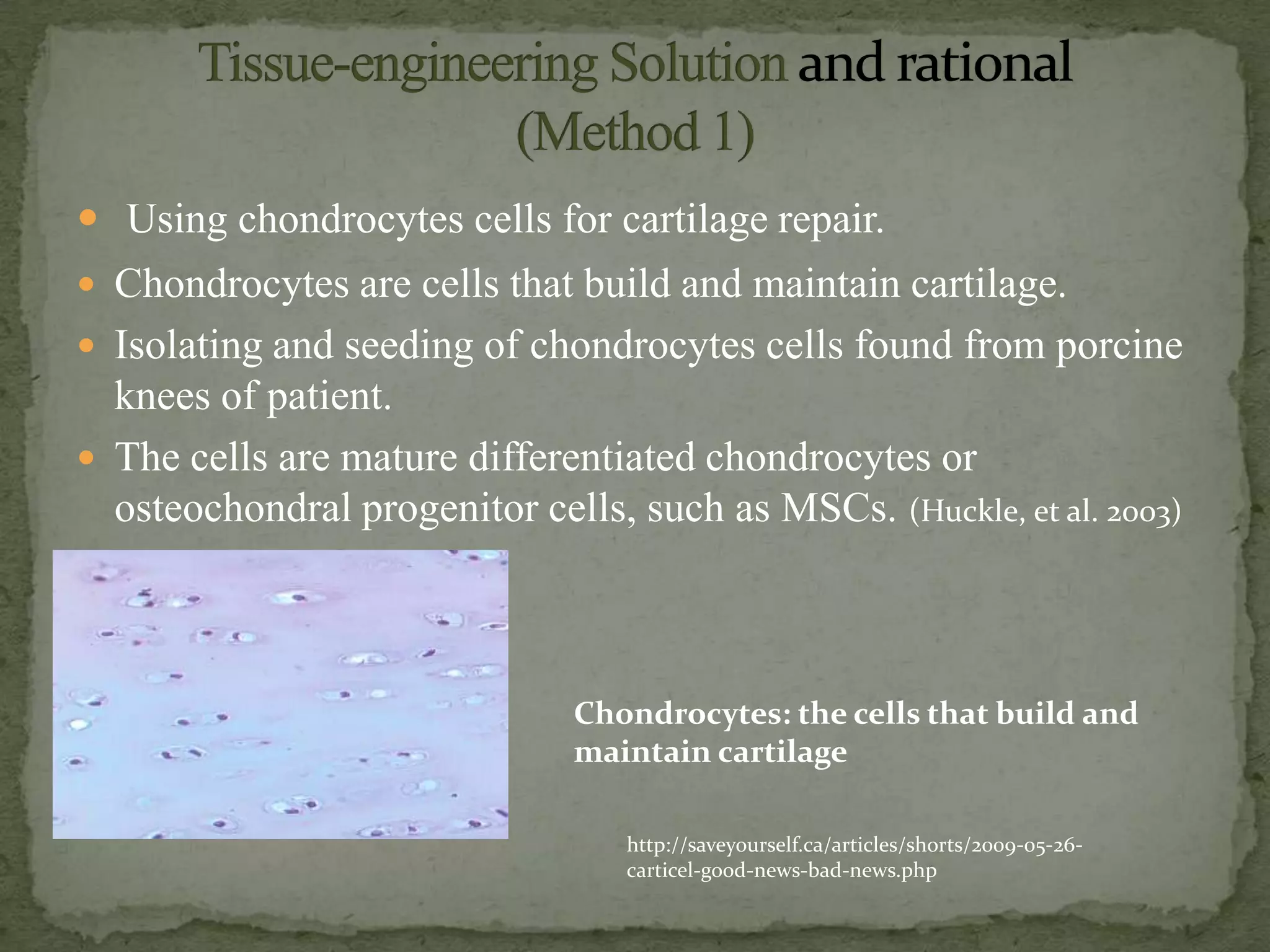
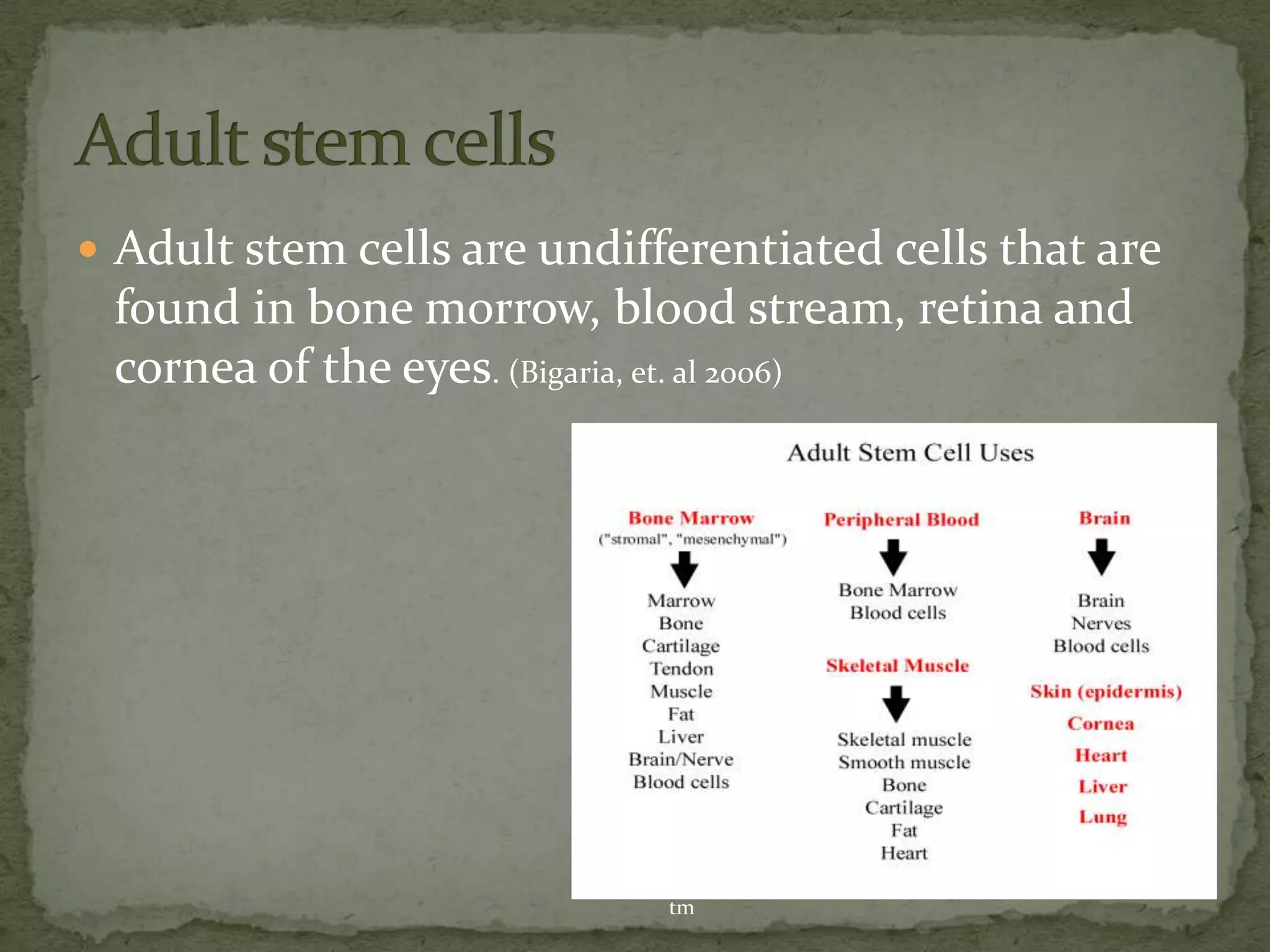
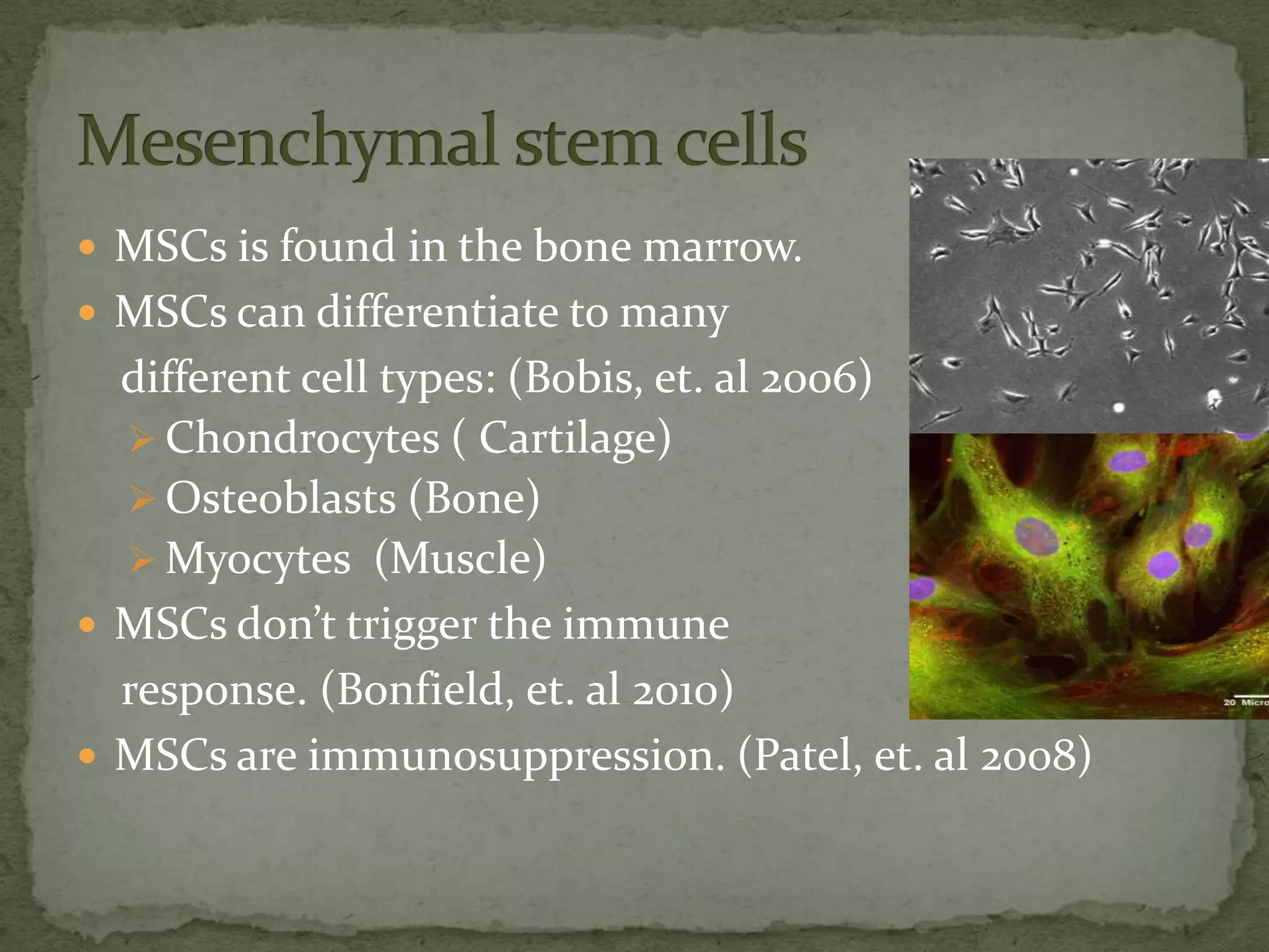
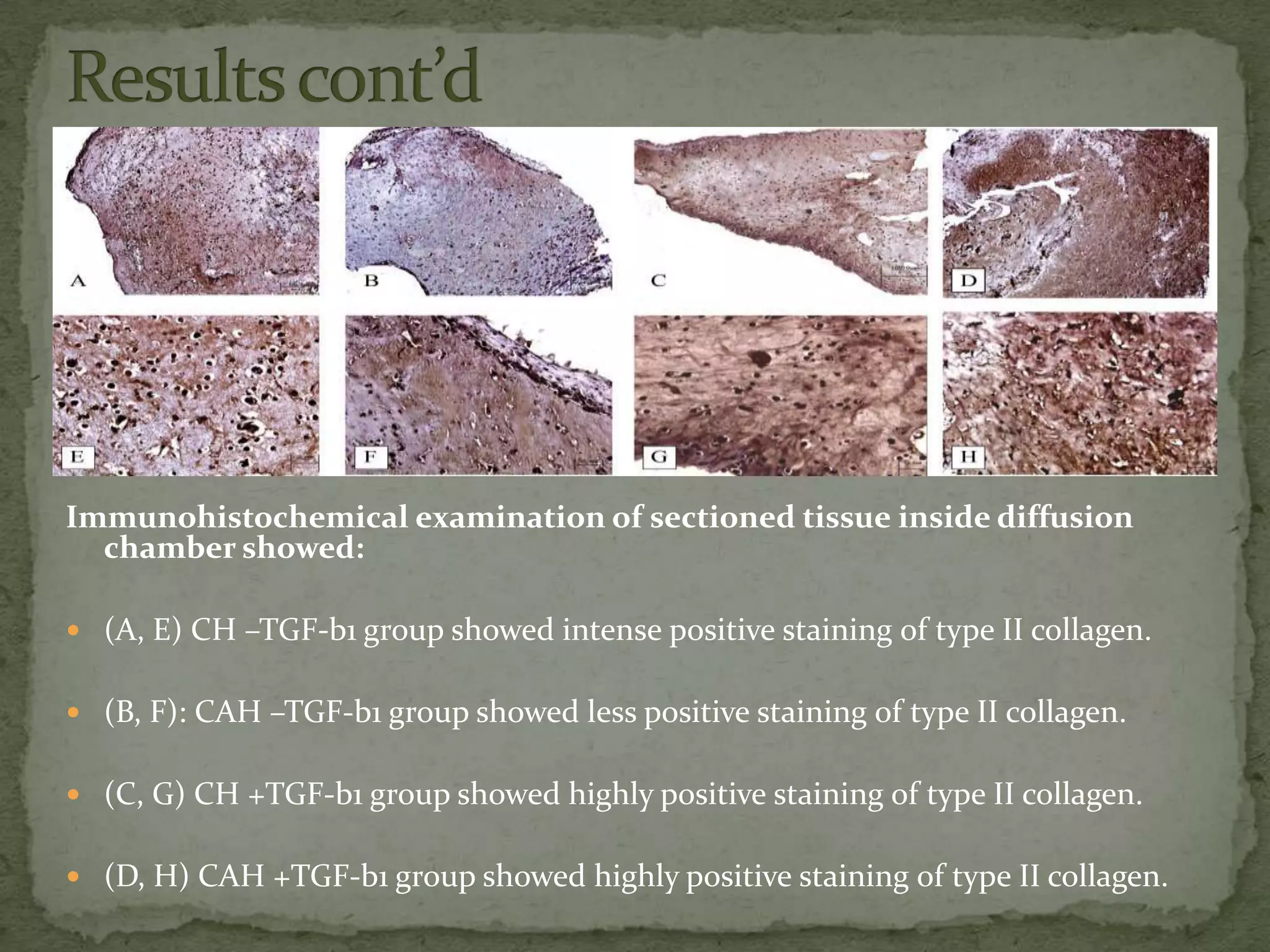
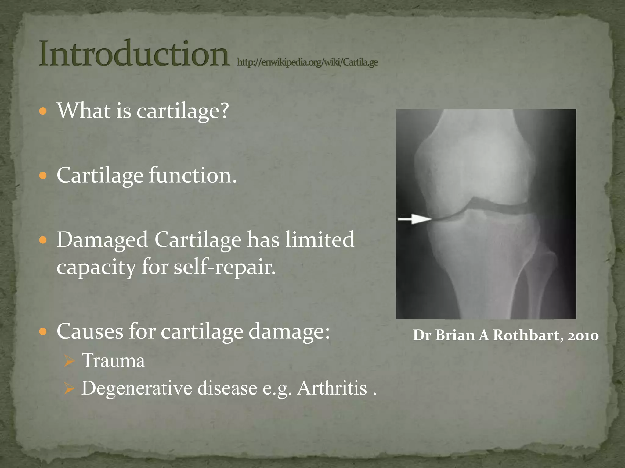
SlideShare a Scribd company logo
 What is cartilage?


 Cartilage function.


 Damaged Cartilage has limited
 capacity for self-repair.

 Causes for cartilage damage:              Dr Brian A Rothbart, 2010
   Trauma
   Degenerative disease e.g. Arthritis .
 Arthritis is a joint inflammation disorder.
 Inflammation happens due to
  cartilage degradation.
 No cure
 All treatments are to decrease
  symptoms or delay progress.
 Cartilage degradation may happens
  due to collagen degradation.
  o Collagen puts the ECM
     together.
 17.1% of the disabilities in USA because of arthritis.
  [Healthy people]
 350 million people
  worldwide have arthritis.
 37 million people in USA.
                             wrongdiagnosis.com

 Health care: $81 billion annually in USA. abcnews.go.com
 Using chondrocytes cells for cartilage repair.
 Chondrocytes are cells that build and maintain cartilage.
 Isolating and seeding of chondrocytes cells found from porcine
  knees of patient.
 The cells are mature differentiated chondrocytes or
  osteochondral progenitor cells, such as MSCs. (Huckle, et al. 2003)


                               Chondrocytes: the cells that build and
                               maintain cartilage

                                  http://saveyourself.ca/articles/shorts/2009-05-26-
                                  carticel-good-news-bad-news.php
 Isolating and seeding of chondrocytes: The cartilage tissue
  was harvested from porcine knees.

 Preparation of protein-loaded chitosan microsphere:
  Solution mixed with FITC-BSA (fluorescein isothiocyanate
  bovine serum albumin) or TGF-β1(transforming growth factor
  –beta1).

 Preparation of chitosan scaffold containing MS-TGF

 SEM (scanning electron microscopy) measurement:-
  Chondrocytes were growing on the scaffold.
            ( Sung Eun Kim, et al. 2006)
 Chitosan microsphere loaded with proteins, BSA (bovine
  serum albumin) and TGF (transforming growth factor)in
  the presence of TPP (Thiamine pyrophosphate)
 The size of microspheres was reduced by the protein.
 Diffusion of TPP into microspheres is time dependent and
  critical to maintain the shape of microsphere .
 The cross-linking time should be longer than 20 min.
                                         (Sung Eun Kim, et al. 2006)
Results cont’d
In vitro protein release from
  microspheres:
 The proteins, BSA (bovine serum
    albumin) and TGF (transforming
    growth factor) were released in
    biphasic manner.
   implies that initially the rate of
    release is fast and later the release is
    slower.
   BSA and TGF released at initial
    phase, 48.8% and 14.5% respectively.
   Release rate of TGF-beta 1 from                 Release profiles of FITC–
    microspheres was lower than BSA                 BSA (0) and TGF-β1 from
    over the whole experiment                       chitosan microspheres .
    BSA (90.1%) released within 5 days.
   Release amount of TGF- beta1 only
    44.9% for 7 days.      ( Sung Eun Kim, et al.
                            2006)
Results cont’d
Chondrocytes growth on chitosan scaffolds without
microsphere-TGF
  SEM (scanning electron microscopy) used for evaluation cell
   morphology and proliferation.
  After 4 days, chondrocytes were present in the superficial area of the
   scaffold and maintain a spherical morphology.
  After 7 days, chondrocytes were slightly proliferated without spreading.


                                                     ( Sung Eun Kim, et al.
                                                     2006)


Scanning electron micrographs of chondrocytes grown on chitosan scaffold for (a)
4 days and (b) 7 days after seeding.
Results cont’d


After 4 days , chondrocytes cells grow on chitosan scaffold
with MS-TGF, there was a significant increase in the number.
After 7 days , chondrocytes were proliferated and open pores
of scaffold occupied by the numbers of aggregates, composed
of several chondrocytes. ( Sung Eun Kim, et al. 2006)


Scanning electron micrographs of chondrocytes grown on chitosan scaffold, seeded with
chitosan microsphere containing TGF-β1. (a) Four days after seeding; the cells were
merged to form large aggregates adherent to the scaffold. (b) Magnified view of a
representative chondrocyte grown for 4 days. (c) Seven days after seeding
 TGF-β1, has promote the protein synthesis and cell
  proliferation in articular cartilage. It acts as an
  autocrine regulator of chondrocytes.
 It also inhibits the actions of matrix metalloproteinase
  that play an important role in the digestion of
  the ECM in both normal and degenerative articular
  cartilage.   ( Sung Eun Kim, et al. 2006).
 Using MSCs stem cells for cartilage repair.
 Implant MSCs encapsulated collagen based scaffold.
 Stem cells found in all multi cellular organisms. They
  can renew themselves and differentiate to different cell
  types.
 Two types of stem cells:
   Embryonic stem cells
   Adult stem cells
 Adult stem cells are undifferentiated cells that are
 found in bone morrow, blood stream, retina and
 cornea of the eyes. (Bigaria, et. al 2006)


                             http://www.stemcellresearch.org/testimony/prentice.h
                             tm
 MSCs is found in the bone marrow.
 MSCs can differentiate to many
  different cell types: (Bobis, et. al 2006)
   Chondrocytes ( Cartilage)
   Osteoblasts (Bone)
   Myocytes (Muscle)
 MSCs don’t trigger the immune
  response. (Bonfield, et. al 2010)
 MSCs are immunosuppression. (Patel, et. al 2008)
 Implanting MSCs in a rabbit cartilage defect.
 MSCs encapsulated in collagen hydrogel (CH) and collagen
  alginate hydrogel (CAH) scaffolds.
 The scaffold encapsulated in five groups of champers:
      CH + TGF-Beta1
      CH - TGF-Beta1
      CAH + TGF-Beta1
      CAH - TGF-Beta1
      MSCs without any scaffolds nor growth factors

 The champers implanted in the rabbits for 8 weeks.


 After 8m weeks, the rabbits sacrificed and champers collected.
 MSCs without any scaffolds, no tissue found.

HE staining of sectioned tissue inside diffusion
chamber showed:

(A) CH –TGF-b1 group showed cells of
   round shape and high cell density.

(B) CAH –TGF-b1 group showed lower
    cell density of round shape.


(C) CH +TGF-b1 group showed cells of
 round shape and high cell density.

(D) CAH +TGF-b1 group showed cells of
   round shape and high cell density.
Immunohistochemical examination of sectioned tissue inside diffusion
  chamber showed:

 (A, E) CH –TGF-b1 group showed intense positive staining of type II collagen.


 (B, F): CAH –TGF-b1 group showed less positive staining of type II collagen.


 (C, G) CH +TGF-b1 group showed highly positive staining of type II collagen.


 (D, H) CAH +TGF-b1 group showed highly positive staining of type II collagen.
 MSCs can differentiate to chondrocytes without
 interact with body fluids.

 A scaffold must be used for chondrogenesis to happen.


 CH support more chondrogenesis than CAH.


 TGF-b1 is important for MSCs differentiation.
 Tissue engineered chondrocytes can be used to treat cartilage
  damage.
 MSCs can be used to treat cartilage damage:
    Reduce the cost of healthcare for arthritis patients.
    Give arthritis patients definitive treatment.
    Open the way to treat other diseases.
 Limitation:
    TGF-β1 appears to be a powerful molecule to repair
     damaged cartilage. But high dose of intra-articular injection may
     induce chemotaxis and activation of inflammatory
     cells, resulting in fibrosis and osteophyte formation in cartilage
     defects.
    Number of surgeries that should be done to cover all the affected
     areas.
 Which better to use chondrocytes or MSCs?


 Do you think using these methods will be
 effective to treat arthritis?
 Bagaria, V., et al., Stem cells in orthopedics: current concepts and possible future
    applications. Indian J Med Sci, 2006. 60(4): p. 162-9.
   Bobis, S., D. Jarocha, and M. Majka, Mesenchymal stem cells: characteristics and
    clinical applications. Folia Histochem Cytobiol, 2006. 44(4): p. 215-30.
   Bonfield, T.L., et al., Defining human mesenchymal stem cell efficacy in vivo. J
    Inflamm (Lond), 2010. 7: p. 51.
   Patel, S.A., et al., Immunological properties of mesenchymal stem cells and clinical
    implications. Arch Immunol Ther Exp (Warsz), 2008. 56(1): p. 1-8.
   Zheng, L., et al., Chondrogenic differentiation of mesenchymal stem cells induced by
    collagen-based hydrogel: an in vivo study. J Biomed Mater Res A, 2010. 93(2): p.
    783-92.
    sung eun kim , et al.Porous chitson scaffold containing microspheres loaded with
    transforming growth factors-beta1: implications for cartilage tissue engineering:
    Biomedical Research Center, 19 May 2003.
   Huckle, et al. Differentiated chondrocytes for cartilage tissue engineering: 2003.

More Related Content

What's hot

Biomaterials for tissue engineering slideshare
Biomaterials for tissue engineering slideshareBiomaterials for tissue engineering slideshare
Biomaterials for tissue engineering slideshare
Bukar Abdullahi
 
Bioreactors in tissue engineering
Bioreactors in tissue engineeringBioreactors in tissue engineering
Bioreactors in tissue engineering
Vidya Kalaivani Rajkumar
 
Bio-engineering
Bio-engineeringBio-engineering
Tissue engineering
Tissue engineeringTissue engineering
Tissue engineeringrajatgothi
 
bone and_cartilage_tissue_engineering by Sumit
 bone and_cartilage_tissue_engineering by Sumit bone and_cartilage_tissue_engineering by Sumit
bone and_cartilage_tissue_engineering by Sumit
Dcrust
 
Bone tissue engineering
Bone tissue engineeringBone tissue engineering
Bone tissue engineering
Mehdi Chamani
 
Cartilage Repair using Stem cell & Orthobiologics
Cartilage Repair using Stem cell & OrthobiologicsCartilage Repair using Stem cell & Orthobiologics
Cartilage Repair using Stem cell & Orthobiologics
Vaibhav Bagaria
 
Polymeric and metallic scaffolds for tissue engineering
Polymeric and metallic scaffolds for tissue engineeringPolymeric and metallic scaffolds for tissue engineering
Polymeric and metallic scaffolds for tissue engineering
Mohamed M. Abdul-Monem
 
Basics of Tissue engineering
Basics of Tissue engineeringBasics of Tissue engineering
Basics of Tissue engineeringMahmoud Hamda
 
Mesenchymal stem cells in Orthopaedics
Mesenchymal stem cells in OrthopaedicsMesenchymal stem cells in Orthopaedics
Mesenchymal stem cells in Orthopaedics
Dr. Bushu Harna
 
Tissue engineering
Tissue engineeringTissue engineering
Principles of tissue engineering
Principles of tissue engineeringPrinciples of tissue engineering
Principles of tissue engineering
RobertMbaluka
 
Bone Marrow Stromal Stem cells (bMSCs) therapy for musculoskeletal problems ...
Bone Marrow Stromal Stem cells (bMSCs) therapy for musculoskeletal problems ...Bone Marrow Stromal Stem cells (bMSCs) therapy for musculoskeletal problems ...
Bone Marrow Stromal Stem cells (bMSCs) therapy for musculoskeletal problems ...Asosiasi Sel Punca Indonesia
 
Biomaterials for tissue engineering
Biomaterials for tissue engineeringBiomaterials for tissue engineering
Biomaterials for tissue engineering
Vidya Kalaivani Rajkumar
 
tissue engineering
tissue engineering tissue engineering
tissue engineering
boris saha
 
TISSUE DEVELOPMENT WITH TISSUE ENGINEERING APPROACH
TISSUE DEVELOPMENT WITH TISSUE ENGINEERING APPROACHTISSUE DEVELOPMENT WITH TISSUE ENGINEERING APPROACH
TISSUE DEVELOPMENT WITH TISSUE ENGINEERING APPROACH
Felix Obi
 
Skin tissue engineering
Skin tissue engineering Skin tissue engineering
Skin tissue engineering
Sam Higginbottom University
 
Tissue ingineering biot
Tissue ingineering biotTissue ingineering biot
Tissue ingineering biot
tanzeel Rehman
 
Tissue Engineering & Regenerative Medicine
Tissue Engineering & Regenerative MedicineTissue Engineering & Regenerative Medicine
Tissue Engineering & Regenerative Medicine
Mohamed Labadi
 

What's hot (20)

Biomaterials for tissue engineering slideshare
Biomaterials for tissue engineering slideshareBiomaterials for tissue engineering slideshare
Biomaterials for tissue engineering slideshare
 
Bioreactors in tissue engineering
Bioreactors in tissue engineeringBioreactors in tissue engineering
Bioreactors in tissue engineering
 
Bio-engineering
Bio-engineeringBio-engineering
Bio-engineering
 
Tissue engineering
Tissue engineeringTissue engineering
Tissue engineering
 
bone and_cartilage_tissue_engineering by Sumit
 bone and_cartilage_tissue_engineering by Sumit bone and_cartilage_tissue_engineering by Sumit
bone and_cartilage_tissue_engineering by Sumit
 
Bone tissue engineering
Bone tissue engineeringBone tissue engineering
Bone tissue engineering
 
Cartilage Repair using Stem cell & Orthobiologics
Cartilage Repair using Stem cell & OrthobiologicsCartilage Repair using Stem cell & Orthobiologics
Cartilage Repair using Stem cell & Orthobiologics
 
Polymeric and metallic scaffolds for tissue engineering
Polymeric and metallic scaffolds for tissue engineeringPolymeric and metallic scaffolds for tissue engineering
Polymeric and metallic scaffolds for tissue engineering
 
Basics of Tissue engineering
Basics of Tissue engineeringBasics of Tissue engineering
Basics of Tissue engineering
 
Mesenchymal stem cells in Orthopaedics
Mesenchymal stem cells in OrthopaedicsMesenchymal stem cells in Orthopaedics
Mesenchymal stem cells in Orthopaedics
 
Tissue engineering
Tissue engineeringTissue engineering
Tissue engineering
 
Principles of tissue engineering
Principles of tissue engineeringPrinciples of tissue engineering
Principles of tissue engineering
 
Bone Marrow Stromal Stem cells (bMSCs) therapy for musculoskeletal problems ...
Bone Marrow Stromal Stem cells (bMSCs) therapy for musculoskeletal problems ...Bone Marrow Stromal Stem cells (bMSCs) therapy for musculoskeletal problems ...
Bone Marrow Stromal Stem cells (bMSCs) therapy for musculoskeletal problems ...
 
Tissue engineering
Tissue engineeringTissue engineering
Tissue engineering
 
Biomaterials for tissue engineering
Biomaterials for tissue engineeringBiomaterials for tissue engineering
Biomaterials for tissue engineering
 
tissue engineering
tissue engineering tissue engineering
tissue engineering
 
TISSUE DEVELOPMENT WITH TISSUE ENGINEERING APPROACH
TISSUE DEVELOPMENT WITH TISSUE ENGINEERING APPROACHTISSUE DEVELOPMENT WITH TISSUE ENGINEERING APPROACH
TISSUE DEVELOPMENT WITH TISSUE ENGINEERING APPROACH
 
Skin tissue engineering
Skin tissue engineering Skin tissue engineering
Skin tissue engineering
 
Tissue ingineering biot
Tissue ingineering biotTissue ingineering biot
Tissue ingineering biot
 
Tissue Engineering & Regenerative Medicine
Tissue Engineering & Regenerative MedicineTissue Engineering & Regenerative Medicine
Tissue Engineering & Regenerative Medicine
 

Viewers also liked

Bone and Cartilage regeneration with cells and tissue engineering products - ...
Bone and Cartilage regeneration with cells and tissue engineering products - ...Bone and Cartilage regeneration with cells and tissue engineering products - ...
Bone and Cartilage regeneration with cells and tissue engineering products - ...
Enric Caceres
 
CARTILAGE
CARTILAGECARTILAGE
CARTILAGE
Ladi Anudeep
 
Cartilage
CartilageCartilage
Cartilage
surajitkundu
 
2 mechanical properties of bone
2 mechanical properties of bone2 mechanical properties of bone
2 mechanical properties of bone
Lisa Benson
 
Biomechanics of-the-articular-cartilage 2
Biomechanics of-the-articular-cartilage 2Biomechanics of-the-articular-cartilage 2
Biomechanics of-the-articular-cartilage 2
Susanta85
 
Articular cartilage
Articular cartilageArticular cartilage
Articular cartilageorthoprince
 
Osteoarthritis ppt
Osteoarthritis pptOsteoarthritis ppt
Osteoarthritis pptRupika Sodhi
 

Viewers also liked (10)

Bone and Cartilage regeneration with cells and tissue engineering products - ...
Bone and Cartilage regeneration with cells and tissue engineering products - ...Bone and Cartilage regeneration with cells and tissue engineering products - ...
Bone and Cartilage regeneration with cells and tissue engineering products - ...
 
CARTILAGE
CARTILAGECARTILAGE
CARTILAGE
 
Cartilage
CartilageCartilage
Cartilage
 
2 mechanical properties of bone
2 mechanical properties of bone2 mechanical properties of bone
2 mechanical properties of bone
 
Osteoarthritis
OsteoarthritisOsteoarthritis
Osteoarthritis
 
Biomechanics of Bones
Biomechanics of BonesBiomechanics of Bones
Biomechanics of Bones
 
Biomechanics of-the-articular-cartilage 2
Biomechanics of-the-articular-cartilage 2Biomechanics of-the-articular-cartilage 2
Biomechanics of-the-articular-cartilage 2
 
Articular cartilage
Articular cartilageArticular cartilage
Articular cartilage
 
Bones
BonesBones
Bones
 
Osteoarthritis ppt
Osteoarthritis pptOsteoarthritis ppt
Osteoarthritis ppt
 

Similar to Cartilage Tissue Engineering

Stem cell research
Stem cell researchStem cell research
Stem cell research
Tariq Mohammed
 
Murine Mesenchymal Stem Cell Isolation Method Comparison
Murine Mesenchymal Stem Cell Isolation Method ComparisonMurine Mesenchymal Stem Cell Isolation Method Comparison
Murine Mesenchymal Stem Cell Isolation Method Comparison
Jake Turner
 
Icrs poster 2
Icrs poster  2Icrs poster  2
Icrs poster 2
Tariq Mohammed
 
Stem cells in regenerative biology and medicine
Stem cells in regenerative biology and medicineStem cells in regenerative biology and medicine
Stem cells in regenerative biology and medicine
Pasteur_Tunis
 
Stem sel pada hewan dan aplikasinya
Stem sel pada hewan dan aplikasinyaStem sel pada hewan dan aplikasinya
Stem sel pada hewan dan aplikasinya
raditio ghifiardi
 
Stem cell therapy in veterinary medicine
Stem cell therapy in veterinary medicineStem cell therapy in veterinary medicine
Stem cell therapy in veterinary medicine
Ajith Y
 
CTE ppt on CARTILAGE TISSUE ENGINEERING.pdf
CTE ppt on CARTILAGE TISSUE ENGINEERING.pdfCTE ppt on CARTILAGE TISSUE ENGINEERING.pdf
CTE ppt on CARTILAGE TISSUE ENGINEERING.pdf
Khushbu
 
刊2013Q405-CYTOO專刊(確定版)
刊2013Q405-CYTOO專刊(確定版)刊2013Q405-CYTOO專刊(確定版)
刊2013Q405-CYTOO專刊(確定版)Shelly Shen
 
Research ArticleAging and rejuvenation of human muscle stem .docx
Research ArticleAging and rejuvenation of human muscle stem .docxResearch ArticleAging and rejuvenation of human muscle stem .docx
Research ArticleAging and rejuvenation of human muscle stem .docx
debishakespeare
 
Sessio formacio bst terapies avancades
Sessio formacio bst terapies avancadesSessio formacio bst terapies avancades
Sessio formacio bst terapies avancades
Banc de Sang i Teixits
 
Bone Morphogenetic Proteins - Role in Periodontal Regeneration
Bone Morphogenetic Proteins - Role in Periodontal RegenerationBone Morphogenetic Proteins - Role in Periodontal Regeneration
Bone Morphogenetic Proteins - Role in Periodontal Regeneration
Dr.Shraddha Kode
 
Psoralen Promotes Myogenic Differentiation of Muscle Cells to Repair Fracture
Psoralen Promotes Myogenic Differentiation of Muscle Cells to Repair Fracture Psoralen Promotes Myogenic Differentiation of Muscle Cells to Repair Fracture
Psoralen Promotes Myogenic Differentiation of Muscle Cells to Repair Fracture
Associate Professor in VSB Coimbatore
 
Effect of Seeding Density of Mesenchymal Stem Cells on Cell Proliferation and...
Effect of Seeding Density of Mesenchymal Stem Cells on Cell Proliferation and...Effect of Seeding Density of Mesenchymal Stem Cells on Cell Proliferation and...
Effect of Seeding Density of Mesenchymal Stem Cells on Cell Proliferation and...Jaideep Sahni
 
Transplantation journal
Transplantation journalTransplantation journal
Transplantation journal
Scidoc Publishers
 
Stem cells journal
Stem cells  journalStem cells  journal
Stem cells journal
Scidoc Publishers
 
Journal of stem cells research
Journal of stem cells researchJournal of stem cells research
Journal of stem cells research
Scidoc Publishers
 
Tissue Engineering JC.ppt
Tissue Engineering JC.pptTissue Engineering JC.ppt
Tissue Engineering JC.ppt
Rinisha Sinha
 
Amphiregulin and Muscle Stem Cells
Amphiregulin and Muscle Stem CellsAmphiregulin and Muscle Stem Cells
Amphiregulin and Muscle Stem Cells
MichaelShadpour
 

Similar to Cartilage Tissue Engineering (20)

Stem cell research
Stem cell researchStem cell research
Stem cell research
 
Murine Mesenchymal Stem Cell Isolation Method Comparison
Murine Mesenchymal Stem Cell Isolation Method ComparisonMurine Mesenchymal Stem Cell Isolation Method Comparison
Murine Mesenchymal Stem Cell Isolation Method Comparison
 
Icrs poster 2
Icrs poster  2Icrs poster  2
Icrs poster 2
 
Introductiondata
IntroductiondataIntroductiondata
Introductiondata
 
Stem cells in regenerative biology and medicine
Stem cells in regenerative biology and medicineStem cells in regenerative biology and medicine
Stem cells in regenerative biology and medicine
 
Stem sel pada hewan dan aplikasinya
Stem sel pada hewan dan aplikasinyaStem sel pada hewan dan aplikasinya
Stem sel pada hewan dan aplikasinya
 
Irmi unige
Irmi unigeIrmi unige
Irmi unige
 
Stem cell therapy in veterinary medicine
Stem cell therapy in veterinary medicineStem cell therapy in veterinary medicine
Stem cell therapy in veterinary medicine
 
CTE ppt on CARTILAGE TISSUE ENGINEERING.pdf
CTE ppt on CARTILAGE TISSUE ENGINEERING.pdfCTE ppt on CARTILAGE TISSUE ENGINEERING.pdf
CTE ppt on CARTILAGE TISSUE ENGINEERING.pdf
 
刊2013Q405-CYTOO專刊(確定版)
刊2013Q405-CYTOO專刊(確定版)刊2013Q405-CYTOO專刊(確定版)
刊2013Q405-CYTOO專刊(確定版)
 
Research ArticleAging and rejuvenation of human muscle stem .docx
Research ArticleAging and rejuvenation of human muscle stem .docxResearch ArticleAging and rejuvenation of human muscle stem .docx
Research ArticleAging and rejuvenation of human muscle stem .docx
 
Sessio formacio bst terapies avancades
Sessio formacio bst terapies avancadesSessio formacio bst terapies avancades
Sessio formacio bst terapies avancades
 
Bone Morphogenetic Proteins - Role in Periodontal Regeneration
Bone Morphogenetic Proteins - Role in Periodontal RegenerationBone Morphogenetic Proteins - Role in Periodontal Regeneration
Bone Morphogenetic Proteins - Role in Periodontal Regeneration
 
Psoralen Promotes Myogenic Differentiation of Muscle Cells to Repair Fracture
Psoralen Promotes Myogenic Differentiation of Muscle Cells to Repair Fracture Psoralen Promotes Myogenic Differentiation of Muscle Cells to Repair Fracture
Psoralen Promotes Myogenic Differentiation of Muscle Cells to Repair Fracture
 
Effect of Seeding Density of Mesenchymal Stem Cells on Cell Proliferation and...
Effect of Seeding Density of Mesenchymal Stem Cells on Cell Proliferation and...Effect of Seeding Density of Mesenchymal Stem Cells on Cell Proliferation and...
Effect of Seeding Density of Mesenchymal Stem Cells on Cell Proliferation and...
 
Transplantation journal
Transplantation journalTransplantation journal
Transplantation journal
 
Stem cells journal
Stem cells  journalStem cells  journal
Stem cells journal
 
Journal of stem cells research
Journal of stem cells researchJournal of stem cells research
Journal of stem cells research
 
Tissue Engineering JC.ppt
Tissue Engineering JC.pptTissue Engineering JC.ppt
Tissue Engineering JC.ppt
 
Amphiregulin and Muscle Stem Cells
Amphiregulin and Muscle Stem CellsAmphiregulin and Muscle Stem Cells
Amphiregulin and Muscle Stem Cells
 

Cartilage Tissue Engineering

  • 1.
  • 2.  What is cartilage?  Cartilage function.  Damaged Cartilage has limited capacity for self-repair.  Causes for cartilage damage: Dr Brian A Rothbart, 2010  Trauma  Degenerative disease e.g. Arthritis .
  • 3.  Arthritis is a joint inflammation disorder.  Inflammation happens due to cartilage degradation.  No cure  All treatments are to decrease symptoms or delay progress.  Cartilage degradation may happens due to collagen degradation. o Collagen puts the ECM together.
  • 4.  17.1% of the disabilities in USA because of arthritis. [Healthy people]  350 million people worldwide have arthritis.  37 million people in USA. wrongdiagnosis.com  Health care: $81 billion annually in USA. abcnews.go.com
  • 5.  Using chondrocytes cells for cartilage repair.  Chondrocytes are cells that build and maintain cartilage.  Isolating and seeding of chondrocytes cells found from porcine knees of patient.  The cells are mature differentiated chondrocytes or osteochondral progenitor cells, such as MSCs. (Huckle, et al. 2003) Chondrocytes: the cells that build and maintain cartilage http://saveyourself.ca/articles/shorts/2009-05-26- carticel-good-news-bad-news.php
  • 6.  Isolating and seeding of chondrocytes: The cartilage tissue was harvested from porcine knees.  Preparation of protein-loaded chitosan microsphere: Solution mixed with FITC-BSA (fluorescein isothiocyanate bovine serum albumin) or TGF-β1(transforming growth factor –beta1).  Preparation of chitosan scaffold containing MS-TGF  SEM (scanning electron microscopy) measurement:- Chondrocytes were growing on the scaffold. ( Sung Eun Kim, et al. 2006)
  • 7.  Chitosan microsphere loaded with proteins, BSA (bovine serum albumin) and TGF (transforming growth factor)in the presence of TPP (Thiamine pyrophosphate)  The size of microspheres was reduced by the protein.  Diffusion of TPP into microspheres is time dependent and critical to maintain the shape of microsphere .  The cross-linking time should be longer than 20 min. (Sung Eun Kim, et al. 2006)
  • 8. Results cont’d In vitro protein release from microspheres:  The proteins, BSA (bovine serum albumin) and TGF (transforming growth factor) were released in biphasic manner.  implies that initially the rate of release is fast and later the release is slower.  BSA and TGF released at initial phase, 48.8% and 14.5% respectively.  Release rate of TGF-beta 1 from Release profiles of FITC– microspheres was lower than BSA BSA (0) and TGF-β1 from over the whole experiment chitosan microspheres .  BSA (90.1%) released within 5 days.  Release amount of TGF- beta1 only 44.9% for 7 days. ( Sung Eun Kim, et al. 2006)
  • 9. Results cont’d Chondrocytes growth on chitosan scaffolds without microsphere-TGF  SEM (scanning electron microscopy) used for evaluation cell morphology and proliferation.  After 4 days, chondrocytes were present in the superficial area of the scaffold and maintain a spherical morphology.  After 7 days, chondrocytes were slightly proliferated without spreading. ( Sung Eun Kim, et al. 2006) Scanning electron micrographs of chondrocytes grown on chitosan scaffold for (a) 4 days and (b) 7 days after seeding.
  • 10. Results cont’d After 4 days , chondrocytes cells grow on chitosan scaffold with MS-TGF, there was a significant increase in the number. After 7 days , chondrocytes were proliferated and open pores of scaffold occupied by the numbers of aggregates, composed of several chondrocytes. ( Sung Eun Kim, et al. 2006) Scanning electron micrographs of chondrocytes grown on chitosan scaffold, seeded with chitosan microsphere containing TGF-β1. (a) Four days after seeding; the cells were merged to form large aggregates adherent to the scaffold. (b) Magnified view of a representative chondrocyte grown for 4 days. (c) Seven days after seeding
  • 11.  TGF-β1, has promote the protein synthesis and cell proliferation in articular cartilage. It acts as an autocrine regulator of chondrocytes.  It also inhibits the actions of matrix metalloproteinase that play an important role in the digestion of the ECM in both normal and degenerative articular cartilage. ( Sung Eun Kim, et al. 2006).
  • 12.  Using MSCs stem cells for cartilage repair.  Implant MSCs encapsulated collagen based scaffold.  Stem cells found in all multi cellular organisms. They can renew themselves and differentiate to different cell types.  Two types of stem cells:  Embryonic stem cells  Adult stem cells
  • 13.  Adult stem cells are undifferentiated cells that are found in bone morrow, blood stream, retina and cornea of the eyes. (Bigaria, et. al 2006) http://www.stemcellresearch.org/testimony/prentice.h tm
  • 14.  MSCs is found in the bone marrow.  MSCs can differentiate to many different cell types: (Bobis, et. al 2006)  Chondrocytes ( Cartilage)  Osteoblasts (Bone)  Myocytes (Muscle)  MSCs don’t trigger the immune response. (Bonfield, et. al 2010)  MSCs are immunosuppression. (Patel, et. al 2008)
  • 15.  Implanting MSCs in a rabbit cartilage defect.  MSCs encapsulated in collagen hydrogel (CH) and collagen alginate hydrogel (CAH) scaffolds.  The scaffold encapsulated in five groups of champers:  CH + TGF-Beta1  CH - TGF-Beta1  CAH + TGF-Beta1  CAH - TGF-Beta1  MSCs without any scaffolds nor growth factors  The champers implanted in the rabbits for 8 weeks.  After 8m weeks, the rabbits sacrificed and champers collected.
  • 16.  MSCs without any scaffolds, no tissue found. HE staining of sectioned tissue inside diffusion chamber showed: (A) CH –TGF-b1 group showed cells of round shape and high cell density. (B) CAH –TGF-b1 group showed lower cell density of round shape. (C) CH +TGF-b1 group showed cells of round shape and high cell density. (D) CAH +TGF-b1 group showed cells of round shape and high cell density.
  • 17. Immunohistochemical examination of sectioned tissue inside diffusion chamber showed:  (A, E) CH –TGF-b1 group showed intense positive staining of type II collagen.  (B, F): CAH –TGF-b1 group showed less positive staining of type II collagen.  (C, G) CH +TGF-b1 group showed highly positive staining of type II collagen.  (D, H) CAH +TGF-b1 group showed highly positive staining of type II collagen.
  • 18.  MSCs can differentiate to chondrocytes without interact with body fluids.  A scaffold must be used for chondrogenesis to happen.  CH support more chondrogenesis than CAH.  TGF-b1 is important for MSCs differentiation.
  • 19.  Tissue engineered chondrocytes can be used to treat cartilage damage.  MSCs can be used to treat cartilage damage:  Reduce the cost of healthcare for arthritis patients.  Give arthritis patients definitive treatment.  Open the way to treat other diseases.  Limitation:  TGF-β1 appears to be a powerful molecule to repair damaged cartilage. But high dose of intra-articular injection may induce chemotaxis and activation of inflammatory cells, resulting in fibrosis and osteophyte formation in cartilage defects.  Number of surgeries that should be done to cover all the affected areas.
  • 20.  Which better to use chondrocytes or MSCs?  Do you think using these methods will be effective to treat arthritis?
  • 21.  Bagaria, V., et al., Stem cells in orthopedics: current concepts and possible future applications. Indian J Med Sci, 2006. 60(4): p. 162-9.  Bobis, S., D. Jarocha, and M. Majka, Mesenchymal stem cells: characteristics and clinical applications. Folia Histochem Cytobiol, 2006. 44(4): p. 215-30.  Bonfield, T.L., et al., Defining human mesenchymal stem cell efficacy in vivo. J Inflamm (Lond), 2010. 7: p. 51.  Patel, S.A., et al., Immunological properties of mesenchymal stem cells and clinical implications. Arch Immunol Ther Exp (Warsz), 2008. 56(1): p. 1-8.  Zheng, L., et al., Chondrogenic differentiation of mesenchymal stem cells induced by collagen-based hydrogel: an in vivo study. J Biomed Mater Res A, 2010. 93(2): p. 783-92.  sung eun kim , et al.Porous chitson scaffold containing microspheres loaded with transforming growth factors-beta1: implications for cartilage tissue engineering: Biomedical Research Center, 19 May 2003.  Huckle, et al. Differentiated chondrocytes for cartilage tissue engineering: 2003.