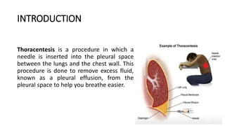
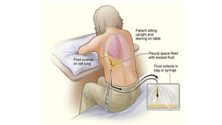
THORACENTESIS
PRESENTED BY:
MR. ABHAY RAJPOOT
INTRODUCTION
Thoracentesis is a procedure in which a
needle is inserted into the pleural space
between the lungs and the chest wall. This
procedure is done to remove excess fluid,
known as a pleural effusion, from the
pleural space to help you breathe easier.
DEFINITION
• Thoracentesis is a procedure to remove fluid from the space between
the lungs and the chest wall called the pleural space.
OR
• Thoracentesis is a procedure that removes an abnormal accumulation
of fluid or air from the chest through a needle or tube.
PURPOSE
• To determine the cause of abnormal accumulation of fluid in the
pleural space.
• Relieve shortness of breath and pain
• As a diagnostic or treatment procedure
• To drain large amounts of pleural fluid
• To equalize pressure on both sides of the thoracic cavity
INDICATION
• Traumatic pneumothorax
• Hemopneumothorax
• Spontaneous pneumothorax
• Bronchopleural fistula
• Pleural effusion
CONTRAINDICATION
• An uncooperative patient
• Coagulation disorder
• Atelectasis
• Only one functioning lung
• Emphysema (pulmonary enlargement)
• Severe cough or hiccups
COMPLICATION
• Pulmonary edema
• Respiratory distress
• Air embolism
• Cardiac tamponade (fluid build up in the space between myocardium
and pericardium)
• Bleeding
• Infection
• Dyspnea and cough
• Atelectasis
NURSING RESPONSIBILITY:
• Explain the purpose, risks/benefits, and steps of the procedure and
obtain consent from the patient or appropriate legal design.
• Take Medical History such as
• Trouble in breathing, coughing, and hiccups
• Had heart disease
• Smoked
• Travelled to places where may have been exposed to tuberculosis
PREPARE EQUIPMENT:
Dressing set
Abraham’s needle
Connecting tubing
Syringe 50ml and 5ml
Scalpel blade and blade 11
Needles (18 and 23 gauge)
Sterile Glove
Mask
Povidone / Alcohol
Local anaesthetic, e.g. lignocaine (lidocaine) 1% or 2%
Formalin bottle
Urine bottle x2
C+S bottle
3-way stopcock
Jug
BEFORE THE PROCEDURE:
Check platelet count and/or presence of coagulopathy. If platelet
count is < 20,000, or there is known coagulopathy as to whether
platelet transfusion or other intervention is needed.
Place patient upright / cardiac position and help patient maintain
position during procedure. R: the upright position ensures that the
diaphragm is more dependent and facilitates the removal of fluid that
usually localizes at the base of the chest.
Explain that he/she will receive a local anesthetic
Clean patient skin with antiseptic soap
Observe patient respiration rate and breathing pattern.
Conti…
Monitor saturation R: To prevent hypoxia
Inform doctor if any changes of the patient
Obtain a chest x-ray to evaluate the fluid level.
For specimen handling, fill the tubes with the required amount of
pleural fluid
Check that each bottle is correctly labelled by checking patient
identifiers- full name, date of birth and/or medical record number
then send to the lab tests
AFTER PROCEDURE:
Document the procedure, patient’s response, characteristics of fluid
and amount, and patient response to follow-up.
Provide post-procedural analgesics as needed.
Rest in bed for about 2 hours after the procedure
Blood pressure and breathing will be checked for up to a few hours
May remove dressing/bandage another day, or replace it if it
becomes soiled or wet
Resume patient regular diet.
Thoracentesis

Thoracentesis

  • 1.
  • 2.
    INTRODUCTION Thoracentesis is aprocedure in which a needle is inserted into the pleural space between the lungs and the chest wall. This procedure is done to remove excess fluid, known as a pleural effusion, from the pleural space to help you breathe easier.
  • 3.
    DEFINITION • Thoracentesis isa procedure to remove fluid from the space between the lungs and the chest wall called the pleural space. OR • Thoracentesis is a procedure that removes an abnormal accumulation of fluid or air from the chest through a needle or tube.
  • 5.
    PURPOSE • To determinethe cause of abnormal accumulation of fluid in the pleural space. • Relieve shortness of breath and pain • As a diagnostic or treatment procedure • To drain large amounts of pleural fluid • To equalize pressure on both sides of the thoracic cavity
  • 6.
    INDICATION • Traumatic pneumothorax •Hemopneumothorax • Spontaneous pneumothorax • Bronchopleural fistula • Pleural effusion
  • 7.
    CONTRAINDICATION • An uncooperativepatient • Coagulation disorder • Atelectasis • Only one functioning lung • Emphysema (pulmonary enlargement) • Severe cough or hiccups
  • 8.
    COMPLICATION • Pulmonary edema •Respiratory distress • Air embolism • Cardiac tamponade (fluid build up in the space between myocardium and pericardium) • Bleeding • Infection • Dyspnea and cough • Atelectasis
  • 9.
    NURSING RESPONSIBILITY: • Explainthe purpose, risks/benefits, and steps of the procedure and obtain consent from the patient or appropriate legal design. • Take Medical History such as • Trouble in breathing, coughing, and hiccups • Had heart disease • Smoked • Travelled to places where may have been exposed to tuberculosis
  • 10.
    PREPARE EQUIPMENT: Dressing set Abraham’sneedle Connecting tubing Syringe 50ml and 5ml Scalpel blade and blade 11 Needles (18 and 23 gauge) Sterile Glove Mask Povidone / Alcohol Local anaesthetic, e.g. lignocaine (lidocaine) 1% or 2% Formalin bottle Urine bottle x2 C+S bottle 3-way stopcock Jug
  • 11.
    BEFORE THE PROCEDURE: Checkplatelet count and/or presence of coagulopathy. If platelet count is < 20,000, or there is known coagulopathy as to whether platelet transfusion or other intervention is needed. Place patient upright / cardiac position and help patient maintain position during procedure. R: the upright position ensures that the diaphragm is more dependent and facilitates the removal of fluid that usually localizes at the base of the chest. Explain that he/she will receive a local anesthetic Clean patient skin with antiseptic soap Observe patient respiration rate and breathing pattern.
  • 12.
    Conti… Monitor saturation R:To prevent hypoxia Inform doctor if any changes of the patient Obtain a chest x-ray to evaluate the fluid level. For specimen handling, fill the tubes with the required amount of pleural fluid Check that each bottle is correctly labelled by checking patient identifiers- full name, date of birth and/or medical record number then send to the lab tests
  • 13.
    AFTER PROCEDURE: Document theprocedure, patient’s response, characteristics of fluid and amount, and patient response to follow-up. Provide post-procedural analgesics as needed. Rest in bed for about 2 hours after the procedure Blood pressure and breathing will be checked for up to a few hours May remove dressing/bandage another day, or replace it if it becomes soiled or wet Resume patient regular diet.