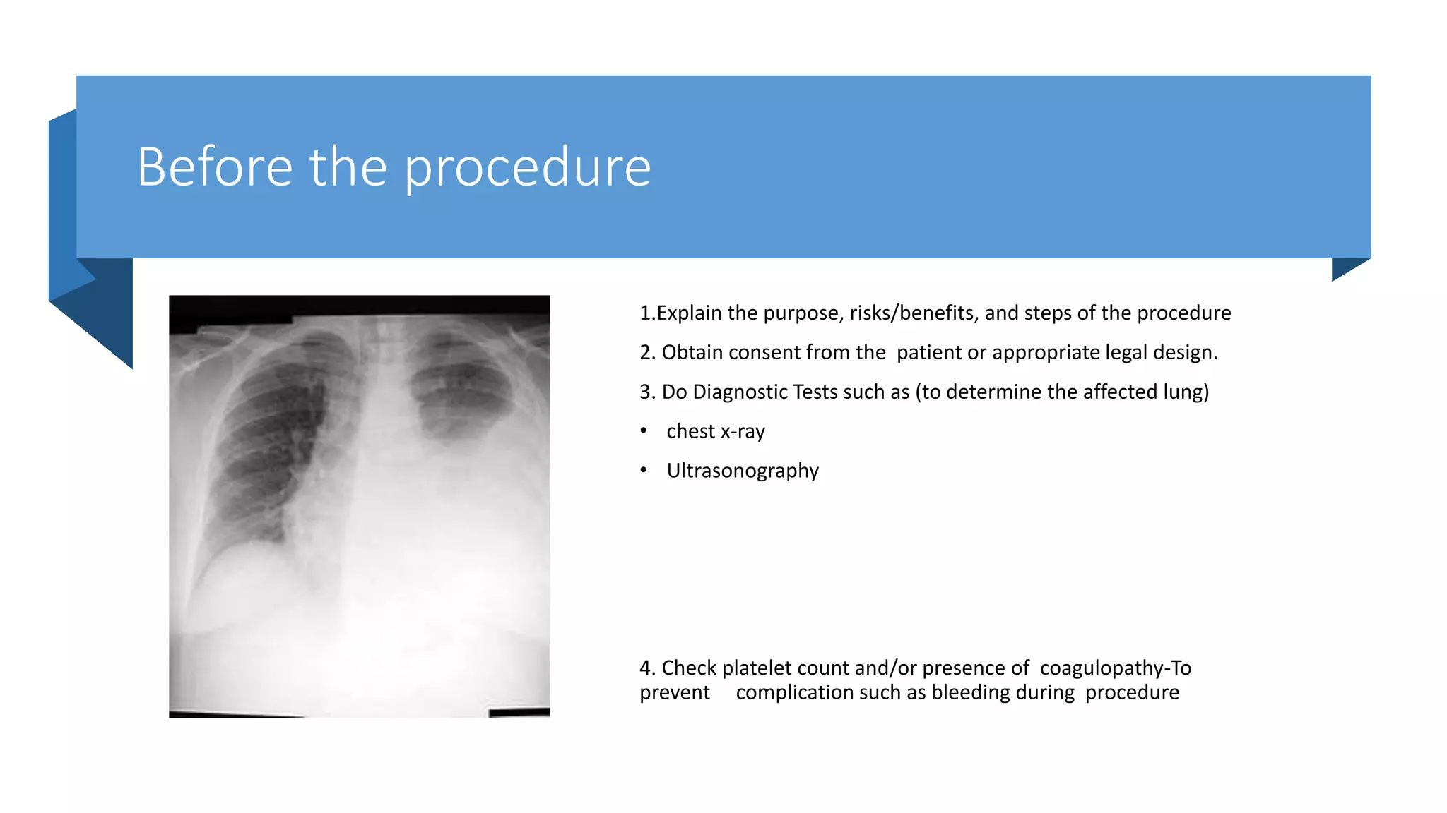
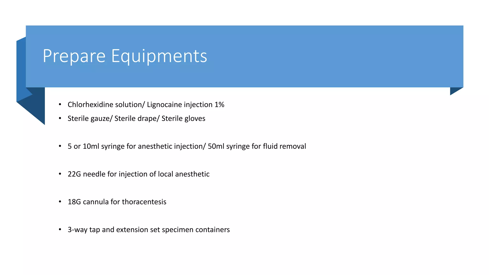
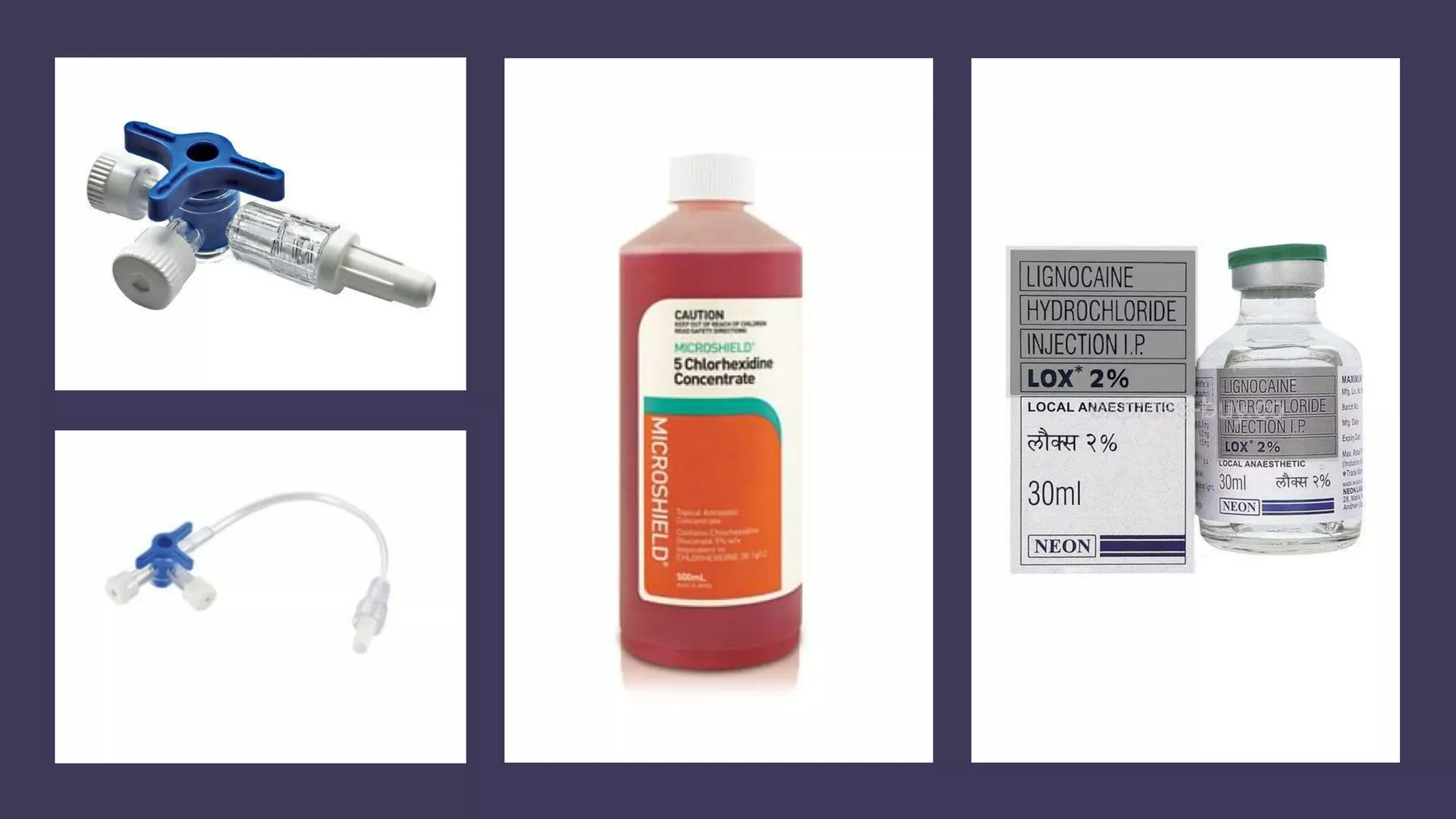
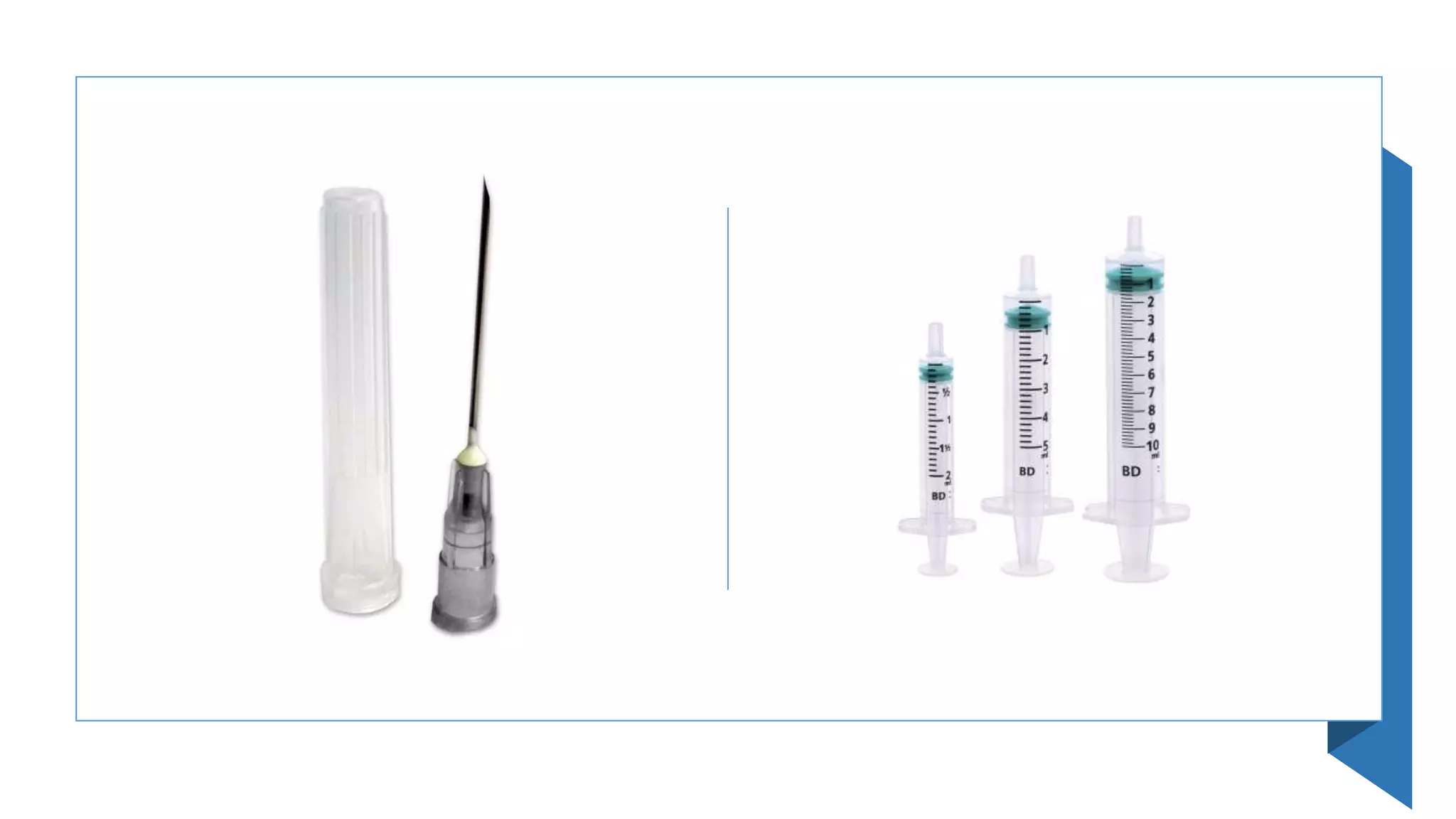
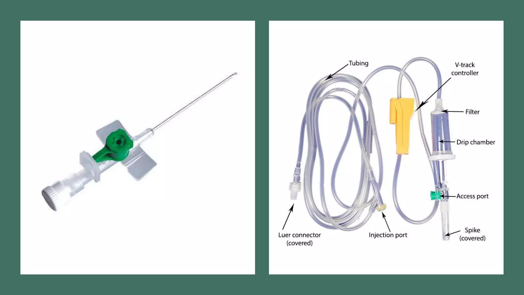
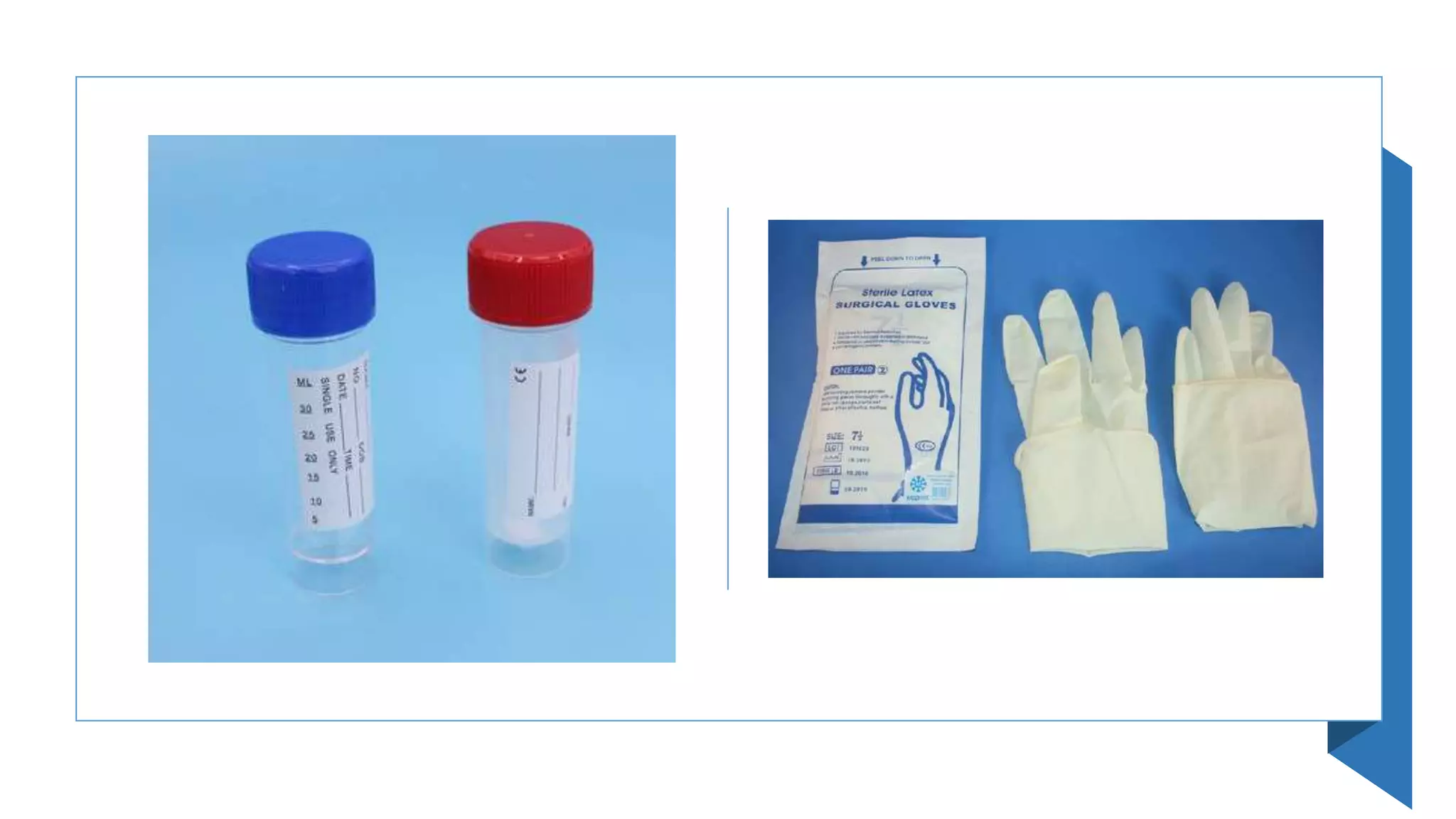
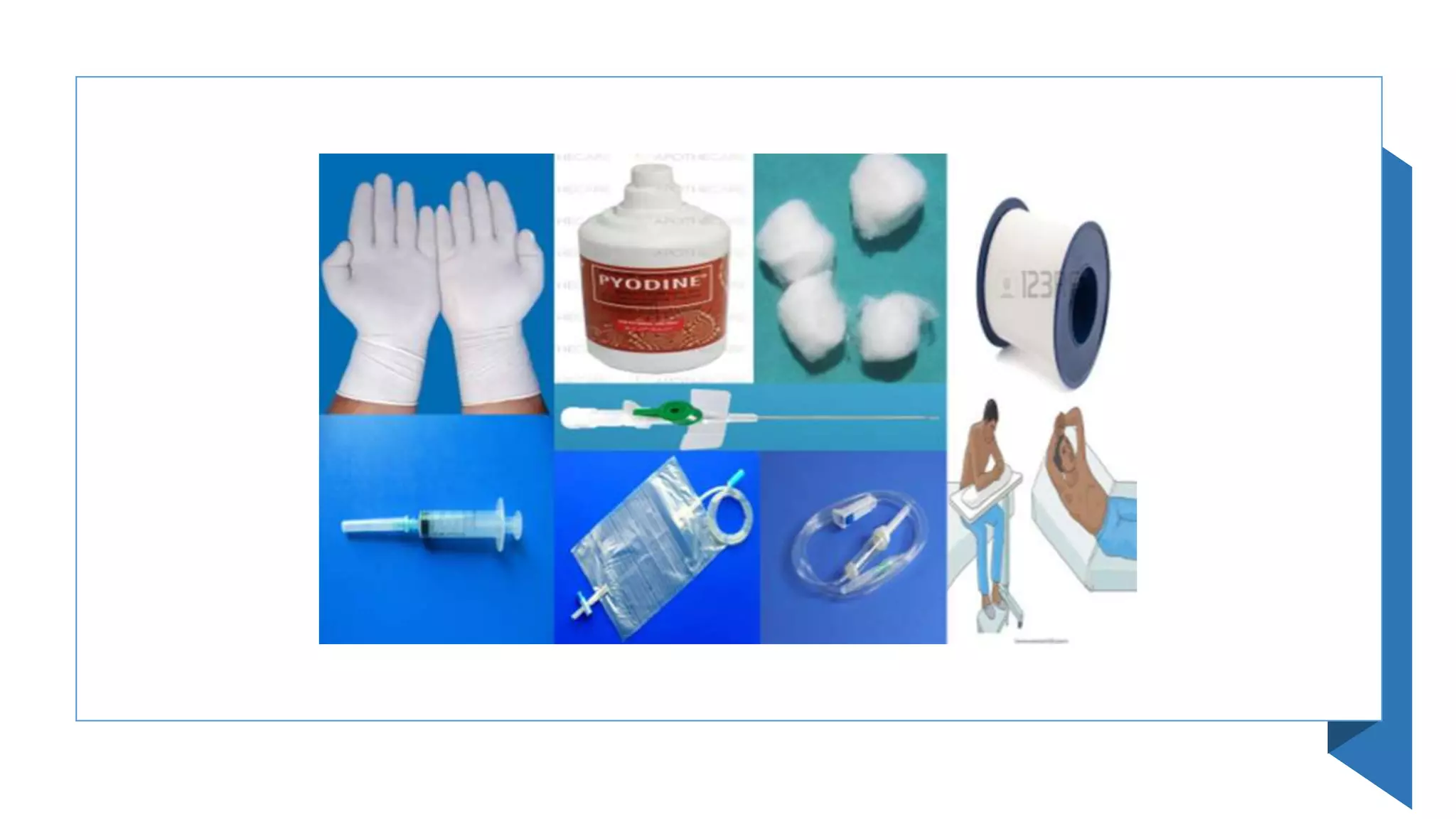
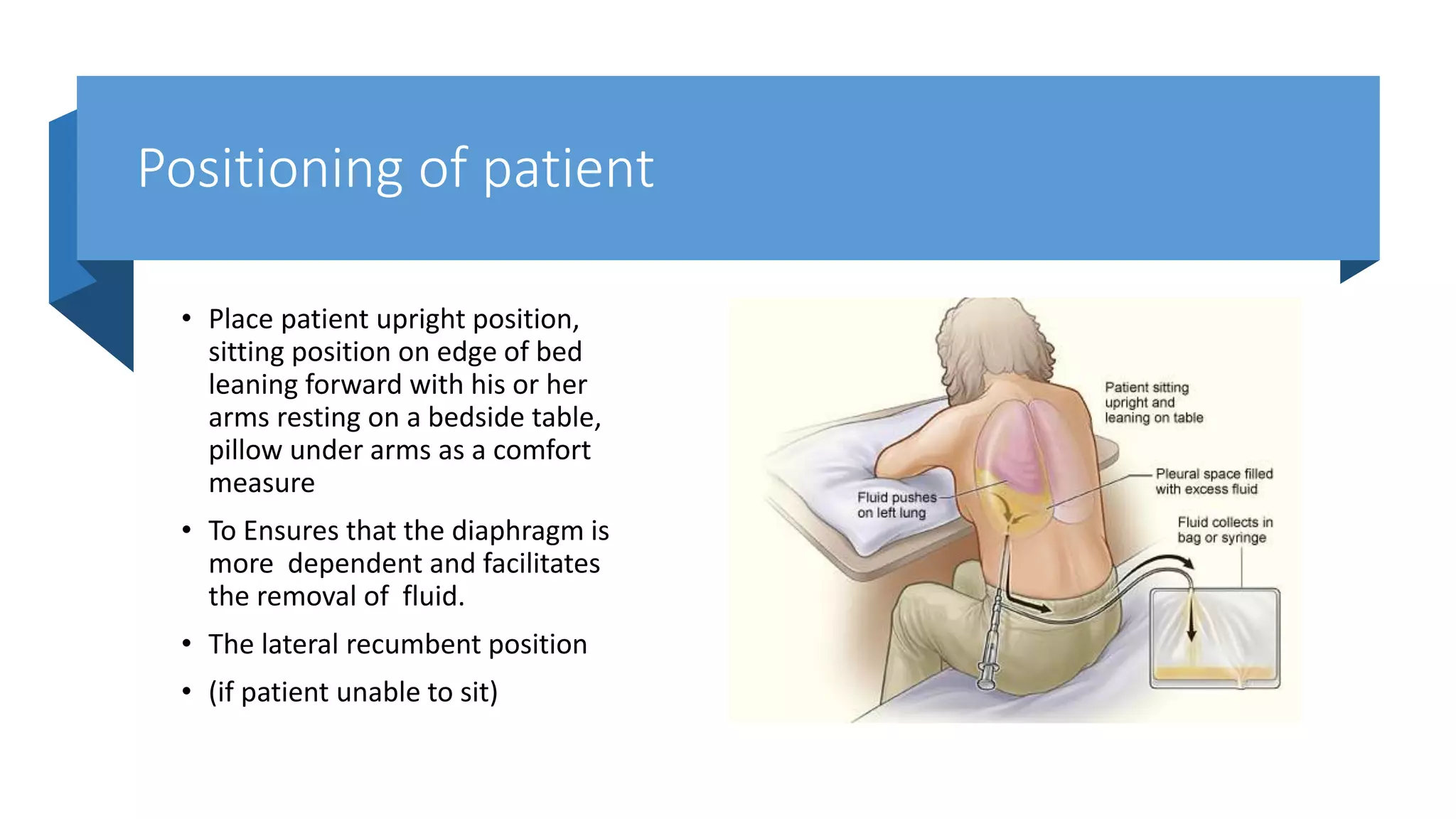
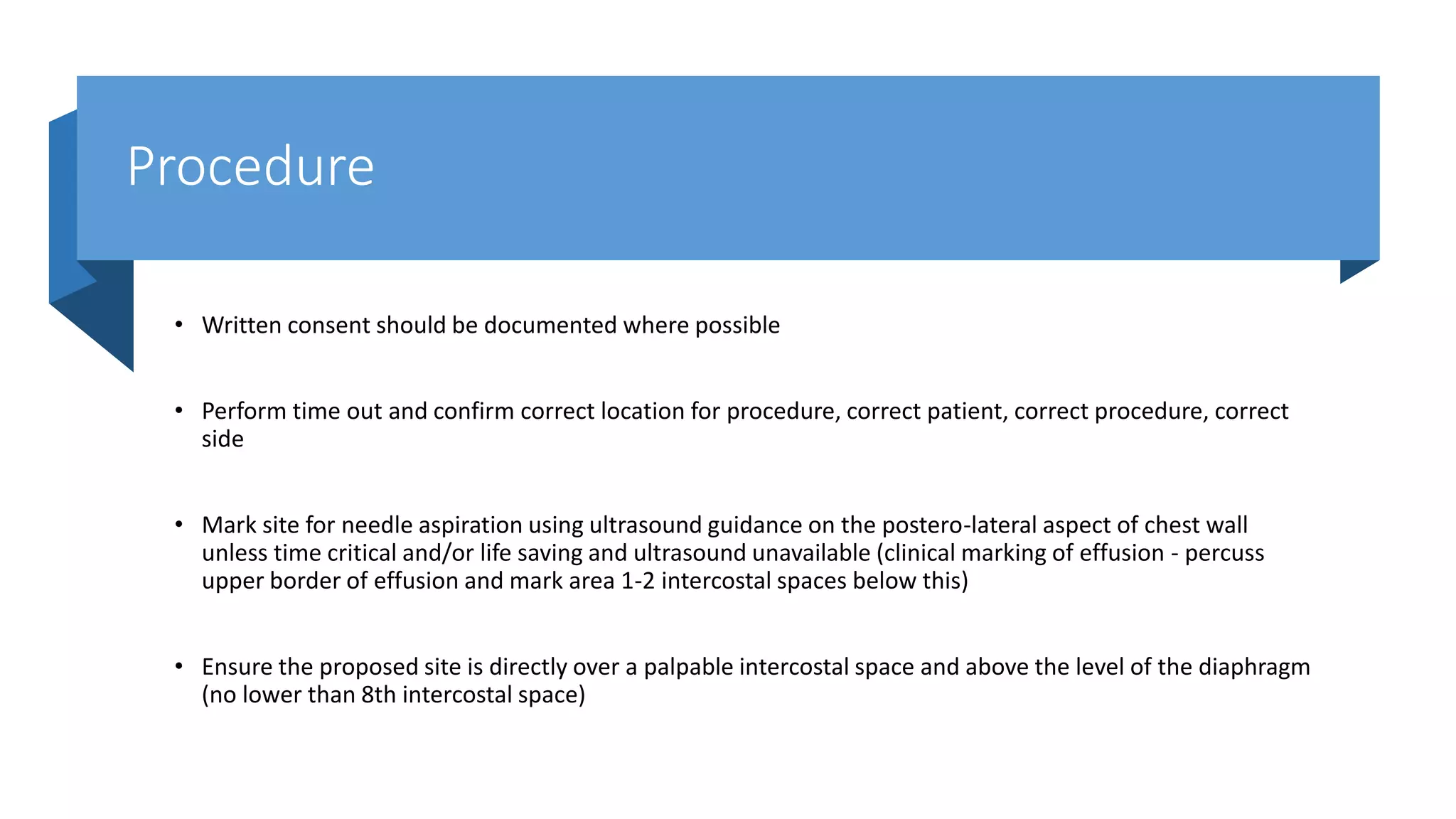
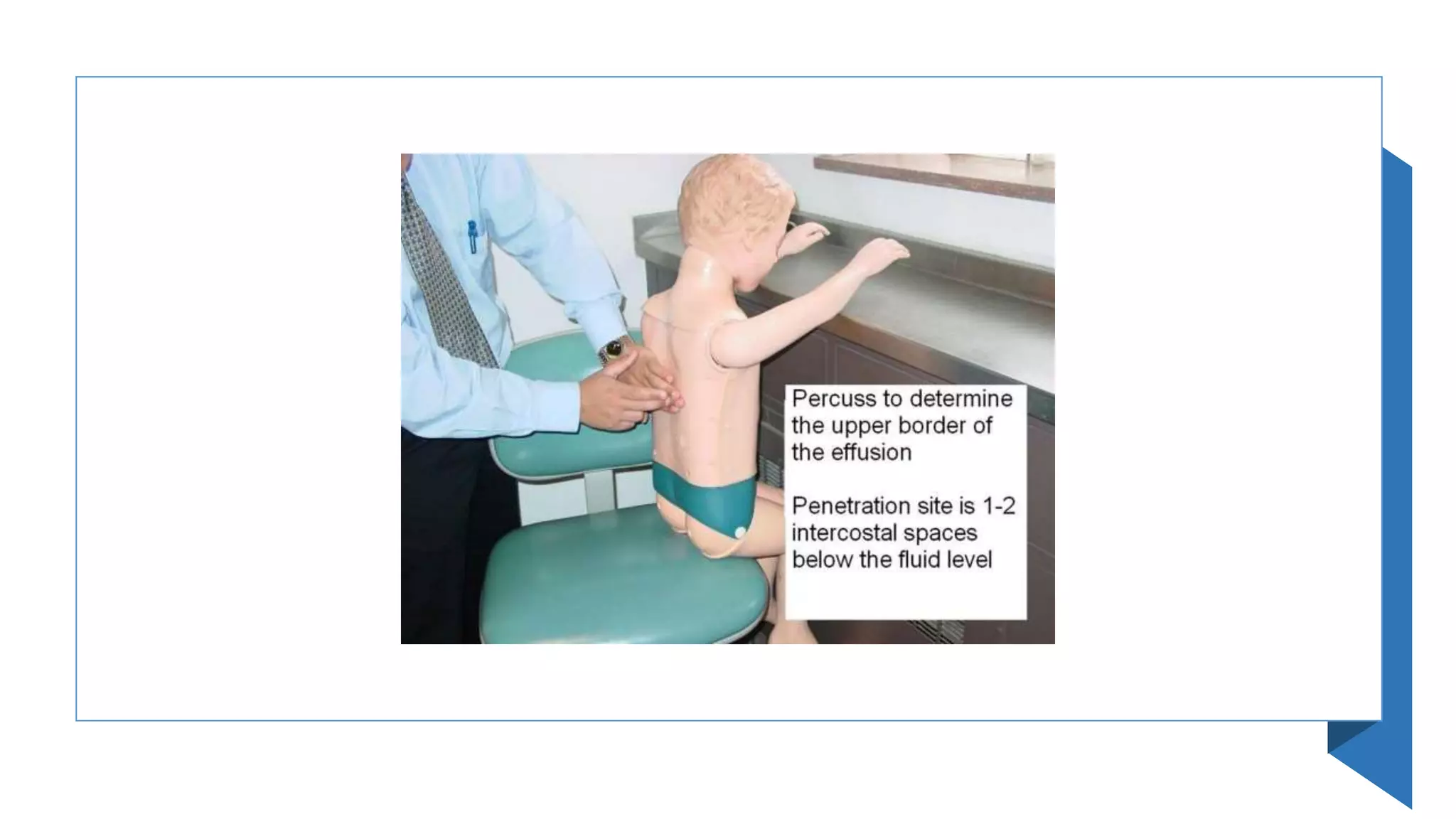
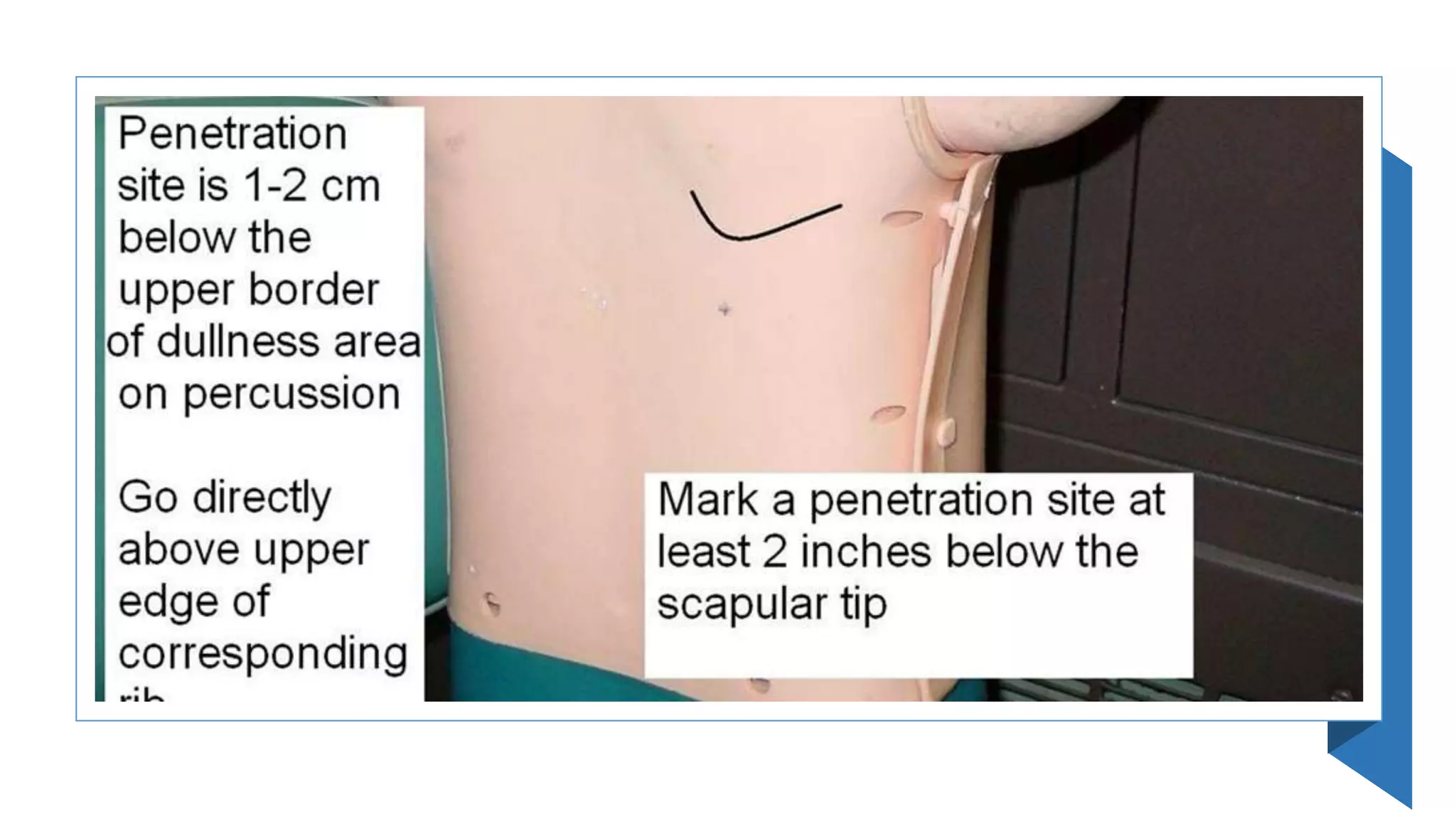
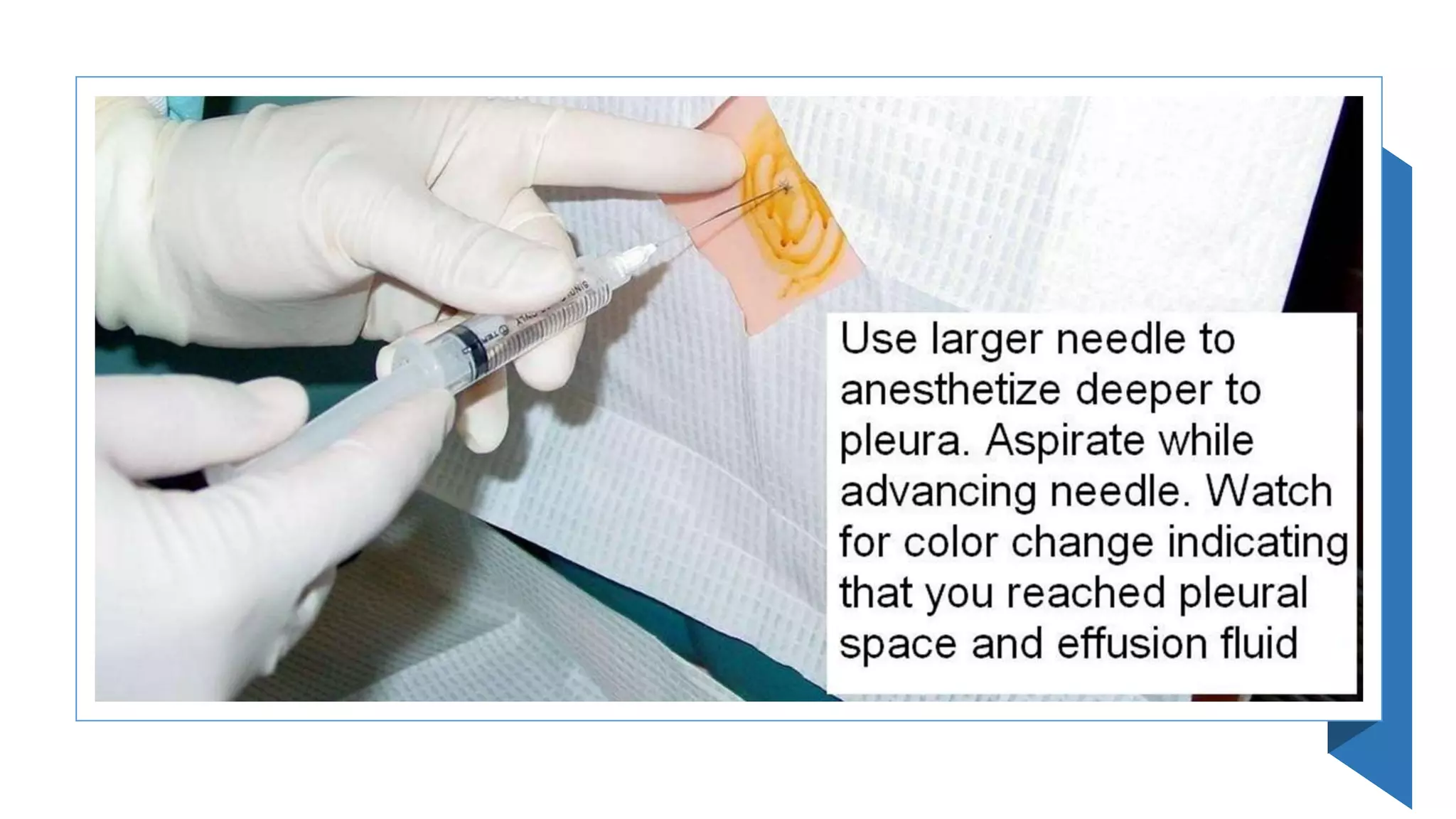
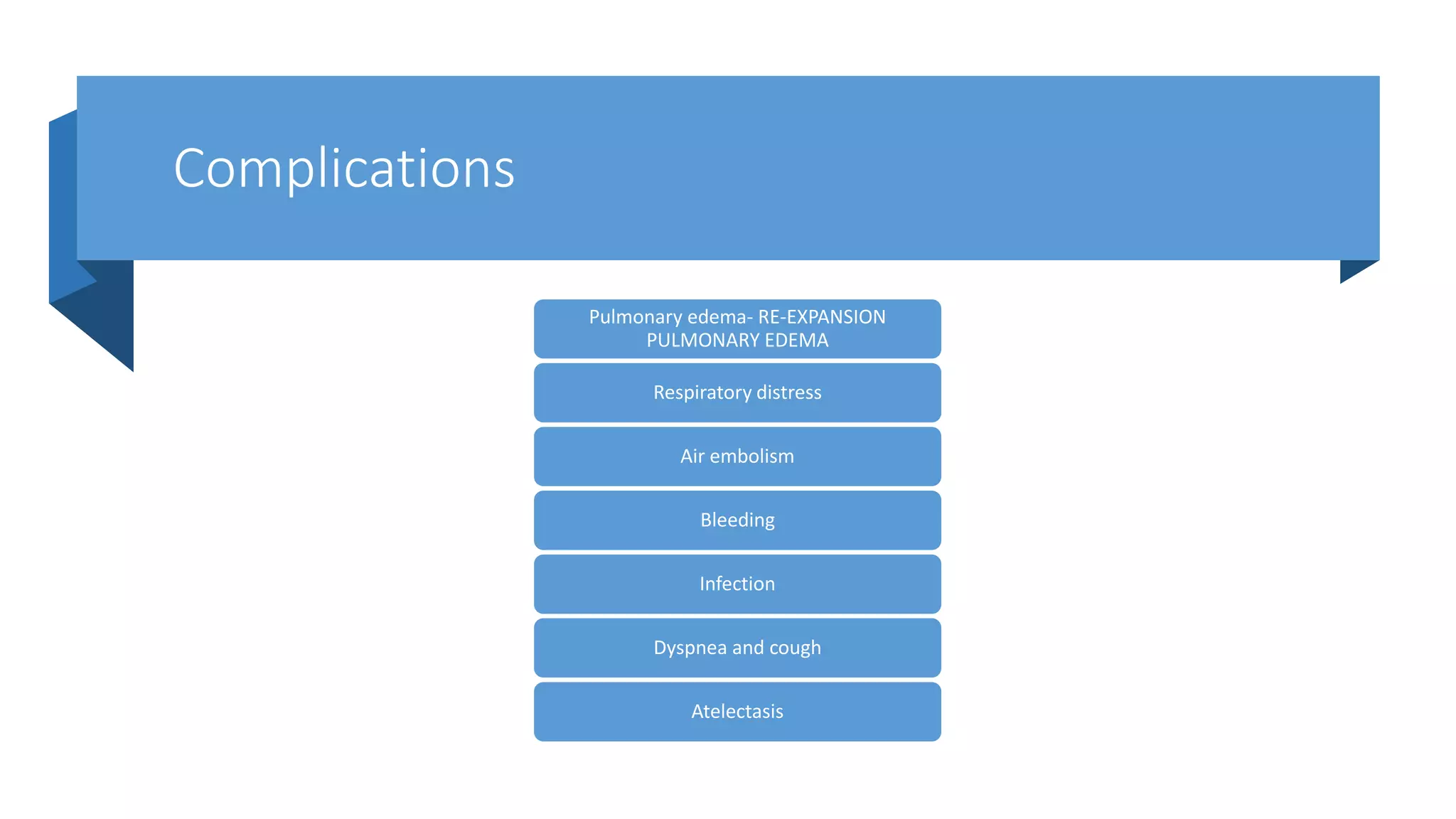
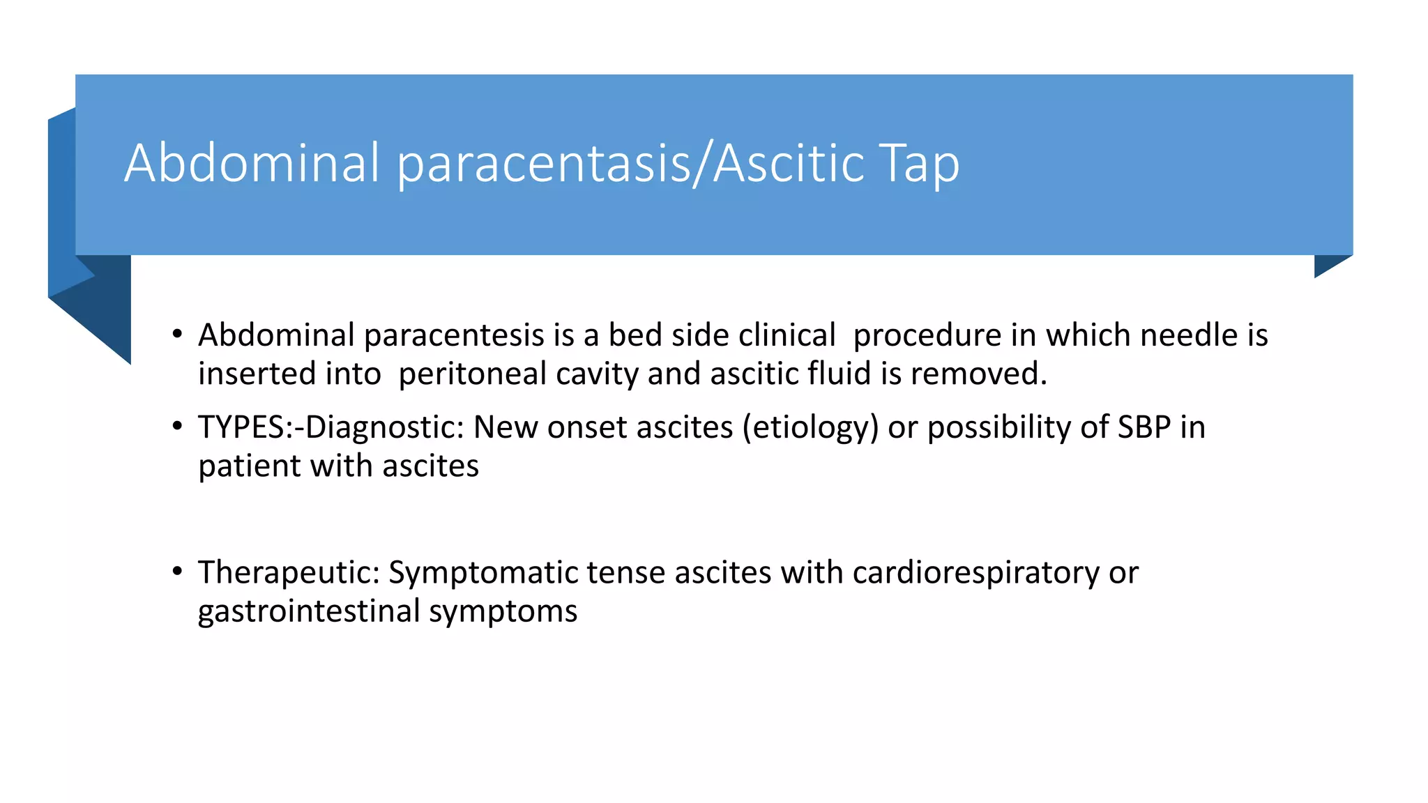
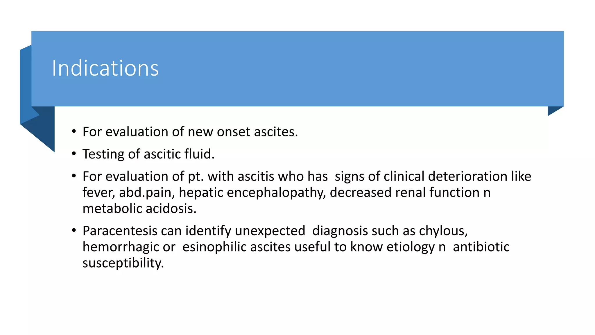
SlideShare a Scribd company logo
CLINICAL
PROCEDURE
PART-1
Dr. Ramesh Chand
Meena
Assistant
Professor Medicine
PLEURAL TAP/THORACOCENTASIS
• Thoracentesis also known as thoracocentesis ,pleural tap, pleural fluid
aspiration, needle thoracotomy,
• Thoracentesis is an invasive percutaneous procedure during which a
needle/cannula is inserted into the pleural space and pleural fluid is
removed, generally after administration of local anesthesia
Indications-
Diagnostic-
• Unilateral pleural effusion
• Bilateral pleural effusions where aetiology unclear
• Hemo-pneumothorax
• Empyema
Therapeutic-
• Symptomatic pleural effusions/ large pleural effusion
• Failed initial medical management (diuresis, treatment of underlying cause)
Contraindication-
• Coagulation disorder
• Active skin infection
• Atelectasis
• Only one functioning lung
• Emphysema
• Severe cough or hiccups
• An uncooperative patient
Before the procedure
1.Explain the purpose, risks/benefits, and steps of the procedure
2. Obtain consent from the patient or appropriate legal design.
3. Do Diagnostic Tests such as (to determine the affected lung)
• chest x-ray
• Ultrasonography
4. Check platelet count and/or presence of coagulopathy-To
prevent complication such as bleeding during procedure
Prepare Equipments
• Chlorhexidine solution/ Lignocaine injection 1%
• Sterile gauze/ Sterile drape/ Sterile gloves
• 5 or 10ml syringe for anesthetic injection/ 50ml syringe for fluid removal
• 22G needle for injection of local anesthetic
• 18G cannula for thoracentesis
• 3-way tap and extension set specimen containers
Positioning of patient
• Place patient upright position,
sitting position on edge of bed
leaning forward with his or her
arms resting on a bedside table,
pillow under arms as a comfort
measure
• To Ensures that the diaphragm is
more dependent and facilitates
the removal of fluid.
• The lateral recumbent position
• (if patient unable to sit)
Procedure
• Written consent should be documented where possible
• Perform time out and confirm correct location for procedure, correct patient, correct procedure, correct
side
• Mark site for needle aspiration using ultrasound guidance on the postero-lateral aspect of chest wall
unless time critical and/or life saving and ultrasound unavailable (clinical marking of effusion - percuss
upper border of effusion and mark area 1-2 intercostal spaces below this)
• Ensure the proposed site is directly over a palpable intercostal space and above the level of the diaphragm
(no lower than 8th intercostal space)
• Anaesthetise the epidermis overlying the superior edge of the rib that lies
below the selected intercostal space using 1% or 2% lignocaine and a 22G
needle
• Now insert that same needle perpendicular to the skin until you hit the rib
and then ‘walk’ it along the superior edge of the rib, alternately injecting
anaesthetic and pulling back on the plunger every 2 or 3mm to rule out
intravascular placement and to check for proper intrapleural placement.
• Once pleural fluid is aspirated, stop advancing the needle and inject additional lignocaine to anaesthetise
the highly sensitive parietal pleura. Note the depth of penetration before withdrawing the needle.
• Attach an 18G cannula to a syringe and advance the needle along in the same plane as the local
anaesthetic was injected, ensuring that you continuously pull back on the plunger
• Once you obtain pleural fluid then remove the needle leaving the cannula in place
• REMEMBER: Cover the open hub of the catheter with a finger to prevent the entry of air into the pleural
cavity
• Attach a 50ml syringe with a 3 way tap to the catheter hub and open the
tap to the patient and syringe to aspirate 50ml of pleural fluid for
diagnostic analysis
• Close the 3 way tap to the patient as you distribute the fluid into relevant
specimen containers
• If performing therapeutic thoracentesis, then attach the extension set to
the third port with the free end in a container to collect the pleural fluid
• Remove fluid in the following fashion with the syringe re-attached to the 3 way tap:
1. Open tap to patient and syringe
2. Withdraw 50ml fluid
3. Close tap to patient and ensure it is open to syringe and extension tubing
4. Empty the syringe whilst it is still attached by pushing the plunger and re-routing the fluid down the
extension set into the container
5. Repeat above steps
Do not remove more than 1500ml fluid due to risk of re-expansion oedema
When procedure is complete remove catheter with patient at end expiration (i.e. ask the patient to take
deep breath in, blow fully out and then hold their breath, then remove the cannula during the breath hold)
and cover site with an occlusive dressing
During the procedure
• Observe patient respiration rate and breathing pattern-R: to provide base
line data to estimate patient tolerance of procedure
• Assess patient vital sign such as B/P, pulse- To prevent any complication
such as hypovolemic shock during procedure
• Observe patient level of consciousness and give- emotional support(to
reduce patients anxiety)
• Monitor O2 saturation (to prevent hypoxia)
• Drain max 1.5 L in one sitting( to avoid Re-expansion pulmonary edema)
Post procedure care
• Perform a post aspiration chest x-ray- to rule out pneumothorax or
condition of lungs, fluid levels
• Provide appropriate analgesia
• Monitor for evidence of any complications: pneumothorax, post expansion
pulmonary oedema, bleeding, intra-abdominal organ injury (rare),
infection (delayed and rare)
After the procedure
• Document the procedure, patient’s response, characteristics of fluid and
amount, and patient response to follow-up.
• Rest in bed for about 2 hours after the procedure -To minimize patient
activity due to complication such as dyspnea.
• Blood pressure and breathing will be checked for up to a few hours
-to make sure don't have complications
Send fluid for:
• Cytology, MC&S, LDH, Protein
• pH (put some fluid in an ABG syringe and run through the ABG machine if
necessary)
• Consider TB culture (acid-fast bacilli)/ gene x-pert if clinically indicated
• Consider glucose or cholesterol (if concerned about chylothorax)
• Send serum blood samples for LDH and protein.
Complications
Pulmonary edema- RE-EXPANSION
PULMONARY EDEMA
Respiratory distress
Air embolism
Bleeding
Infection
Dyspnea and cough
Atelectasis
ABDOMINAL
PARACENTASIS
Abdominal paracentasis/Ascitic Tap
• Abdominal paracentesis is a bed side clinical procedure in which needle is
inserted into peritoneal cavity and ascitic fluid is removed.
• TYPES:-Diagnostic: New onset ascites (etiology) or possibility of SBP in
patient with ascites
• Therapeutic: Symptomatic tense ascites with cardiorespiratory or
gastrointestinal symptoms
Indications
• For evaluation of new onset ascites.
• Testing of ascitic fluid.
• For evaluation of pt. with ascitis who has signs of clinical deterioration like
fever, abd.pain, hepatic encephalopathy, decreased renal function n
metabolic acidosis.
• Paracentesis can identify unexpected diagnosis such as chylous,
hemorrhagic or esinophilic ascites useful to know etiology n antibiotic
susceptibility.
Contraindications
• Absolute-overlying site infection
• Relative- coagulopathy (INR>1.5, Plt<50) although routine INR and FBC not
indicated prior to procedure and replacement only if signs of bleeding;
• Anatomical – adhesions, neuropathic bladder, suggest US prior to needle insertion
to identify areas of anatomical concern.
• Massive ileus with bowel distension.
• Near the surgical scar because scars are asso. With tethering of bowel to abd.wall
n will cause bowel perforation.
• Shock
• Abnormal coagulation studies like increased INR n Thrombocytopenia are
not contraindications.
• 70% pts with Ascites have abnormal PT but risk of bleeding is low.
• Pt who bleed had renal failure suggesting qualitative platelet dysfunction
asso. With renal failure. Here desmopressin may be used before
paracentesis in pts with cirrhosis and renal failure.
Patient preparation-
• Explain the procedure & Obtain Consent
• No fasting before Procedure
EQUIPMENT & STAFF
• Clinician & Assistant
• Bottles should be labelled for tests prior doing paracentesis
• Bacterial culture is done in pts with SBP
Choice of needles-
• DIAGNOSTIC: 1.5 Inch, 22 Gauge needle For Obese :3.5 Inch, 22 Gauge
spinal needle
• THERAPEUTIC: 15/ 16 Gauge needle to speed up the removal
Position of patient-
Supine posture ,head may be elevated 30degree,
Knee elbow position for removal of minimal fluid in dependent area
EQUIPMENTS
• PPE – sterile gloves
• Antiseptic
• Local anaesthetic - lignocaine+/-adrenaline 1% 10mls
• Paracentesis kit - drape, syringe for local(5mls), syringe for sample(20mls), needle for LA
injection (25G or 23G)
• Sample collection – EDTA tube, blood culture bottles, urine container
• Leur lock drainage bag
• 12F Dwellcath needle
• 20% albumin if LVP planned
Site-
• Lt lower Quadrant (Dullness on percussion)
• 3cm medial & 2cm above the ant. Sup. Iliac spine
• Not near umbilicus because of presence of collateral vessels
• Surgical scars & visible veins should be avoided.
WHY LEFT???
• Abd. Wall is thinner.
• Pool of fluid is more.
• Pt can be rolled easily to left for drainage
WHY NOT RIGHT???
• Appendicectomy scar, caecum filled with gas in pts taking lactulose.
• Care must be taken not to injure inferior epigastic artery which bleeds
massively & which is located near pubic tubercle
SKIN STERLISATION
• Mark the site as “X” &
positions 12, 3, 6, 9 a few
centimeters from “X”
• Sterilise with Iodine or
Chlorhexidine Solution
starting from X using
widening circular motions.
LOCAL ANESTHESIA
• Anaesthetise using 3- 5 ml of 1% Lignocaine Solution in a “Z” track
technique.
• Needle used for it is 1.5inch which is sufficiently long.
• Choose the site & pass the needle tangentially, raising a wheal with
Lignocaine.
• “Z” track creates a non linear pathway b/n Skin& Ascitic fluid & minimise
the chance of leakage.
• With one hand pull the abdominal wall n with other hand operate the syringe.
Hand on the abd.wall should not be removed untill the needle enters the fluid.
• Insert the needle n syringe 5mm deep
• pull the plunger back with each advancement to see if any blood is aspirated.
• then inject the lignocaine sol.
• Cont. the same procedure until the needle enters fluid.
• Aspiration should be intermittent not continuous.
• Cont. may pull the bowel or omentum onto needle tip,occluding the tip.
• Yellow color fluid indicates needle is in the peritoneal cavity.
• NEEDLE INSERTION:
• Needle is inserted along anesthetised pathway after nick is given with 11
no. blade. Fliud should drip from the hub of the needle.
• Larger the nick greater the post paracentesis leak.
• Ultrasound guidance can be used to guide the procedure.
• During laproscopy parietal peritoneum may form tenting over needle n fluid doesn’t come.
• Operator cant see this n may mis interpret as DRY TAP.
• Rotating the needle for 90 degrees or more will pierce the peritoneum n help the drainage.
• Small amount of fluid may be difficult to drain bcoz omentum/bowel may block the end of needle. So
multi hole needles are helpful.
• Misconception of poor flow is LOCULATION.
• True loculation is seen in peritoneal carcinomatosis with malignant adhesions or bowel rupture with
surgical peritonitis.
• Loculation never occur in cirrhosis or heart failure with ascites or SBP.
• Stable needle n depth of penetration of needle are crucial for successful
paracentesis.
TESTING- 25 ml fluid is enough for cell count,diff count,chemical testing n
bacterial culture.
In TB 50ml for cytology
50ml for smear n culture
LARGE VOLUME PARACENTASIS
• It is removal of >5 lit of fluid.
• In refractory ascites,removal of as much fluid as possible with
sod.restricted diet n diuretics will extend the interval to next paracentesis.
• REMOVAL OF NEEDLE:
• Needle is removed with one rapid smooth withdrawal motion.
• Distract the pt by asking him to cough bcoz cough will prevent pain
sensation
Tips and tricks
• Check (fluid amount and adhesions) the insertion point with USG prior to
commencement with patient in position
• Dot advance the cannula once filed non-sterile
• Use the Z-line insertion technique to decrease leakage
Complications
Common
Local- Leakage of ascitic fluid(single suture)
Haematoma
Systemic- Post paracentesis circulatory dysfunction(PCD) following LVP(large volume paracentesis >5L)
• Uncommon
secondary bacterial peritonitis-Infection (local or intraperitoneal) rare<0.2%
• Bowel perforation - rare with US
• Vessel perforation - mitigated by point of entry
LUMBAR PUNCTURE
Lumbar puncture
• Diagnostic lumbar puncture is the insertion of a hollow needle into the
subarachnoid space of the spinal canal for collection of cerebrospinal fluid
(CSF). The procedure may include measurement of CSF pressure; however
this measurement is not routine in every Emergency Department.
• LP is considered an invasive procedure, and where possible, written
consent should be sought from the patient.
Indication
Emergency Department indications are clinical suspicion of:
• CNS infection (meningitis, encephalitis)
• Subarachnoid Haemorrhage (SAH)
Contraindications
• Skin infection surrounding the site
• Evidence of raised intracranial pressure
• Platelet count less than 50 ×10^9/L
• INR >1.5
• Unfractionated Heparin or LMWH in the past 24 hours
• Known coagulopathy e.g. Haemophila, von Willebrand disease
• Trauma to lumbar vertebrae
• Unstable patient/ALOC
Equipments
• A standard kit might contain:
• Sterile gloves
• Chlorhexidine 2%, cotton balls and forceps
• Drape
• Lignocaine 1%, 23g needle, syringe
• Spinal needle (see below)
• Pressure measuring tube and three way tap (see below)
• 3 or 4 collection tubes
Patient positioning
• The spinal cord ends at L1/L2 level in adults.
• Place one hand on each ASIS and move medially to the midline, locating the spine. This is
L4 and LP may be performed through the 2 spaces above (L3/L4 or L2/L3) or one space
below (L4/L5).
• LP can be performed with the patient either lying down or sitting up. Choose the position
that you and the patient are most comfortable with.
• Optimise patient position to increase the interspinous distance as much as possible: flex
the spine and hips.
• Perform LP on infants and unconscious patients, with the patient lying down, and an
assistant maintaining spinal flexion.
Lying down
• Patient should be lying in the lateral decubitus position.
• Ensure the vertical plane of the patients back is
perpendicular to the bed.
• Flex knees and hips so that knees are close to the chest.
• While flexion of the neck is often taught as important,
evidence suggests that this has no effect of the size of
the interspinous opening and may be uncomfortable for
the patient.
• Postioning correctly is all about increasing the
interspinous distance as much as possible.
Sitting-
• This position generally makes it easier to identify anatomical landmarks and
planes.
• Sit the patient on a firm surface.
• Patient should lean forward, and can hug a pillow on a table at the right height to
flex (not extend) the back.
• Lift the patient’s legs by putting their feet on a stool or chair and flex the hips to
above 90 degrees.
• If pressure measuring is required, lie the patient on their side (after successful
needle insertion) with assistance and then open the tap to measure the pressure.
Preparation
• Wash hands and use sterile gloves.
• Prepare the skin with povidone-iodine or chlorhexidine.
• Apply sterile drape(s).
• Uncap specimen tubes.
• Infiltrate skin with local anaesthetic.
• Use introducer needle on syringe with LA, insert in the plane for the LP
• Aim in between spinous processes, towards the umbilicus (ie slightly cephalad).
• Inject LA as you go, remove syringe and leave introducer needle in situ.
puncture
• Use the spinal needle (with stylet in) to pierce the skin ( or where you have used an introducer needle
insert) over the selected interspinous space in the midline.
• Aim in between spinous processes, towards the umbilicus (ie slightly cephalad).
• As the needle passes through the interspinous ligament, there is likely to be increased resistance.
• Advance the needle through the ligament until there is a decrease in resistance. The needle tip is now
likely to be near or within the subarachnoid space.
• In a 70 kg male the ligamentum flavum sits at around 4.5 cm, use this distance or a measured distance
from ultrasound.
• Remove the stylet to check for CSF.If there is no flow of CSF, replace the stylet and advance the needle
slightly before checking again for CSF.
Cont.
• Alternatively, remove the stylet once the needle is in the interspinous ligament and advance the open
needle slowly, watching for flow of CSF.
• If the needle meets hard (bony) resistance, withdraw the open needle slowly, watching for flow of CSF. If
there is no flow once the needle tip has backed into the ligament, try advancing in a different direction
using either technique.
• When there is reliable flow of CSF, attach a 3-way-tap if measuring pressures, or collect a sample directly
into the specimen containers.
• Collect 10 to 20 drops (5 to 10 for children) in each container, keeping track of the order in which
containers are filled (they may already be numbered).
• After collection, replace the stylet and remove the needle and stylet together.
• Apply a sterile dressing to the puncture site.
COMPLICATIONS
• Failure to obtain CSF
• Post-lumbar-puncture headache (increased risk with:
large bore needle; cutting needle; multiple attempt;
excessive CSF removal; dehydration; female patient)
• Epidural haematoma / local haemorrhage
• Epidural abscess
• Transtentorial herniation (view the video 'Lumbar
Puncture Procedure and Interpretation' to see a
discussion on whether this is the risk we think it is)
• Back pain
IV
CANNULATION
Dr. Ramesh Chand
Meena
Assistant Professor
Medicine
Definition-
• Intravenous (IV) Cannulation Is a technique in which a cannula is placed
inside a vein to provide venous access
PURPOSES-
To administer intravenous injection
To administer intravenous fluids.
To administer radiological contrast agent prior to CT/MRI
Blood sampling/ transfusion of blood and blood products, chemotherapy
Equipments-
Articles A tray containing :
• Sterile gloves
• Iv cannula
• Tourniquet
• Cotton swabs
• Adhesive tape
• Spirit
• I.V. stand ,I.V. set
Parts of cannula
Types
Why veins are suitable for iv cannulation ?
• Superficial
• Palpable
• Visible
• Blood flow at low pressure
• Relatively large internal diameter
• Tough vascular wall-able to form seal around cannula
• Offer rapid route to systemic circulation
• Many choices remote from sensitive structures.
Point to remember while selecting the vein
• Make use of the most distal part of the vein
• Avoid puncturing the vein on joint . It will hinder the normal movement of
joint
• Avoid puncturing the vein in close proximity to an infected wound
• Avoid puncturing the vein on extremity having a shunt
• Try avoid puncturing the vein of lower extremities in adults . Because of
the high risk of development of deep vein thrombosis
Procedure-
• To start an Iv , first prepare all of equipment . This will include the IV bag , with
containing tubing , with all air flushed out of tubing
• Place a tourniquet around the arm . This should be tight enough to block venous
blood flow back to the heart , but not so tight that it obstructs the arterial flow
• Wait long enough for the veins in the hands & arm to fill & become tight . In a
normal person , this may take 2-5 minutes .In a dehydrated person or someone in
shock , it may take longer
• Cleanse the skin of injection site with spirit cotton balls . If spirit is not available ,
use any antiseptic or skin cleanser
Continue-
• Use your left thumb to hold the vein in place while you insert the IV needle at a
shallow angle ( about a 20 degree angle ) through the skin & into the vein
• As you enter the vein , you will feel a slight “pop”. You will know you are in the
vein when you see blood returning in the “flash back “ chamber
• Keep the needle in place with one hand while you push the catheter (which
surrounds the needle ) further into the vein . This will thread it upstream ,
securing it into the vein
• Once that catheter is completely inserted , hold it in place with one hand while
you release the tourniquet & pull the needle straight out with the other hand
Procedure continue-
• Pressing down the skin where the catheter tip is located will prevent blood from
flowing back out the IV catheter before you have a chance to connect the IV
tubing's
• Connect the IV tubing's & run In the IV fluids briskly , at first . Then slow it down
to a steady drip
• If the fluid does not flow freely at the beginning, check the IV tubing's to see if
there are any valves or other obstruction to flow .If the tubing is wide open , but
the IV is dripping only very slowly , you are probably not in vein.
• Try again -Observe the site the edema , swelling , redness. After administering IV
fluids , flush the I.V. line with NS & lock it with stopper
Vein seek
App
Vein finders
• A Vein Finder
• Is exactly what it sounds like, It is a device that is used to reduce the
amount of time it takes to find a vein on a patient
• How Do Vein Finders Work? An infrared vein finder uses a specific type of
light, which is able to penetrate the skin by a
• few millimetres to illuminate the targeted veins
• Other types of vein finders, usually in the form of glasses, which work by
altering the color of the light visible to the eye
Complications of IV cannulation
• Thrombophlebitis
• Local and systemic infection
• Bleeding/ injury to artery
• Extravasation
• Air embolism
Thanking you

More Related Content

What's hot

Lower Gastrointestinal Bleed
Lower Gastrointestinal BleedLower Gastrointestinal Bleed
Lower Gastrointestinal Bleed
Dr. Kiran Pandey
 
Stemi by dr.mehelina
Stemi by dr.mehelinaStemi by dr.mehelina
Stemi by dr.mehelina
Nizam Uddin
 
Amoebic colitis
Amoebic colitisAmoebic colitis
Amoebic colitis
santusan
 
Septic shock management (1)
Septic shock management (1)Septic shock management (1)
Septic shock management (1)
shashank agrawal
 
Signs of aortic regurgitation
Signs of aortic regurgitationSigns of aortic regurgitation
Signs of aortic regurgitationKurian Joseph
 
Acute coronary syndrome
Acute coronary syndromeAcute coronary syndrome
Acute coronary syndrome
BALASUBRAMANIAM IYER
 
Acute coronary syndrome (acs)
Acute coronary syndrome (acs)Acute coronary syndrome (acs)
Acute coronary syndrome (acs)
farranajwa
 
Pericardial diseases
Pericardial  diseasesPericardial  diseases
Pericardial diseases
ikramdr01
 
Atherosclerosis
AtherosclerosisAtherosclerosis
Atherosclerosis
AJAY MANDAL
 
Acute coronary syndrome NSTEMI
Acute coronary syndrome NSTEMIAcute coronary syndrome NSTEMI
Acute coronary syndrome NSTEMI
Jackie San
 
Aortic anurysm
Aortic anurysmAortic anurysm
Aortic anurysm
Monika Devi NR
 
Transposition of great arteries
Transposition of great arteriesTransposition of great arteries
Transposition of great arteriesPriya Dharshini
 
Left Bundle Branch Block (LBBB)
Left Bundle Branch Block (LBBB)Left Bundle Branch Block (LBBB)
Left Bundle Branch Block (LBBB)
Kerolus Shehata
 
Aortic regurgitation
Aortic regurgitationAortic regurgitation
Aortic regurgitation
Vitrag Shah
 
Peripheral artery disease
Peripheral artery diseasePeripheral artery disease
Peripheral artery disease
Dr Dinesh Pandey
 
Septic shock Pathophysiology
Septic shock Pathophysiology Septic shock Pathophysiology
Septic shock Pathophysiology
Society for Microbiology and Infection care
 
Chronic limb ischemia
Chronic limb ischemia Chronic limb ischemia
Chronic limb ischemia
Kundan Singh
 
Deep vein thrombosis (DVT)
Deep vein thrombosis (DVT)Deep vein thrombosis (DVT)
Deep vein thrombosis (DVT)
pankaj rana
 

What's hot (20)

Pulmonary Embolism
Pulmonary EmbolismPulmonary Embolism
Pulmonary Embolism
 
Lower Gastrointestinal Bleed
Lower Gastrointestinal BleedLower Gastrointestinal Bleed
Lower Gastrointestinal Bleed
 
Stemi by dr.mehelina
Stemi by dr.mehelinaStemi by dr.mehelina
Stemi by dr.mehelina
 
Amoebic colitis
Amoebic colitisAmoebic colitis
Amoebic colitis
 
Septic shock management (1)
Septic shock management (1)Septic shock management (1)
Septic shock management (1)
 
Signs of aortic regurgitation
Signs of aortic regurgitationSigns of aortic regurgitation
Signs of aortic regurgitation
 
Acute coronary syndrome
Acute coronary syndromeAcute coronary syndrome
Acute coronary syndrome
 
Acute coronary syndrome (acs)
Acute coronary syndrome (acs)Acute coronary syndrome (acs)
Acute coronary syndrome (acs)
 
Pericardial diseases
Pericardial  diseasesPericardial  diseases
Pericardial diseases
 
Septic shock
Septic shockSeptic shock
Septic shock
 
Atherosclerosis
AtherosclerosisAtherosclerosis
Atherosclerosis
 
Acute coronary syndrome NSTEMI
Acute coronary syndrome NSTEMIAcute coronary syndrome NSTEMI
Acute coronary syndrome NSTEMI
 
Aortic anurysm
Aortic anurysmAortic anurysm
Aortic anurysm
 
Transposition of great arteries
Transposition of great arteriesTransposition of great arteries
Transposition of great arteries
 
Left Bundle Branch Block (LBBB)
Left Bundle Branch Block (LBBB)Left Bundle Branch Block (LBBB)
Left Bundle Branch Block (LBBB)
 
Aortic regurgitation
Aortic regurgitationAortic regurgitation
Aortic regurgitation
 
Peripheral artery disease
Peripheral artery diseasePeripheral artery disease
Peripheral artery disease
 
Septic shock Pathophysiology
Septic shock Pathophysiology Septic shock Pathophysiology
Septic shock Pathophysiology
 
Chronic limb ischemia
Chronic limb ischemia Chronic limb ischemia
Chronic limb ischemia
 
Deep vein thrombosis (DVT)
Deep vein thrombosis (DVT)Deep vein thrombosis (DVT)
Deep vein thrombosis (DVT)
 

Similar to Clinical procedure part 1

MAL.Z PRESENTS THORACENTESIS.pptx
MAL.Z PRESENTS THORACENTESIS.pptxMAL.Z PRESENTS THORACENTESIS.pptx
MAL.Z PRESENTS THORACENTESIS.pptx
Mal Z
 
Thoracocentesis
ThoracocentesisThoracocentesis
Thoracocentesis
Saint Vincent Hospital
 
thoracocentesis-141
thoracocentesis-141thoracocentesis-141
thoracocentesis-141
SyidaAhmat
 
Physiotherapy in surgery in abdominal and thoracic surgery
Physiotherapy in surgery in abdominal and thoracic surgeryPhysiotherapy in surgery in abdominal and thoracic surgery
Physiotherapy in surgery in abdominal and thoracic surgery
DrKhushbooBhattPT
 
Intravenous catheterisation powerpoint presentation
Intravenous catheterisation powerpoint presentationIntravenous catheterisation powerpoint presentation
Intravenous catheterisation powerpoint presentation
drkeerthana812
 
Abdominal Paracentesis - Nursing Role
Abdominal Paracentesis - Nursing RoleAbdominal Paracentesis - Nursing Role
Abdominal Paracentesis - Nursing Role
Ahmad Thanin
 
HEPATOBILIARY STUDIES.pptx
HEPATOBILIARY STUDIES.pptxHEPATOBILIARY STUDIES.pptx
HEPATOBILIARY STUDIES.pptx
Josephmwanika
 
Thoracentesis
Thoracentesis Thoracentesis
Thoracentesis
Ramzan Ali
 
Chest Tube In-Service
Chest Tube In-ServiceChest Tube In-Service
Chest Tube In-Service
Dene W. Daugherty
 
INTRAVENOUS FLUID THERAPY jsvsb with babban mbsvkst
INTRAVENOUS FLUID THERAPY jsvsb with babban mbsvkstINTRAVENOUS FLUID THERAPY jsvsb with babban mbsvkst
INTRAVENOUS FLUID THERAPY jsvsb with babban mbsvkst
AshishS82
 
Iv infusion DR. Parshant
Iv infusion DR. ParshantIv infusion DR. Parshant
Iv infusion DR. Parshant
Ps Nadda
 
Thoracic surgery and it's management
Thoracic surgery and it's managementThoracic surgery and it's management
Thoracic surgery and it's management
RakhiYadav53
 
Tonsillectomy.pptx
Tonsillectomy.pptxTonsillectomy.pptx
Tonsillectomy.pptx
Satishray9
 
laryngoscopy thoracoscopy thoracocentesis pulmonary angiograpy.pptx
laryngoscopy thoracoscopy thoracocentesis pulmonary angiograpy.pptxlaryngoscopy thoracoscopy thoracocentesis pulmonary angiograpy.pptx
laryngoscopy thoracoscopy thoracocentesis pulmonary angiograpy.pptx
RoshnaAdhikari1
 
SPECIMEN COLLECTION LAB SAFETY B.pptx111
SPECIMEN COLLECTION LAB SAFETY B.pptx111SPECIMEN COLLECTION LAB SAFETY B.pptx111
SPECIMEN COLLECTION LAB SAFETY B.pptx111
akoeljames8543
 
2015-09-18_IV_therapy_and_IV_access_Part_2.pptx
2015-09-18_IV_therapy_and_IV_access_Part_2.pptx2015-09-18_IV_therapy_and_IV_access_Part_2.pptx
2015-09-18_IV_therapy_and_IV_access_Part_2.pptx
CatherineMonana2
 
Thoracentesis 12 june 2020
Thoracentesis 12 june 2020Thoracentesis 12 june 2020
Thoracentesis 12 june 2020
seema varghese
 
Therapeutic procedures
Therapeutic proceduresTherapeutic procedures
Therapeutic procedures
Dr Durgesh Kumar
 
2.EVD CARE,TRACHEOSTOMY TUBE CARE AND CHEST.pptx
2.EVD CARE,TRACHEOSTOMY TUBE CARE AND CHEST.pptx2.EVD CARE,TRACHEOSTOMY TUBE CARE AND CHEST.pptx
2.EVD CARE,TRACHEOSTOMY TUBE CARE AND CHEST.pptx
Polytitus Laurent
 

Similar to Clinical procedure part 1 (20)

MAL.Z PRESENTS THORACENTESIS.pptx
MAL.Z PRESENTS THORACENTESIS.pptxMAL.Z PRESENTS THORACENTESIS.pptx
MAL.Z PRESENTS THORACENTESIS.pptx
 
Thoracocentesis
ThoracocentesisThoracocentesis
Thoracocentesis
 
thoracocentesis-141
thoracocentesis-141thoracocentesis-141
thoracocentesis-141
 
Physiotherapy in surgery in abdominal and thoracic surgery
Physiotherapy in surgery in abdominal and thoracic surgeryPhysiotherapy in surgery in abdominal and thoracic surgery
Physiotherapy in surgery in abdominal and thoracic surgery
 
Intravenous catheterisation powerpoint presentation
Intravenous catheterisation powerpoint presentationIntravenous catheterisation powerpoint presentation
Intravenous catheterisation powerpoint presentation
 
Abdominal Paracentesis - Nursing Role
Abdominal Paracentesis - Nursing RoleAbdominal Paracentesis - Nursing Role
Abdominal Paracentesis - Nursing Role
 
HEPATOBILIARY STUDIES.pptx
HEPATOBILIARY STUDIES.pptxHEPATOBILIARY STUDIES.pptx
HEPATOBILIARY STUDIES.pptx
 
Thoracentesis
Thoracentesis Thoracentesis
Thoracentesis
 
Chest Tube In-Service
Chest Tube In-ServiceChest Tube In-Service
Chest Tube In-Service
 
INTRAVENOUS FLUID THERAPY jsvsb with babban mbsvkst
INTRAVENOUS FLUID THERAPY jsvsb with babban mbsvkstINTRAVENOUS FLUID THERAPY jsvsb with babban mbsvkst
INTRAVENOUS FLUID THERAPY jsvsb with babban mbsvkst
 
Iv infusion DR. Parshant
Iv infusion DR. ParshantIv infusion DR. Parshant
Iv infusion DR. Parshant
 
Thoracic surgery and it's management
Thoracic surgery and it's managementThoracic surgery and it's management
Thoracic surgery and it's management
 
Tonsillectomy.pptx
Tonsillectomy.pptxTonsillectomy.pptx
Tonsillectomy.pptx
 
laryngoscopy thoracoscopy thoracocentesis pulmonary angiograpy.pptx
laryngoscopy thoracoscopy thoracocentesis pulmonary angiograpy.pptxlaryngoscopy thoracoscopy thoracocentesis pulmonary angiograpy.pptx
laryngoscopy thoracoscopy thoracocentesis pulmonary angiograpy.pptx
 
SPECIMEN COLLECTION LAB SAFETY B.pptx111
SPECIMEN COLLECTION LAB SAFETY B.pptx111SPECIMEN COLLECTION LAB SAFETY B.pptx111
SPECIMEN COLLECTION LAB SAFETY B.pptx111
 
2015-09-18_IV_therapy_and_IV_access_Part_2.pptx
2015-09-18_IV_therapy_and_IV_access_Part_2.pptx2015-09-18_IV_therapy_and_IV_access_Part_2.pptx
2015-09-18_IV_therapy_and_IV_access_Part_2.pptx
 
Thoracentesis 12 june 2020
Thoracentesis 12 june 2020Thoracentesis 12 june 2020
Thoracentesis 12 june 2020
 
Therapeutic procedures
Therapeutic proceduresTherapeutic procedures
Therapeutic procedures
 
2.EVD CARE,TRACHEOSTOMY TUBE CARE AND CHEST.pptx
2.EVD CARE,TRACHEOSTOMY TUBE CARE AND CHEST.pptx2.EVD CARE,TRACHEOSTOMY TUBE CARE AND CHEST.pptx
2.EVD CARE,TRACHEOSTOMY TUBE CARE AND CHEST.pptx
 
Bedside Procedure
Bedside ProcedureBedside Procedure
Bedside Procedure
 

Recently uploaded

POST OPERATIVE OLIGURIA and its management
POST OPERATIVE OLIGURIA and its managementPOST OPERATIVE OLIGURIA and its management
POST OPERATIVE OLIGURIA and its management
touseefaziz1
 
Hemodialysis: Chapter 3, Dialysis Water Unit - Dr.Gawad
Hemodialysis: Chapter 3, Dialysis Water Unit - Dr.GawadHemodialysis: Chapter 3, Dialysis Water Unit - Dr.Gawad
Hemodialysis: Chapter 3, Dialysis Water Unit - Dr.Gawad
NephroTube - Dr.Gawad
 
KDIGO 2024 guidelines for diabetologists
KDIGO 2024 guidelines for diabetologistsKDIGO 2024 guidelines for diabetologists
KDIGO 2024 guidelines for diabetologists
د.محمود نجيب
 
New Directions in Targeted Therapeutic Approaches for Older Adults With Mantl...
New Directions in Targeted Therapeutic Approaches for Older Adults With Mantl...New Directions in Targeted Therapeutic Approaches for Older Adults With Mantl...
New Directions in Targeted Therapeutic Approaches for Older Adults With Mantl...
i3 Health
 
Surgical Site Infections, pathophysiology, and prevention.pptx
Surgical Site Infections, pathophysiology, and prevention.pptxSurgical Site Infections, pathophysiology, and prevention.pptx
Surgical Site Infections, pathophysiology, and prevention.pptx
jval Landero
 
Physiology of Chemical Sensation of smell.pdf
Physiology of Chemical Sensation of smell.pdfPhysiology of Chemical Sensation of smell.pdf
Physiology of Chemical Sensation of smell.pdf
MedicoseAcademics
 
Ophthalmology Clinical Tests for OSCE exam
Ophthalmology Clinical Tests for OSCE examOphthalmology Clinical Tests for OSCE exam
Ophthalmology Clinical Tests for OSCE exam
KafrELShiekh University
 
Surat @ℂall @Girls ꧁❤8527049040❤꧂@ℂall @Girls Service Vip Top Model Safe
Surat @ℂall @Girls ꧁❤8527049040❤꧂@ℂall @Girls Service Vip Top Model SafeSurat @ℂall @Girls ꧁❤8527049040❤꧂@ℂall @Girls Service Vip Top Model Safe
Surat @ℂall @Girls ꧁❤8527049040❤꧂@ℂall @Girls Service Vip Top Model Safe
Savita Shen $i11
 
micro teaching on communication m.sc nursing.pdf
micro teaching on communication m.sc nursing.pdfmicro teaching on communication m.sc nursing.pdf
micro teaching on communication m.sc nursing.pdf
Anurag Sharma
 
HOT NEW PRODUCT! BIG SALES FAST SHIPPING NOW FROM CHINA!! EU KU DB BK substit...
HOT NEW PRODUCT! BIG SALES FAST SHIPPING NOW FROM CHINA!! EU KU DB BK substit...HOT NEW PRODUCT! BIG SALES FAST SHIPPING NOW FROM CHINA!! EU KU DB BK substit...
HOT NEW PRODUCT! BIG SALES FAST SHIPPING NOW FROM CHINA!! EU KU DB BK substit...
GL Anaacs
 
Couples presenting to the infertility clinic- Do they really have infertility...
Couples presenting to the infertility clinic- Do they really have infertility...Couples presenting to the infertility clinic- Do they really have infertility...
Couples presenting to the infertility clinic- Do they really have infertility...
Sujoy Dasgupta
 
Antiulcer drugs Advance Pharmacology .pptx
Antiulcer drugs Advance Pharmacology .pptxAntiulcer drugs Advance Pharmacology .pptx
Antiulcer drugs Advance Pharmacology .pptx
Rohit chaurpagar
 
24 Upakrama.pptx class ppt useful in all
24 Upakrama.pptx class ppt useful in all24 Upakrama.pptx class ppt useful in all
24 Upakrama.pptx class ppt useful in all
DrSathishMS1
 
Evaluation of antidepressant activity of clitoris ternatea in animals
Evaluation of antidepressant activity of clitoris ternatea in animalsEvaluation of antidepressant activity of clitoris ternatea in animals
Evaluation of antidepressant activity of clitoris ternatea in animals
Shweta
 
Lung Cancer: Artificial Intelligence, Synergetics, Complex System Analysis, S...
Lung Cancer: Artificial Intelligence, Synergetics, Complex System Analysis, S...Lung Cancer: Artificial Intelligence, Synergetics, Complex System Analysis, S...
Lung Cancer: Artificial Intelligence, Synergetics, Complex System Analysis, S...
Oleg Kshivets
 
ARTHROLOGY PPT NCISM SYLLABUS AYURVEDA STUDENTS
ARTHROLOGY PPT NCISM SYLLABUS AYURVEDA STUDENTSARTHROLOGY PPT NCISM SYLLABUS AYURVEDA STUDENTS
ARTHROLOGY PPT NCISM SYLLABUS AYURVEDA STUDENTS
Dr. Vinay Pareek
 
Maxilla, Mandible & Hyoid Bone & Clinical Correlations by Dr. RIG.pptx
Maxilla, Mandible & Hyoid Bone & Clinical Correlations by Dr. RIG.pptxMaxilla, Mandible & Hyoid Bone & Clinical Correlations by Dr. RIG.pptx
Maxilla, Mandible & Hyoid Bone & Clinical Correlations by Dr. RIG.pptx
Dr. Rabia Inam Gandapore
 
The POPPY STUDY (Preconception to post-partum cardiovascular function in prim...
The POPPY STUDY (Preconception to post-partum cardiovascular function in prim...The POPPY STUDY (Preconception to post-partum cardiovascular function in prim...
The POPPY STUDY (Preconception to post-partum cardiovascular function in prim...
Catherine Liao
 
ANATOMY AND PHYSIOLOGY OF URINARY SYSTEM.pptx
ANATOMY AND PHYSIOLOGY OF URINARY SYSTEM.pptxANATOMY AND PHYSIOLOGY OF URINARY SYSTEM.pptx
ANATOMY AND PHYSIOLOGY OF URINARY SYSTEM.pptx
Swetaba Besh
 
THOA 2.ppt Human Organ Transplantation Act
THOA 2.ppt Human Organ Transplantation ActTHOA 2.ppt Human Organ Transplantation Act
THOA 2.ppt Human Organ Transplantation Act
DrSathishMS1
 

Recently uploaded (20)

POST OPERATIVE OLIGURIA and its management
POST OPERATIVE OLIGURIA and its managementPOST OPERATIVE OLIGURIA and its management
POST OPERATIVE OLIGURIA and its management
 
Hemodialysis: Chapter 3, Dialysis Water Unit - Dr.Gawad
Hemodialysis: Chapter 3, Dialysis Water Unit - Dr.GawadHemodialysis: Chapter 3, Dialysis Water Unit - Dr.Gawad
Hemodialysis: Chapter 3, Dialysis Water Unit - Dr.Gawad
 
KDIGO 2024 guidelines for diabetologists
KDIGO 2024 guidelines for diabetologistsKDIGO 2024 guidelines for diabetologists
KDIGO 2024 guidelines for diabetologists
 
New Directions in Targeted Therapeutic Approaches for Older Adults With Mantl...
New Directions in Targeted Therapeutic Approaches for Older Adults With Mantl...New Directions in Targeted Therapeutic Approaches for Older Adults With Mantl...
New Directions in Targeted Therapeutic Approaches for Older Adults With Mantl...
 
Surgical Site Infections, pathophysiology, and prevention.pptx
Surgical Site Infections, pathophysiology, and prevention.pptxSurgical Site Infections, pathophysiology, and prevention.pptx
Surgical Site Infections, pathophysiology, and prevention.pptx
 
Physiology of Chemical Sensation of smell.pdf
Physiology of Chemical Sensation of smell.pdfPhysiology of Chemical Sensation of smell.pdf
Physiology of Chemical Sensation of smell.pdf
 
Ophthalmology Clinical Tests for OSCE exam
Ophthalmology Clinical Tests for OSCE examOphthalmology Clinical Tests for OSCE exam
Ophthalmology Clinical Tests for OSCE exam
 
Surat @ℂall @Girls ꧁❤8527049040❤꧂@ℂall @Girls Service Vip Top Model Safe
Surat @ℂall @Girls ꧁❤8527049040❤꧂@ℂall @Girls Service Vip Top Model SafeSurat @ℂall @Girls ꧁❤8527049040❤꧂@ℂall @Girls Service Vip Top Model Safe
Surat @ℂall @Girls ꧁❤8527049040❤꧂@ℂall @Girls Service Vip Top Model Safe
 
micro teaching on communication m.sc nursing.pdf
micro teaching on communication m.sc nursing.pdfmicro teaching on communication m.sc nursing.pdf
micro teaching on communication m.sc nursing.pdf
 
HOT NEW PRODUCT! BIG SALES FAST SHIPPING NOW FROM CHINA!! EU KU DB BK substit...
HOT NEW PRODUCT! BIG SALES FAST SHIPPING NOW FROM CHINA!! EU KU DB BK substit...HOT NEW PRODUCT! BIG SALES FAST SHIPPING NOW FROM CHINA!! EU KU DB BK substit...
HOT NEW PRODUCT! BIG SALES FAST SHIPPING NOW FROM CHINA!! EU KU DB BK substit...
 
Couples presenting to the infertility clinic- Do they really have infertility...
Couples presenting to the infertility clinic- Do they really have infertility...Couples presenting to the infertility clinic- Do they really have infertility...
Couples presenting to the infertility clinic- Do they really have infertility...
 
Antiulcer drugs Advance Pharmacology .pptx
Antiulcer drugs Advance Pharmacology .pptxAntiulcer drugs Advance Pharmacology .pptx
Antiulcer drugs Advance Pharmacology .pptx
 
24 Upakrama.pptx class ppt useful in all
24 Upakrama.pptx class ppt useful in all24 Upakrama.pptx class ppt useful in all
24 Upakrama.pptx class ppt useful in all
 
Evaluation of antidepressant activity of clitoris ternatea in animals
Evaluation of antidepressant activity of clitoris ternatea in animalsEvaluation of antidepressant activity of clitoris ternatea in animals
Evaluation of antidepressant activity of clitoris ternatea in animals
 
Lung Cancer: Artificial Intelligence, Synergetics, Complex System Analysis, S...
Lung Cancer: Artificial Intelligence, Synergetics, Complex System Analysis, S...Lung Cancer: Artificial Intelligence, Synergetics, Complex System Analysis, S...
Lung Cancer: Artificial Intelligence, Synergetics, Complex System Analysis, S...
 
ARTHROLOGY PPT NCISM SYLLABUS AYURVEDA STUDENTS
ARTHROLOGY PPT NCISM SYLLABUS AYURVEDA STUDENTSARTHROLOGY PPT NCISM SYLLABUS AYURVEDA STUDENTS
ARTHROLOGY PPT NCISM SYLLABUS AYURVEDA STUDENTS
 
Maxilla, Mandible & Hyoid Bone & Clinical Correlations by Dr. RIG.pptx
Maxilla, Mandible & Hyoid Bone & Clinical Correlations by Dr. RIG.pptxMaxilla, Mandible & Hyoid Bone & Clinical Correlations by Dr. RIG.pptx
Maxilla, Mandible & Hyoid Bone & Clinical Correlations by Dr. RIG.pptx
 
The POPPY STUDY (Preconception to post-partum cardiovascular function in prim...
The POPPY STUDY (Preconception to post-partum cardiovascular function in prim...The POPPY STUDY (Preconception to post-partum cardiovascular function in prim...
The POPPY STUDY (Preconception to post-partum cardiovascular function in prim...
 
ANATOMY AND PHYSIOLOGY OF URINARY SYSTEM.pptx
ANATOMY AND PHYSIOLOGY OF URINARY SYSTEM.pptxANATOMY AND PHYSIOLOGY OF URINARY SYSTEM.pptx
ANATOMY AND PHYSIOLOGY OF URINARY SYSTEM.pptx
 
THOA 2.ppt Human Organ Transplantation Act
THOA 2.ppt Human Organ Transplantation ActTHOA 2.ppt Human Organ Transplantation Act
THOA 2.ppt Human Organ Transplantation Act
 

Clinical procedure part 1

  • 2. PLEURAL TAP/THORACOCENTASIS • Thoracentesis also known as thoracocentesis ,pleural tap, pleural fluid aspiration, needle thoracotomy, • Thoracentesis is an invasive percutaneous procedure during which a needle/cannula is inserted into the pleural space and pleural fluid is removed, generally after administration of local anesthesia
  • 3. Indications- Diagnostic- • Unilateral pleural effusion • Bilateral pleural effusions where aetiology unclear • Hemo-pneumothorax • Empyema Therapeutic- • Symptomatic pleural effusions/ large pleural effusion • Failed initial medical management (diuresis, treatment of underlying cause)
  • 4. Contraindication- • Coagulation disorder • Active skin infection • Atelectasis • Only one functioning lung • Emphysema • Severe cough or hiccups • An uncooperative patient
  • 5. Before the procedure 1.Explain the purpose, risks/benefits, and steps of the procedure 2. Obtain consent from the patient or appropriate legal design. 3. Do Diagnostic Tests such as (to determine the affected lung) • chest x-ray • Ultrasonography 4. Check platelet count and/or presence of coagulopathy-To prevent complication such as bleeding during procedure
  • 6. Prepare Equipments • Chlorhexidine solution/ Lignocaine injection 1% • Sterile gauze/ Sterile drape/ Sterile gloves • 5 or 10ml syringe for anesthetic injection/ 50ml syringe for fluid removal • 22G needle for injection of local anesthetic • 18G cannula for thoracentesis • 3-way tap and extension set specimen containers
  • 7.
  • 8.
  • 9.
  • 10.
  • 11.
  • 12. Positioning of patient • Place patient upright position, sitting position on edge of bed leaning forward with his or her arms resting on a bedside table, pillow under arms as a comfort measure • To Ensures that the diaphragm is more dependent and facilitates the removal of fluid. • The lateral recumbent position • (if patient unable to sit)
  • 13.
  • 14. Procedure • Written consent should be documented where possible • Perform time out and confirm correct location for procedure, correct patient, correct procedure, correct side • Mark site for needle aspiration using ultrasound guidance on the postero-lateral aspect of chest wall unless time critical and/or life saving and ultrasound unavailable (clinical marking of effusion - percuss upper border of effusion and mark area 1-2 intercostal spaces below this) • Ensure the proposed site is directly over a palpable intercostal space and above the level of the diaphragm (no lower than 8th intercostal space)
  • 15.
  • 16.
  • 17.
  • 18. • Anaesthetise the epidermis overlying the superior edge of the rib that lies below the selected intercostal space using 1% or 2% lignocaine and a 22G needle • Now insert that same needle perpendicular to the skin until you hit the rib and then ‘walk’ it along the superior edge of the rib, alternately injecting anaesthetic and pulling back on the plunger every 2 or 3mm to rule out intravascular placement and to check for proper intrapleural placement.
  • 19. • Once pleural fluid is aspirated, stop advancing the needle and inject additional lignocaine to anaesthetise the highly sensitive parietal pleura. Note the depth of penetration before withdrawing the needle. • Attach an 18G cannula to a syringe and advance the needle along in the same plane as the local anaesthetic was injected, ensuring that you continuously pull back on the plunger • Once you obtain pleural fluid then remove the needle leaving the cannula in place • REMEMBER: Cover the open hub of the catheter with a finger to prevent the entry of air into the pleural cavity
  • 20. • Attach a 50ml syringe with a 3 way tap to the catheter hub and open the tap to the patient and syringe to aspirate 50ml of pleural fluid for diagnostic analysis • Close the 3 way tap to the patient as you distribute the fluid into relevant specimen containers • If performing therapeutic thoracentesis, then attach the extension set to the third port with the free end in a container to collect the pleural fluid
  • 21. • Remove fluid in the following fashion with the syringe re-attached to the 3 way tap: 1. Open tap to patient and syringe 2. Withdraw 50ml fluid 3. Close tap to patient and ensure it is open to syringe and extension tubing 4. Empty the syringe whilst it is still attached by pushing the plunger and re-routing the fluid down the extension set into the container 5. Repeat above steps Do not remove more than 1500ml fluid due to risk of re-expansion oedema When procedure is complete remove catheter with patient at end expiration (i.e. ask the patient to take deep breath in, blow fully out and then hold their breath, then remove the cannula during the breath hold) and cover site with an occlusive dressing
  • 22. During the procedure • Observe patient respiration rate and breathing pattern-R: to provide base line data to estimate patient tolerance of procedure • Assess patient vital sign such as B/P, pulse- To prevent any complication such as hypovolemic shock during procedure • Observe patient level of consciousness and give- emotional support(to reduce patients anxiety) • Monitor O2 saturation (to prevent hypoxia) • Drain max 1.5 L in one sitting( to avoid Re-expansion pulmonary edema)
  • 23. Post procedure care • Perform a post aspiration chest x-ray- to rule out pneumothorax or condition of lungs, fluid levels • Provide appropriate analgesia • Monitor for evidence of any complications: pneumothorax, post expansion pulmonary oedema, bleeding, intra-abdominal organ injury (rare), infection (delayed and rare)
  • 24. After the procedure • Document the procedure, patient’s response, characteristics of fluid and amount, and patient response to follow-up. • Rest in bed for about 2 hours after the procedure -To minimize patient activity due to complication such as dyspnea. • Blood pressure and breathing will be checked for up to a few hours -to make sure don't have complications
  • 25. Send fluid for: • Cytology, MC&S, LDH, Protein • pH (put some fluid in an ABG syringe and run through the ABG machine if necessary) • Consider TB culture (acid-fast bacilli)/ gene x-pert if clinically indicated • Consider glucose or cholesterol (if concerned about chylothorax) • Send serum blood samples for LDH and protein.
  • 26. Complications Pulmonary edema- RE-EXPANSION PULMONARY EDEMA Respiratory distress Air embolism Bleeding Infection Dyspnea and cough Atelectasis
  • 28. Abdominal paracentasis/Ascitic Tap • Abdominal paracentesis is a bed side clinical procedure in which needle is inserted into peritoneal cavity and ascitic fluid is removed. • TYPES:-Diagnostic: New onset ascites (etiology) or possibility of SBP in patient with ascites • Therapeutic: Symptomatic tense ascites with cardiorespiratory or gastrointestinal symptoms
  • 29. Indications • For evaluation of new onset ascites. • Testing of ascitic fluid. • For evaluation of pt. with ascitis who has signs of clinical deterioration like fever, abd.pain, hepatic encephalopathy, decreased renal function n metabolic acidosis. • Paracentesis can identify unexpected diagnosis such as chylous, hemorrhagic or esinophilic ascites useful to know etiology n antibiotic susceptibility.
  • 30. Contraindications • Absolute-overlying site infection • Relative- coagulopathy (INR>1.5, Plt<50) although routine INR and FBC not indicated prior to procedure and replacement only if signs of bleeding; • Anatomical – adhesions, neuropathic bladder, suggest US prior to needle insertion to identify areas of anatomical concern. • Massive ileus with bowel distension. • Near the surgical scar because scars are asso. With tethering of bowel to abd.wall n will cause bowel perforation. • Shock
  • 31. • Abnormal coagulation studies like increased INR n Thrombocytopenia are not contraindications. • 70% pts with Ascites have abnormal PT but risk of bleeding is low. • Pt who bleed had renal failure suggesting qualitative platelet dysfunction asso. With renal failure. Here desmopressin may be used before paracentesis in pts with cirrhosis and renal failure.
  • 32. Patient preparation- • Explain the procedure & Obtain Consent • No fasting before Procedure EQUIPMENT & STAFF • Clinician & Assistant • Bottles should be labelled for tests prior doing paracentesis • Bacterial culture is done in pts with SBP
  • 33. Choice of needles- • DIAGNOSTIC: 1.5 Inch, 22 Gauge needle For Obese :3.5 Inch, 22 Gauge spinal needle • THERAPEUTIC: 15/ 16 Gauge needle to speed up the removal Position of patient- Supine posture ,head may be elevated 30degree, Knee elbow position for removal of minimal fluid in dependent area
  • 34. EQUIPMENTS • PPE – sterile gloves • Antiseptic • Local anaesthetic - lignocaine+/-adrenaline 1% 10mls • Paracentesis kit - drape, syringe for local(5mls), syringe for sample(20mls), needle for LA injection (25G or 23G) • Sample collection – EDTA tube, blood culture bottles, urine container • Leur lock drainage bag • 12F Dwellcath needle • 20% albumin if LVP planned
  • 35. Site- • Lt lower Quadrant (Dullness on percussion) • 3cm medial & 2cm above the ant. Sup. Iliac spine • Not near umbilicus because of presence of collateral vessels • Surgical scars & visible veins should be avoided.
  • 36.
  • 37. WHY LEFT??? • Abd. Wall is thinner. • Pool of fluid is more. • Pt can be rolled easily to left for drainage WHY NOT RIGHT??? • Appendicectomy scar, caecum filled with gas in pts taking lactulose. • Care must be taken not to injure inferior epigastic artery which bleeds massively & which is located near pubic tubercle
  • 38. SKIN STERLISATION • Mark the site as “X” & positions 12, 3, 6, 9 a few centimeters from “X” • Sterilise with Iodine or Chlorhexidine Solution starting from X using widening circular motions.
  • 39. LOCAL ANESTHESIA • Anaesthetise using 3- 5 ml of 1% Lignocaine Solution in a “Z” track technique. • Needle used for it is 1.5inch which is sufficiently long. • Choose the site & pass the needle tangentially, raising a wheal with Lignocaine. • “Z” track creates a non linear pathway b/n Skin& Ascitic fluid & minimise the chance of leakage.
  • 40.
  • 41. • With one hand pull the abdominal wall n with other hand operate the syringe. Hand on the abd.wall should not be removed untill the needle enters the fluid. • Insert the needle n syringe 5mm deep • pull the plunger back with each advancement to see if any blood is aspirated. • then inject the lignocaine sol. • Cont. the same procedure until the needle enters fluid.
  • 42. • Aspiration should be intermittent not continuous. • Cont. may pull the bowel or omentum onto needle tip,occluding the tip. • Yellow color fluid indicates needle is in the peritoneal cavity. • NEEDLE INSERTION: • Needle is inserted along anesthetised pathway after nick is given with 11 no. blade. Fliud should drip from the hub of the needle. • Larger the nick greater the post paracentesis leak.
  • 43. • Ultrasound guidance can be used to guide the procedure. • During laproscopy parietal peritoneum may form tenting over needle n fluid doesn’t come. • Operator cant see this n may mis interpret as DRY TAP. • Rotating the needle for 90 degrees or more will pierce the peritoneum n help the drainage. • Small amount of fluid may be difficult to drain bcoz omentum/bowel may block the end of needle. So multi hole needles are helpful. • Misconception of poor flow is LOCULATION. • True loculation is seen in peritoneal carcinomatosis with malignant adhesions or bowel rupture with surgical peritonitis. • Loculation never occur in cirrhosis or heart failure with ascites or SBP.
  • 44. • Stable needle n depth of penetration of needle are crucial for successful paracentesis. TESTING- 25 ml fluid is enough for cell count,diff count,chemical testing n bacterial culture. In TB 50ml for cytology 50ml for smear n culture
  • 45. LARGE VOLUME PARACENTASIS • It is removal of >5 lit of fluid. • In refractory ascites,removal of as much fluid as possible with sod.restricted diet n diuretics will extend the interval to next paracentesis. • REMOVAL OF NEEDLE: • Needle is removed with one rapid smooth withdrawal motion. • Distract the pt by asking him to cough bcoz cough will prevent pain sensation
  • 46. Tips and tricks • Check (fluid amount and adhesions) the insertion point with USG prior to commencement with patient in position • Dot advance the cannula once filed non-sterile • Use the Z-line insertion technique to decrease leakage
  • 47. Complications Common Local- Leakage of ascitic fluid(single suture) Haematoma Systemic- Post paracentesis circulatory dysfunction(PCD) following LVP(large volume paracentesis >5L) • Uncommon secondary bacterial peritonitis-Infection (local or intraperitoneal) rare<0.2% • Bowel perforation - rare with US • Vessel perforation - mitigated by point of entry
  • 49. Lumbar puncture • Diagnostic lumbar puncture is the insertion of a hollow needle into the subarachnoid space of the spinal canal for collection of cerebrospinal fluid (CSF). The procedure may include measurement of CSF pressure; however this measurement is not routine in every Emergency Department. • LP is considered an invasive procedure, and where possible, written consent should be sought from the patient.
  • 50. Indication Emergency Department indications are clinical suspicion of: • CNS infection (meningitis, encephalitis) • Subarachnoid Haemorrhage (SAH) Contraindications • Skin infection surrounding the site • Evidence of raised intracranial pressure • Platelet count less than 50 ×10^9/L • INR >1.5 • Unfractionated Heparin or LMWH in the past 24 hours • Known coagulopathy e.g. Haemophila, von Willebrand disease • Trauma to lumbar vertebrae • Unstable patient/ALOC
  • 51. Equipments • A standard kit might contain: • Sterile gloves • Chlorhexidine 2%, cotton balls and forceps • Drape • Lignocaine 1%, 23g needle, syringe • Spinal needle (see below) • Pressure measuring tube and three way tap (see below) • 3 or 4 collection tubes
  • 52.
  • 53. Patient positioning • The spinal cord ends at L1/L2 level in adults. • Place one hand on each ASIS and move medially to the midline, locating the spine. This is L4 and LP may be performed through the 2 spaces above (L3/L4 or L2/L3) or one space below (L4/L5). • LP can be performed with the patient either lying down or sitting up. Choose the position that you and the patient are most comfortable with. • Optimise patient position to increase the interspinous distance as much as possible: flex the spine and hips. • Perform LP on infants and unconscious patients, with the patient lying down, and an assistant maintaining spinal flexion.
  • 54. Lying down • Patient should be lying in the lateral decubitus position. • Ensure the vertical plane of the patients back is perpendicular to the bed. • Flex knees and hips so that knees are close to the chest. • While flexion of the neck is often taught as important, evidence suggests that this has no effect of the size of the interspinous opening and may be uncomfortable for the patient. • Postioning correctly is all about increasing the interspinous distance as much as possible.
  • 55.
  • 56. Sitting- • This position generally makes it easier to identify anatomical landmarks and planes. • Sit the patient on a firm surface. • Patient should lean forward, and can hug a pillow on a table at the right height to flex (not extend) the back. • Lift the patient’s legs by putting their feet on a stool or chair and flex the hips to above 90 degrees. • If pressure measuring is required, lie the patient on their side (after successful needle insertion) with assistance and then open the tap to measure the pressure.
  • 57.
  • 58. Preparation • Wash hands and use sterile gloves. • Prepare the skin with povidone-iodine or chlorhexidine. • Apply sterile drape(s). • Uncap specimen tubes. • Infiltrate skin with local anaesthetic. • Use introducer needle on syringe with LA, insert in the plane for the LP • Aim in between spinous processes, towards the umbilicus (ie slightly cephalad). • Inject LA as you go, remove syringe and leave introducer needle in situ.
  • 59. puncture • Use the spinal needle (with stylet in) to pierce the skin ( or where you have used an introducer needle insert) over the selected interspinous space in the midline. • Aim in between spinous processes, towards the umbilicus (ie slightly cephalad). • As the needle passes through the interspinous ligament, there is likely to be increased resistance. • Advance the needle through the ligament until there is a decrease in resistance. The needle tip is now likely to be near or within the subarachnoid space. • In a 70 kg male the ligamentum flavum sits at around 4.5 cm, use this distance or a measured distance from ultrasound. • Remove the stylet to check for CSF.If there is no flow of CSF, replace the stylet and advance the needle slightly before checking again for CSF.
  • 60. Cont. • Alternatively, remove the stylet once the needle is in the interspinous ligament and advance the open needle slowly, watching for flow of CSF. • If the needle meets hard (bony) resistance, withdraw the open needle slowly, watching for flow of CSF. If there is no flow once the needle tip has backed into the ligament, try advancing in a different direction using either technique. • When there is reliable flow of CSF, attach a 3-way-tap if measuring pressures, or collect a sample directly into the specimen containers. • Collect 10 to 20 drops (5 to 10 for children) in each container, keeping track of the order in which containers are filled (they may already be numbered). • After collection, replace the stylet and remove the needle and stylet together. • Apply a sterile dressing to the puncture site.
  • 61. COMPLICATIONS • Failure to obtain CSF • Post-lumbar-puncture headache (increased risk with: large bore needle; cutting needle; multiple attempt; excessive CSF removal; dehydration; female patient) • Epidural haematoma / local haemorrhage • Epidural abscess • Transtentorial herniation (view the video 'Lumbar Puncture Procedure and Interpretation' to see a discussion on whether this is the risk we think it is) • Back pain
  • 63. Definition- • Intravenous (IV) Cannulation Is a technique in which a cannula is placed inside a vein to provide venous access PURPOSES- To administer intravenous injection To administer intravenous fluids. To administer radiological contrast agent prior to CT/MRI Blood sampling/ transfusion of blood and blood products, chemotherapy
  • 64. Equipments- Articles A tray containing : • Sterile gloves • Iv cannula • Tourniquet • Cotton swabs • Adhesive tape • Spirit • I.V. stand ,I.V. set
  • 65.
  • 67. Types
  • 68.
  • 69.
  • 70. Why veins are suitable for iv cannulation ? • Superficial • Palpable • Visible • Blood flow at low pressure • Relatively large internal diameter • Tough vascular wall-able to form seal around cannula • Offer rapid route to systemic circulation • Many choices remote from sensitive structures.
  • 71. Point to remember while selecting the vein • Make use of the most distal part of the vein • Avoid puncturing the vein on joint . It will hinder the normal movement of joint • Avoid puncturing the vein in close proximity to an infected wound • Avoid puncturing the vein on extremity having a shunt • Try avoid puncturing the vein of lower extremities in adults . Because of the high risk of development of deep vein thrombosis
  • 72.
  • 73.
  • 74.
  • 75. Procedure- • To start an Iv , first prepare all of equipment . This will include the IV bag , with containing tubing , with all air flushed out of tubing • Place a tourniquet around the arm . This should be tight enough to block venous blood flow back to the heart , but not so tight that it obstructs the arterial flow • Wait long enough for the veins in the hands & arm to fill & become tight . In a normal person , this may take 2-5 minutes .In a dehydrated person or someone in shock , it may take longer • Cleanse the skin of injection site with spirit cotton balls . If spirit is not available , use any antiseptic or skin cleanser
  • 76.
  • 77.
  • 78. Continue- • Use your left thumb to hold the vein in place while you insert the IV needle at a shallow angle ( about a 20 degree angle ) through the skin & into the vein • As you enter the vein , you will feel a slight “pop”. You will know you are in the vein when you see blood returning in the “flash back “ chamber • Keep the needle in place with one hand while you push the catheter (which surrounds the needle ) further into the vein . This will thread it upstream , securing it into the vein • Once that catheter is completely inserted , hold it in place with one hand while you release the tourniquet & pull the needle straight out with the other hand
  • 79.
  • 80. Procedure continue- • Pressing down the skin where the catheter tip is located will prevent blood from flowing back out the IV catheter before you have a chance to connect the IV tubing's • Connect the IV tubing's & run In the IV fluids briskly , at first . Then slow it down to a steady drip • If the fluid does not flow freely at the beginning, check the IV tubing's to see if there are any valves or other obstruction to flow .If the tubing is wide open , but the IV is dripping only very slowly , you are probably not in vein. • Try again -Observe the site the edema , swelling , redness. After administering IV fluids , flush the I.V. line with NS & lock it with stopper
  • 81.
  • 82.
  • 83.
  • 84.
  • 86. Vein finders • A Vein Finder • Is exactly what it sounds like, It is a device that is used to reduce the amount of time it takes to find a vein on a patient • How Do Vein Finders Work? An infrared vein finder uses a specific type of light, which is able to penetrate the skin by a • few millimetres to illuminate the targeted veins • Other types of vein finders, usually in the form of glasses, which work by altering the color of the light visible to the eye
  • 87.
  • 88. Complications of IV cannulation • Thrombophlebitis • Local and systemic infection • Bleeding/ injury to artery • Extravasation • Air embolism