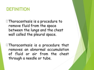
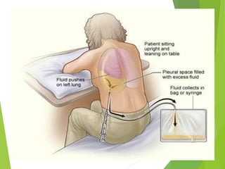
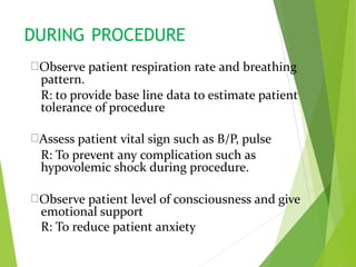
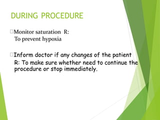
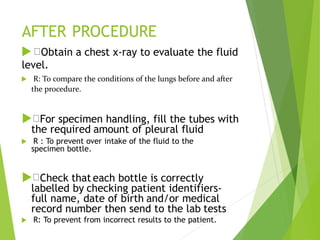
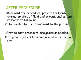
1
Thoracentesis
DEFINITION
Thoracentesis is a procedure to
remove fluid from the space
between the lungs and the chest
wall called the pleural space.
Thoracentesis is a procedure that
removes an abnormal accumulation
of fluid or air from the chest
through a needle or tube.
PURPOSETo determine the cause of abnormal
accumulation of fluid in the pleural
space.
Relieve shortness of breath and pain
As a diagnostic or treatment procedure
To drain large amounts of pleural fluid
To equalize pressure on both sides of
the thoracic cavity
INDICATION
 Traumatic pneumothorax
 Hemopneumothorax
 Spontaneous pneumothorax
 Bronchopleural fistula
 Pleural effusion
CONTRAINDICATION
An uncooperative patient
Coagulation disorder
Atelectasis
Only one functioning lung
Emphysema(pulmonary enlargement)
Severe cough or hiccups
COMPLICATION
▪ Pulmonary edema
▪ Respiratory distress
▪ Air embolism
▪ Cardiac tamponade(fluid
build up in the space
between myocardium and
pericardium)
▪ Bleeding
▪ Infection
▪ Dyspnea and
cough
▪ Atelectasis(lung
collapes)
7
BEFORE THE PROCEDURE
Explain the purpose,
risks/benefits, and steps of the
procedure and obtain consent from
the patient or appropriate legal
design.
• An explanation helps orient the
patient to the procedure assist in
coping and provide an opportunity
to ask question and verbalise
anxiety
CON’T…
Take Medical History such as
➢Trouble in breathing, coughing, and hiccups
➢Had heart disease
➢Smoked
➢Travelled to places where may have been
exposed to tuberculosis
R: to detect any abnormalities regarding
the procedure
9
• Prepare Equipment
 Dressing set
 Abraham’s needle
 Connecting tubing
 Syringe 50ml and
5ml
 Scapel blade and
blade 11
 Needles (18
and 23 gauge)
 Sterile Glove
 Mask
 Povidone / Alcohol
 Local anaesthetic,
e.g. lignocaine
(lidocaine) 1%
 or 2%
 Formalin bottle
 Urine bottle x2
 C+S bottle
 3-way stopcock
 Fenestrated towel
 Jug
BEFORE THE PROCEDURE
 Check platelet count and/or
presence of coagulopathy. If platelet
count is < 20,000, or there is known
coagulopathy as to whether platelet
transfusion or other intervention is
needed
To prevent complication such as
bleeding while during procedure.
Place patient upright / cardiac position and
help patient maintain position during
procedure.
R: the upright position ensures that the
diaphragm is more dependent and facilitates
the removal of fluid that usually localizes at
the base of the chest.
Explain that he/she will receive a
local anesthetic
R: to minimize pain during the procedure.
Clean patient skin with antiseptic soap
R: To prevent infection and maintain
aseptic technique.
DURING PROCEDURE
Observe patient respiration rate and breathing
pattern.
R: to provide base line data to estimate patient
tolerance of procedure
Assess patient vital sign such as B/P, pulse
R: To prevent any complication such as
hypovolemic shock during procedure.
Observe patient level of consciousness and give
emotional support
R: To reduce patient anxiety
DURING PROCEDURE
Monitor saturation R:
To prevent hypoxia
Inform doctor if any changes of the patient
R: To make sure whether need to continue the
procedure or stop immediately.
AFTER PROCEDURE
 Obtain a chest x-ray to evaluate the fluid
level.
 R: To compare the conditions of the lungs before and after
the procedure.
 For specimen handling, fill the tubes with
the required amount of pleural fluid
 R : To prevent over intake of the fluid to the
specimen bottle.
 Check that each bottle is correctly
labelled by checking patient identifiers-
full name, date of birth and/or medical
record number then send to the lab tests
 R: To prevent from incorrect results to the patient.
AFTER PROCEDURE
Document the procedure, patient’s response,
characteristics of fluid and amount, and patient
response to follow-up.
R: To develop further treatment to the patient.
Provide post-procedural analgesics as needed.
R: To prevent patient from pain related to the incision
site.
AFTER PROCEDURE
Rest in bed for about 2 hours after the procedure
R: To minimize patient activity due to complication
such as dyspnea.
Blood pressure and breathing will be checked for up
to a few hours
R: to make sure don't have complications
AFTER PROCEDURE
May remove dressing/bandage another day,
or replace it if it becomes soiled or wet
R: To prevent from getting infection.
Resume patient regular diet.
R: To promote wound healing.
THANK YOU !!!

thoracentesis final

  • 1.
  • 2.
    DEFINITION Thoracentesis is aprocedure to remove fluid from the space between the lungs and the chest wall called the pleural space. Thoracentesis is a procedure that removes an abnormal accumulation of fluid or air from the chest through a needle or tube.
  • 3.
    PURPOSETo determine thecause of abnormal accumulation of fluid in the pleural space. Relieve shortness of breath and pain As a diagnostic or treatment procedure To drain large amounts of pleural fluid To equalize pressure on both sides of the thoracic cavity
  • 4.
    INDICATION  Traumatic pneumothorax Hemopneumothorax  Spontaneous pneumothorax  Bronchopleural fistula  Pleural effusion
  • 5.
    CONTRAINDICATION An uncooperative patient Coagulationdisorder Atelectasis Only one functioning lung Emphysema(pulmonary enlargement) Severe cough or hiccups
  • 6.
    COMPLICATION ▪ Pulmonary edema ▪Respiratory distress ▪ Air embolism ▪ Cardiac tamponade(fluid build up in the space between myocardium and pericardium) ▪ Bleeding ▪ Infection ▪ Dyspnea and cough ▪ Atelectasis(lung collapes)
  • 7.
  • 8.
    BEFORE THE PROCEDURE Explainthe purpose, risks/benefits, and steps of the procedure and obtain consent from the patient or appropriate legal design. • An explanation helps orient the patient to the procedure assist in coping and provide an opportunity to ask question and verbalise anxiety
  • 9.
    CON’T… Take Medical Historysuch as ➢Trouble in breathing, coughing, and hiccups ➢Had heart disease ➢Smoked ➢Travelled to places where may have been exposed to tuberculosis R: to detect any abnormalities regarding the procedure 9
  • 10.
    • Prepare Equipment Dressing set  Abraham’s needle  Connecting tubing  Syringe 50ml and 5ml  Scapel blade and blade 11  Needles (18 and 23 gauge)  Sterile Glove  Mask  Povidone / Alcohol  Local anaesthetic, e.g. lignocaine (lidocaine) 1%  or 2%  Formalin bottle  Urine bottle x2  C+S bottle  3-way stopcock  Fenestrated towel  Jug
  • 11.
    BEFORE THE PROCEDURE Check platelet count and/or presence of coagulopathy. If platelet count is < 20,000, or there is known coagulopathy as to whether platelet transfusion or other intervention is needed To prevent complication such as bleeding while during procedure.
  • 12.
    Place patient upright/ cardiac position and help patient maintain position during procedure. R: the upright position ensures that the diaphragm is more dependent and facilitates the removal of fluid that usually localizes at the base of the chest. Explain that he/she will receive a local anesthetic R: to minimize pain during the procedure. Clean patient skin with antiseptic soap R: To prevent infection and maintain aseptic technique.
  • 14.
    DURING PROCEDURE Observe patientrespiration rate and breathing pattern. R: to provide base line data to estimate patient tolerance of procedure Assess patient vital sign such as B/P, pulse R: To prevent any complication such as hypovolemic shock during procedure. Observe patient level of consciousness and give emotional support R: To reduce patient anxiety
  • 15.
    DURING PROCEDURE Monitor saturationR: To prevent hypoxia Inform doctor if any changes of the patient R: To make sure whether need to continue the procedure or stop immediately.
  • 16.
    AFTER PROCEDURE  Obtaina chest x-ray to evaluate the fluid level.  R: To compare the conditions of the lungs before and after the procedure.  For specimen handling, fill the tubes with the required amount of pleural fluid  R : To prevent over intake of the fluid to the specimen bottle.  Check that each bottle is correctly labelled by checking patient identifiers- full name, date of birth and/or medical record number then send to the lab tests  R: To prevent from incorrect results to the patient.
  • 17.
    AFTER PROCEDURE Document theprocedure, patient’s response, characteristics of fluid and amount, and patient response to follow-up. R: To develop further treatment to the patient. Provide post-procedural analgesics as needed. R: To prevent patient from pain related to the incision site.
  • 18.
    AFTER PROCEDURE Rest inbed for about 2 hours after the procedure R: To minimize patient activity due to complication such as dyspnea. Blood pressure and breathing will be checked for up to a few hours R: to make sure don't have complications
  • 19.
    AFTER PROCEDURE May removedressing/bandage another day, or replace it if it becomes soiled or wet R: To prevent from getting infection. Resume patient regular diet. R: To promote wound healing.
  • 20.