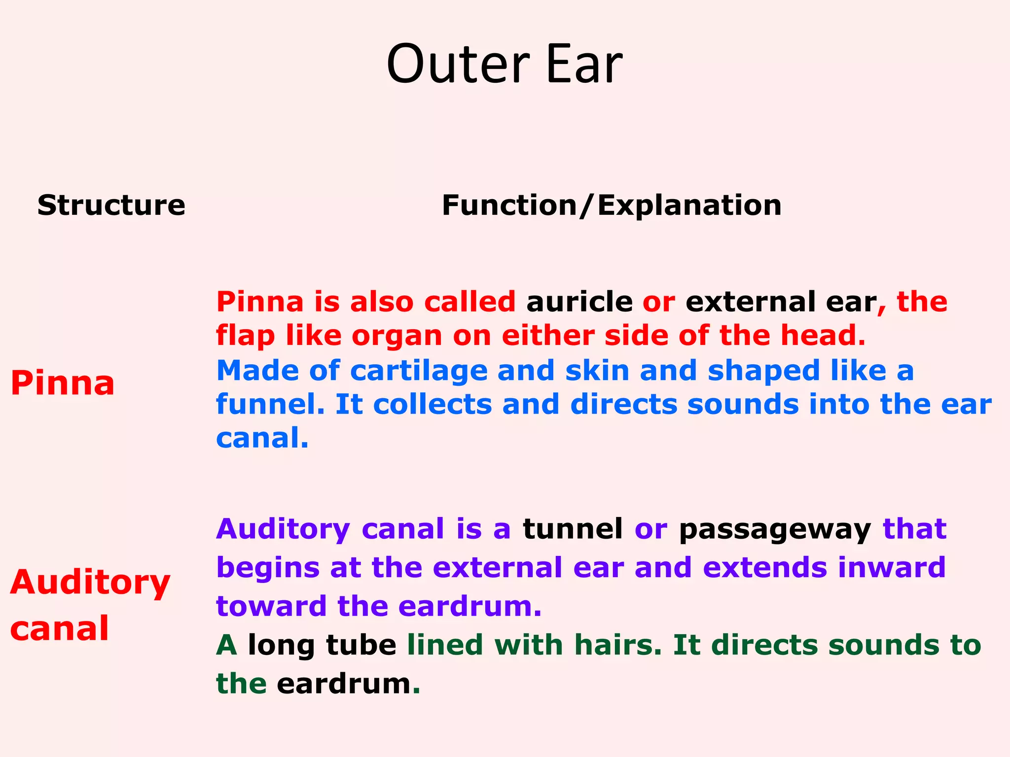
HUMAN EAR
Prepared by
Mr. Abhay Rajpoot
HOD (Dep. of Medical Surgical)
abhayrajpoot5591@gmail.com
Study of Human Ear
The ear is the sensory organ of sound.
The sense of hearing is sensitive to the sound
stimuli.
Every structure of the ear has their own
functions and are very important
Three main parts of the Ear
Outer Ear
Outer Ear
pinna auditory canal
Outer Ear
Structure Function/Explanation
Pinna
Pinna is also called auricle or external ear, the
flap like organ on either side of the head.
Made of cartilage and skin and shaped like a
funnel. It collects and directs sounds into the ear
canal.
Auditory
canal
Auditory canal is a tunnel or passageway that
begins at the external ear and extends inward
toward the eardrum.
A long tube lined with hairs. It directs sounds to
the eardrum.
Middle Ear
Structure
Middle Ear
Function/Explanation
Eardrum
Eardrum also known as the tympanic membrane.
semitransparent thin fibroelastic connective tissue
membrane, covered by epidermis (external side) and a
cuboidal mucous epithielium (inner side) .It vibrates and
transmits sound waves to the ossicles.
Ossicles
Made up of three small bones which is the hammer
(Malleus), the anvil (Incus) and the stirrup (stapes). It intensifies
the vibrations of the sound waves by 22 times before
transmitting to the oval window.
Eustachian
tube
A narrow tube that joins the middle ear to the throat that
balances the air pressure at both sides of the eardrum.
An oval-shaped, thin membrane between the middle ear
Oval window and the inner ear. It transmits sound vibrations from the
middle ear to the inner ear.
Bones & Muscles of Middle ear
Inner ear
Inner Ear or Bony Labyrinth
Structure Function/Explanation
Cochlea
Cochlea (bony labrynth) is a system of coiled tubes
snail-like structure in the inner ear filled with fluid
called perilymph & endolymph.It consists of three
tubes (canals)
1. Scala Vestibuli (Vestibular canal)
2. Scala media (cochlear canal)
3. Scala tympani (tympanic canal)
And ORGAN OF CORTI (electromechanically sensitive hair cells)
Auditory
nerve
It carries the impulses to the brain which then
interprets the impulses as sound.
Semicircular
Canals &
Vestible
For body balance.
SENSORYCELLS FOR HEARING
 Contains Glutamate (transmitter)
 Connected withAuditory nerve fibers
 Reissner’s membrane (Vestibular membrane)
◦ Soft thin membrane seperates scala vestibuli and scala media
which does not obstruct the passage of sound
 Basilar Membrane
◦ Fibrous membrane that separates the scala media from the scala
tympani
 Tectorial membrane
◦ Gel-like membrane is capable of bending hair cells
 Organ of Corti
◦ Receptor organ that generates nerve impulses in response to
vibration of the basilar membrane
◦ Nerve cells:
 Single row of internal hair cells (3500 nos, 12 μm dia)
 three or four rows of external hair cells (12,000 nos , 8 μm dia)
SOUND
 Results from the motion of air molecules which oscillate.
 Sound waves travel in all directions from their source.
 Waves are characterized by frequency and intensity
Frequency
Measured in hertz (cycles per second).
Greater the frequency the higher the pitch.
Intensity:
Directly related to amplitude of sound waves.
Measured in decibels (db).
Physiology
of
hearing
AUDITORY PATHWAY
When sound waves travels through the auditory tunnel,
Channels sound waves to the tympanic membrane and Increases sound wave
intensity .
The ossicles amplify the sound 22 X
Tensor tymphani muscles initiates the vibrations depends upon frequency.
When the foot of the stapes moves inward against the oval window, the round
window must bulge outward because the cochlea is bounded on all sides by bony
walls.
Vibrations of stapes and oval window displace perilymph fluid within scala
vestibuli
 Vibrations pass to the scala tympani.
 As sound frequency increases, pressure waves of the perilymph are
transmitted through the vestibular membrane and through the basilar
membrane.
 sound waves entering at the oval window is to cause the basilar
membrane to bend in the direction of the round window
 Movements of perilymph travel to the base of cochlea where they
displace the round window.
 This makes the cochlear ducts to be displaced which move the
tectorial membrane
 During this a shearing force is created, causes moving and bending
of the stereocilia.
 Ion channels open, depolarizing the hair cells, releasing glutamate
that stimulates the sensory neuron .
 Greater bending of stereocilia, the increased frequency of AP
produced.
 Nerve impulses in cochlear nerve travel to brain stem and on to the
auditory areas of cerebral cortex, where it is interpreted as sound
Pattern of Vibration of the Basilar Membrane for
Different
Sound Frequencies
Vestibular Apparatus and Equilibrium
Vestibular apparatus maintains the body (mainly the head) at
equilibrium (at balance) and stabilizing the eyes relative to the
environment
 Equilibrium has two functional parts
 Static equilibrium
 Receptors in the vestibule
 Maintenance of the position of the body (mainly the
head) relative to the force of gravity
 Dynamic equilibrium
 Receptors in the semicircular canals
 Maintenance of the position of the body (mainly the head)
in response to sudden movements such as rotation,
acceleration, and deceleration
 Semicircular Canals
 Provide information about rotational acceleration.
 Project in 3 different planes.
 Each canal contains a semicircular duct
(Endolymph Movement inside it).
 At the base is the crista ampullaris (enlarged
swellings at base of each canal communicating
with utricles)
Static Equilibrium
 Maculae – receptors within the vestibule
 Report on the position of the head with respect to the
pull of gravity when the body is not moving (static =
rest)
 Anatomy of the maculae
 Hair cells are embedded in the otolithic membrane
 Otoliths (tiny stones) float in a gel around the hair cells
 Movements cause otoliths to bend the hair cells 
sends impulses along the vestibular nerve to the
cerebellum of the brain
Function of Maculae
Dynamic Equilibrium
 Responds to angular or rotatory movements of the
head
 Crista ampullaris – receptors found within the
semicircular canals
 Tuft of hair cells are covered with a gelatinous
cap called the cupula
Human ear

Human ear

  • 1.
    HUMAN EAR Prepared by Mr.Abhay Rajpoot HOD (Dep. of Medical Surgical) abhayrajpoot5591@gmail.com
  • 2.
  • 3.
    The ear isthe sensory organ of sound. The sense of hearing is sensitive to the sound stimuli. Every structure of the ear has their own functions and are very important
  • 4.
    Three main partsof the Ear
  • 5.
  • 6.
    Outer Ear Structure Function/Explanation Pinna Pinnais also called auricle or external ear, the flap like organ on either side of the head. Made of cartilage and skin and shaped like a funnel. It collects and directs sounds into the ear canal. Auditory canal Auditory canal is a tunnel or passageway that begins at the external ear and extends inward toward the eardrum. A long tube lined with hairs. It directs sounds to the eardrum.
  • 7.
  • 8.
    Structure Middle Ear Function/Explanation Eardrum Eardrum alsoknown as the tympanic membrane. semitransparent thin fibroelastic connective tissue membrane, covered by epidermis (external side) and a cuboidal mucous epithielium (inner side) .It vibrates and transmits sound waves to the ossicles. Ossicles Made up of three small bones which is the hammer (Malleus), the anvil (Incus) and the stirrup (stapes). It intensifies the vibrations of the sound waves by 22 times before transmitting to the oval window. Eustachian tube A narrow tube that joins the middle ear to the throat that balances the air pressure at both sides of the eardrum. An oval-shaped, thin membrane between the middle ear Oval window and the inner ear. It transmits sound vibrations from the middle ear to the inner ear.
  • 9.
    Bones & Musclesof Middle ear
  • 10.
  • 11.
    Inner Ear orBony Labyrinth Structure Function/Explanation Cochlea Cochlea (bony labrynth) is a system of coiled tubes snail-like structure in the inner ear filled with fluid called perilymph & endolymph.It consists of three tubes (canals) 1. Scala Vestibuli (Vestibular canal) 2. Scala media (cochlear canal) 3. Scala tympani (tympanic canal) And ORGAN OF CORTI (electromechanically sensitive hair cells) Auditory nerve It carries the impulses to the brain which then interprets the impulses as sound. Semicircular Canals & Vestible For body balance.
  • 13.
    SENSORYCELLS FOR HEARING Contains Glutamate (transmitter)  Connected withAuditory nerve fibers
  • 15.
     Reissner’s membrane(Vestibular membrane) ◦ Soft thin membrane seperates scala vestibuli and scala media which does not obstruct the passage of sound  Basilar Membrane ◦ Fibrous membrane that separates the scala media from the scala tympani  Tectorial membrane ◦ Gel-like membrane is capable of bending hair cells  Organ of Corti ◦ Receptor organ that generates nerve impulses in response to vibration of the basilar membrane ◦ Nerve cells:  Single row of internal hair cells (3500 nos, 12 μm dia)  three or four rows of external hair cells (12,000 nos , 8 μm dia)
  • 16.
    SOUND  Results fromthe motion of air molecules which oscillate.  Sound waves travel in all directions from their source.  Waves are characterized by frequency and intensity Frequency Measured in hertz (cycles per second). Greater the frequency the higher the pitch. Intensity: Directly related to amplitude of sound waves. Measured in decibels (db).
  • 17.
  • 19.
    AUDITORY PATHWAY When soundwaves travels through the auditory tunnel, Channels sound waves to the tympanic membrane and Increases sound wave intensity . The ossicles amplify the sound 22 X Tensor tymphani muscles initiates the vibrations depends upon frequency. When the foot of the stapes moves inward against the oval window, the round window must bulge outward because the cochlea is bounded on all sides by bony walls. Vibrations of stapes and oval window displace perilymph fluid within scala vestibuli
  • 20.
     Vibrations passto the scala tympani.  As sound frequency increases, pressure waves of the perilymph are transmitted through the vestibular membrane and through the basilar membrane.  sound waves entering at the oval window is to cause the basilar membrane to bend in the direction of the round window  Movements of perilymph travel to the base of cochlea where they displace the round window.  This makes the cochlear ducts to be displaced which move the tectorial membrane  During this a shearing force is created, causes moving and bending of the stereocilia.
  • 21.
     Ion channelsopen, depolarizing the hair cells, releasing glutamate that stimulates the sensory neuron .  Greater bending of stereocilia, the increased frequency of AP produced.  Nerve impulses in cochlear nerve travel to brain stem and on to the auditory areas of cerebral cortex, where it is interpreted as sound
  • 22.
    Pattern of Vibrationof the Basilar Membrane for Different Sound Frequencies
  • 23.
    Vestibular Apparatus andEquilibrium Vestibular apparatus maintains the body (mainly the head) at equilibrium (at balance) and stabilizing the eyes relative to the environment  Equilibrium has two functional parts  Static equilibrium  Receptors in the vestibule  Maintenance of the position of the body (mainly the head) relative to the force of gravity  Dynamic equilibrium  Receptors in the semicircular canals  Maintenance of the position of the body (mainly the head) in response to sudden movements such as rotation, acceleration, and deceleration
  • 25.
     Semicircular Canals Provide information about rotational acceleration.  Project in 3 different planes.  Each canal contains a semicircular duct (Endolymph Movement inside it).  At the base is the crista ampullaris (enlarged swellings at base of each canal communicating with utricles)
  • 26.
    Static Equilibrium  Maculae– receptors within the vestibule  Report on the position of the head with respect to the pull of gravity when the body is not moving (static = rest)  Anatomy of the maculae  Hair cells are embedded in the otolithic membrane  Otoliths (tiny stones) float in a gel around the hair cells  Movements cause otoliths to bend the hair cells  sends impulses along the vestibular nerve to the cerebellum of the brain
  • 27.
  • 28.
    Dynamic Equilibrium  Respondsto angular or rotatory movements of the head  Crista ampullaris – receptors found within the semicircular canals  Tuft of hair cells are covered with a gelatinous cap called the cupula