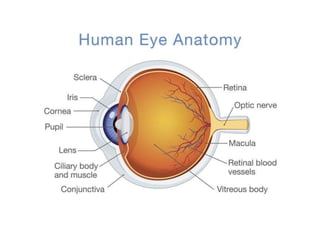
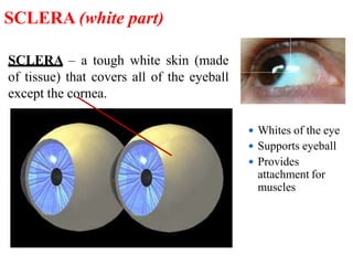
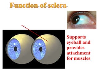
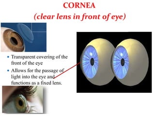
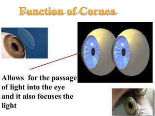
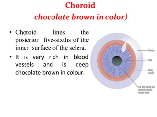
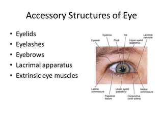
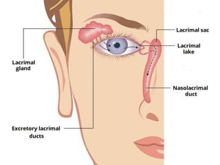
Human Eye
Prepared by
Mr.Abhay Rajpoot
HOD (Dep. of Medical Surgical)
abhayrajpoot5591@gmail.com
• The eye is the organ of sight. It is situated in
the orbital cavity and supplied by the optic
nerve (2nd cranial nerve).
• It is almost spherical in shape and about 2.5
cm in diameter. The space between the eye and
the orbital cavity is occupied by adipose tissue.
The bony walls of the orbit and the fat help to
protect the eye from injury.
1. Outer fibrous layer: sclera and cornea.
2. Middle vascular layer or uveal tract:
consisting of the choroid, ciliary body and
iris.
3. Inner nervous tissue layer: retina.
SCLERA – a tough white skin (made
of tissue) that covers all of the eyeball
except the cornea.
 Whites of the eye
 Supports eyeball
 Provides
attachment for
muscles
SCLERA (white part)
Supports
eyeball and
provides
attachment
for muscles
 Transparent covering of the
front of the eye
 Allows for the passage of
light into the eye and
functions as a fixed lens.
CORNEA
(clear lens in front of eye)
Allows for the passage
of light into the eye
and it also focuses the
light
Choroid
chocolate brown in color)
• Choroid lines the
posterior five-sixths of the
inner surface of the sclera.
• It is very rich
vessels and
in blood
is deep
chocolate brown in colour.
Light enters the eye through the pupil,
stimulates the sensory receptors in the retina
and is then absorbed by the choroid.
 colored part of
eye
 controls light
entering
IRIS
(colored part)
controls the amount of
light entering the eye
Parasympathetic
stimulation constricts the
pupil and sympathetic
stimulation dilates it
Pupil size is
controlled by iris
muscles
 Black hole in iris
 Where light enters
PUPIL (black hole)
The hole where light enters into the eye
PUPIL
When the eye needs
more light to enter
(when it is dark), the
pupils get larger;
allowing more light to
enter the eye
When the eye needs
less light
(when it
bright), the
to enter
is very
pupils
get smaller; allowing
less light to enter the
eye
elastic
 lens is a highly
circular
biconvex body,
lying immediately
behind the pupil
 allows us to see
objects near and
far
LENS
(lens behind pupil)
allows us to see
objects near and
far
 Internal membrane
 Contains light-receptive cells, rods cell (120 million) and
cone cell (6 million)
 Rod cell help[ to see in dim light and cone cell help to
see in bright light.
 Converts light to electrical signals.
RETINA
converts light
waves to electrical
signals
The eye is supplied with arterial blood
by the ciliary arteries and the central
retinal artery. These are branches of the
ophthalmic artery, one of the branches of
the internal carotid artery.
Blood supply to the eye
 Transmits electrical
impulses from retina to the
brain
 Creates blind spot
 Brain takes inverted image
and flips it so we can see
OPTIC NERVE
Transmits electrical
signals from retina to
the brain
Interior of the Eye
The eyeball is not a solid sphere. It contains a large
interior spaced, it is divided into two cavities, anterior
and posterior.
The anterior cavity is further divided into two chambers,
anterior and posterior chamber. The anterior chamber
lies anterior to the iris and posterior to the cornea.
Aqueous humor is the fluid present in the anterior
cavity. The posterior cavity of eye is quite larger than
the anterior. This cavity is present posterior to the lens,
suspensory ligament and ciliary body.
Vitreous humour is the fluid present in the posterior
cavity. This jelly like fluid consists of 99% H2O some
salts and muco protein. It helps to maintain the
intraoccular pressure.
Aqueous humour drains out of the anterior chamber at
the same rate, at which it enters the posterior chamber.
In this way, the amount of fluid remains same. The
normal intraoccular pressure is 16 to 20 mmHg. If the
intraoccular pressure increases, it leads to disease
glaucoma.
These are two arched ridges of the supraorbital
margins of the frontal bone. Numerous hairs
(eyebrows) project obliquely from the surface of the
skin. They protect the eyeball from sweat, dust and
other foreign bodies.
Eyebrows
The eyelids are two movable folds of tissue
situated above and below the front of each eye. On
their free edges there are short curved hairs, the
eyelashes
Eyelids
The eyelids and eyelashes protect the eye from
injury:-
Reflex closure of the lids occurs when the
conjunctiva or eyelashes are touched, when an object
comes close to the eye or when a bright light shines
into the eye – this is called the corneal reflex
blinking at about 3- to 7-second intervals spreads
tears and oily secretions over the cornea, preventing
drying.
Functions of eye lids
This is a fine transparent membrane that
lines the eyelids and the front of the eyeball.
Where it lines the eyelids it consists of highly
vascular columnar epithelium.
Conjunctiva
Each eye this consists of lacrimal gland and its
ducts lacrimal
Lacrimal sac nasolacrimal duct.
The lacrimal glands are exocrine glands situated in
recesses in the frontal bones on the lateral aspect of each
eye just behind the supraorbital margin. Each gland is
approximately the size and shape of an almond, and is
composed of secretory epithelial cells. The glands secrete
tears composed of water, mineral salts, antibodies
(immunoglobulin), and lysozyme, a bactericidal enzyme.
Lacrimal apparatus
-Washing away irritating materials, e.g. dust, grit
-Bactericidal enzyme lysozyme prevents microbial
infection
-Prevents drying of the conjunctiva.
Functions of lacrimal gland
2 Lenses: Cornea and Lens
How Your Lens Focuses
• Your lens has a small depth of field
– You can't see something close and far with both objects
in focus at the same time
• Hold out your thumb about a foot away from your eye
– Then, alternately focus on thumb and me (right above
your thumb)
• Note that you cannot see both me and your thumb sharply
(in focus) at the same time
– You focus on one or the other by changing the bulge of
your lens
teacher is in focus
thumb is out of focus
thumb is in focus
Teacher is out of focus
The human eye

The human eye

  • 1.
    Human Eye Prepared by Mr.AbhayRajpoot HOD (Dep. of Medical Surgical) abhayrajpoot5591@gmail.com
  • 2.
    • The eyeis the organ of sight. It is situated in the orbital cavity and supplied by the optic nerve (2nd cranial nerve). • It is almost spherical in shape and about 2.5 cm in diameter. The space between the eye and the orbital cavity is occupied by adipose tissue. The bony walls of the orbit and the fat help to protect the eye from injury.
  • 3.
    1. Outer fibrouslayer: sclera and cornea. 2. Middle vascular layer or uveal tract: consisting of the choroid, ciliary body and iris. 3. Inner nervous tissue layer: retina.
  • 5.
    SCLERA – atough white skin (made of tissue) that covers all of the eyeball except the cornea.  Whites of the eye  Supports eyeball  Provides attachment for muscles SCLERA (white part)
  • 6.
  • 7.
     Transparent coveringof the front of the eye  Allows for the passage of light into the eye and functions as a fixed lens. CORNEA (clear lens in front of eye)
  • 8.
    Allows for thepassage of light into the eye and it also focuses the light
  • 9.
    Choroid chocolate brown incolor) • Choroid lines the posterior five-sixths of the inner surface of the sclera. • It is very rich vessels and in blood is deep chocolate brown in colour.
  • 10.
    Light enters theeye through the pupil, stimulates the sensory receptors in the retina and is then absorbed by the choroid.
  • 11.
     colored partof eye  controls light entering IRIS (colored part)
  • 12.
    controls the amountof light entering the eye Parasympathetic stimulation constricts the pupil and sympathetic stimulation dilates it
  • 13.
    Pupil size is controlledby iris muscles  Black hole in iris  Where light enters PUPIL (black hole)
  • 14.
    The hole wherelight enters into the eye
  • 15.
    PUPIL When the eyeneeds more light to enter (when it is dark), the pupils get larger; allowing more light to enter the eye
  • 16.
    When the eyeneeds less light (when it bright), the to enter is very pupils get smaller; allowing less light to enter the eye
  • 17.
    elastic  lens isa highly circular biconvex body, lying immediately behind the pupil  allows us to see objects near and far LENS (lens behind pupil)
  • 18.
    allows us tosee objects near and far
  • 19.
     Internal membrane Contains light-receptive cells, rods cell (120 million) and cone cell (6 million)  Rod cell help[ to see in dim light and cone cell help to see in bright light.  Converts light to electrical signals. RETINA
  • 20.
    converts light waves toelectrical signals
  • 21.
    The eye issupplied with arterial blood by the ciliary arteries and the central retinal artery. These are branches of the ophthalmic artery, one of the branches of the internal carotid artery. Blood supply to the eye
  • 22.
     Transmits electrical impulsesfrom retina to the brain  Creates blind spot  Brain takes inverted image and flips it so we can see OPTIC NERVE
  • 23.
  • 25.
    Interior of theEye The eyeball is not a solid sphere. It contains a large interior spaced, it is divided into two cavities, anterior and posterior. The anterior cavity is further divided into two chambers, anterior and posterior chamber. The anterior chamber lies anterior to the iris and posterior to the cornea. Aqueous humor is the fluid present in the anterior cavity. The posterior cavity of eye is quite larger than the anterior. This cavity is present posterior to the lens, suspensory ligament and ciliary body.
  • 26.
    Vitreous humour isthe fluid present in the posterior cavity. This jelly like fluid consists of 99% H2O some salts and muco protein. It helps to maintain the intraoccular pressure. Aqueous humour drains out of the anterior chamber at the same rate, at which it enters the posterior chamber. In this way, the amount of fluid remains same. The normal intraoccular pressure is 16 to 20 mmHg. If the intraoccular pressure increases, it leads to disease glaucoma.
  • 28.
    These are twoarched ridges of the supraorbital margins of the frontal bone. Numerous hairs (eyebrows) project obliquely from the surface of the skin. They protect the eyeball from sweat, dust and other foreign bodies. Eyebrows
  • 29.
    The eyelids aretwo movable folds of tissue situated above and below the front of each eye. On their free edges there are short curved hairs, the eyelashes Eyelids
  • 30.
    The eyelids andeyelashes protect the eye from injury:- Reflex closure of the lids occurs when the conjunctiva or eyelashes are touched, when an object comes close to the eye or when a bright light shines into the eye – this is called the corneal reflex blinking at about 3- to 7-second intervals spreads tears and oily secretions over the cornea, preventing drying. Functions of eye lids
  • 31.
    This is afine transparent membrane that lines the eyelids and the front of the eyeball. Where it lines the eyelids it consists of highly vascular columnar epithelium. Conjunctiva
  • 32.
    Each eye thisconsists of lacrimal gland and its ducts lacrimal Lacrimal sac nasolacrimal duct. The lacrimal glands are exocrine glands situated in recesses in the frontal bones on the lateral aspect of each eye just behind the supraorbital margin. Each gland is approximately the size and shape of an almond, and is composed of secretory epithelial cells. The glands secrete tears composed of water, mineral salts, antibodies (immunoglobulin), and lysozyme, a bactericidal enzyme. Lacrimal apparatus
  • 34.
    -Washing away irritatingmaterials, e.g. dust, grit -Bactericidal enzyme lysozyme prevents microbial infection -Prevents drying of the conjunctiva. Functions of lacrimal gland
  • 35.
  • 36.
    How Your LensFocuses • Your lens has a small depth of field – You can't see something close and far with both objects in focus at the same time • Hold out your thumb about a foot away from your eye – Then, alternately focus on thumb and me (right above your thumb) • Note that you cannot see both me and your thumb sharply (in focus) at the same time – You focus on one or the other by changing the bulge of your lens
  • 37.
    teacher is infocus thumb is out of focus thumb is in focus Teacher is out of focus