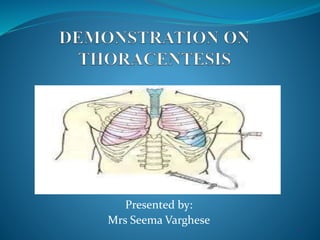
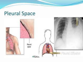
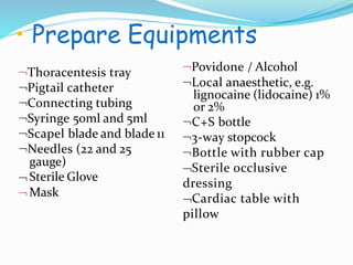
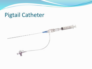
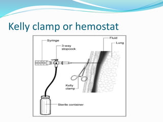
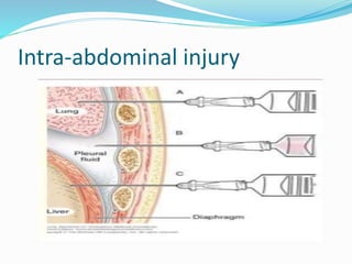
1
Presented by:
Mrs Seema Varghese
Objectives
• Define thoracentesis
• Enlist its purpose, indications
• Enumerate instruments
• Explain pre procedure care
• Explain during procedure care
• Describe post procedure care
• List out complications
Introduction
 Thoracentesis, also known as
thoracocentesis, pleural tap, needle
thoracostomy, or needle decompression.
DEFINITION
Thoracentesis is the insertion of a large bore
needle through the chest wall into the pleural
space to obtain specimen for diagnostic
evaluation or removal of fluid.
.
Pleural Space
PURPOSES
 To determine the cause of abnormal
accumulation of fluid in the pleural space.
Relieve shortness of breath and pain
A s a diagnostic or treatment procedure
To drain large amounts of pleural fluid
INDICATIONS
CONTRAINDICATIONS
Coagulation disorder
A n uncooperative patient
Only one functioninglung
Severe cough or hiccups
Local Infection (Cellulitis/ herpes zoster)
• Prepare Equipments
Thoracentesis tray
Pigtail catheter
Connecting tubing
Syringe 50ml and 5ml
Scapel blade and blade 11
Needles (22 and 25
gauge)


Sterile Glove
Mask
Povidone / Alcohol
Local anaesthetic, e.g.
lignocaine (lidocaine) 1%
or 2%
C+S bottle
3-way stopcock
Bottle with rubber cap
Sterile occlusive
dressing
Cardiac table with
pillow
Pigtail Catheter
Kelly clamp or hemostat
7
Nursing Responsibilities
BEFORE THE PROCEDURE
Assess the condition of the patient
Explain the purpose, risks/benefits, and steps
of the procedure and obtain consent from the
patient or appropriate legal guardian.
R: An explanation helps orient the patient to
the procedure assist in coping and provide an
opportunity to ask question and verbalize
anxiety
Keep ready chest x-ray
Cont…
Using ultrasonography to identify a site for
diagnostic thoracentesis
R- significantly lower risk of pneumothorax
Check platelet count and/or presence of
coagulopathy. If platelet count is < 20,000,
or there is known coagulopathy as to
whether platelet transfusion or other
intervention is needed
R: To prevent complication such as
bleeding while during procedure.
Cont…
Administer a cough suppressant if indicated.
R-Movement and coughing during the
procedure may cause inadvertent damage to the
lung or pleura.
 IV access should be established
Keep Atropine injection ready
R- in case of profound vasovagal response
Administer supplemental oxygen
R- prevents hypoxia
9
Cont…
Position the client upright, leaning forward with
arms and head supported on an anchored overbed
table. Physician spread the sterile drape
R- This position spreads the ribs, enlarging the
intercostal space for needle insertion.
Physician will explain that he/she will receive a
local anesthetic (1%/ 2% lidocaine)
R: to minimize pain during the procedure.
 Physician cleans patient skin with antiseptic
solution
R: To prevent infection and maintain aseptic
technique.
Position
Midclavicular
Anesthetize
DURING PROCEDURE
Observe patient respiration rate and breathing
pattern.
R: to provide base line data to estimate patient
tolerance of procedure
Assess patient vital sign such as B/P, pulse
R: To prevent any complication such as
hypovolemic shock during procedure.
Observe patient level of consciousness and give
emotional support
R: To reduce patient anxiety
Cont…
 A simple assessment, such as listening to the
lungs with a stethoscope and Percussion (tapping
on the lung area with a finger) can indicate extra
fluid.
 R- to locate the area of insertion and to assess
maximum degree of dullness
Locating the site
Cont…
 Marking: 2 inches below from the area of
dullness starts
R- for easy drainage of fluid
 Fluid usually 1000ml -1200 ml of pleural fluid
is removed (per day maximum).
 Monitor for hypotension, hypoxemia
R- avoid rapid removal of fluid
DURING PROCEDURE
Monitor saturation
 R: prevents hypoxia
Inform doctor if any changes in the
condition of patient
 R: To make sure whether need to continue
the procedure or stop immediately.
Post procedure care
Apply a dressing over the puncture site and position
on the unaffected side for 1 hour.
R-This allows the pleural puncture to heal.
Label obtained specimen with name, date, source, and
diagnosis; send specimen to the laboratory for
analysis.
R-Fluid obtained during thoracentesis may be examined
for abnormal cells, bacteria, and other substances to
determine the cause of the pleural effusion.
Cont…
During the first several hours after thoracentesis, frequently
assess and document vital signs; oxygen saturation; respiratory
status and puncture site for bleeding
R-Frequent assessment is important to detect possible
complications of thoracentesis, such as pneumothorax.
Obtain a chest X-ray.
R- Chest X-ray is ordered to detect possible pneumothorax.
Normal activities generally can be resumed after 1-2 hour if no
evidence of pneumothorax or other complication is present.
R- The puncture wound of thoracentesis heals rapidly.
Cont…
Document the procedure, patient’s response,
characteristics of fluid and amount, and
patient response to follow-up.
R: To develop further treatment to the patient.
Provide post-procedural analgesics as needed.
R: To prevent patient from pain related to the
incision site.
Cont…
 May remove dressing/bandage another day,
or replace it if it becomes soiled or wet
R: To prevent from getting infection.
Resume patient regular diet.
R: To promote wound healing.
COMPLICATIONS
Bleeding
Infection
Respiratory distress due to multiple needle
insertion
Pneumothorax
Intra-abdominal injury (liver/ spleen/
diaphragm)
Intra-abdominal injury
Conclusion
 Thoracentesis is a minimally invasive
procedure used to diagnose and treat pleural
effusions, a condition in which there is
excess fluid in the pleural space, also called
the pleural cavity. This space exists between
the outside of the lungs and the inside of the
chest wall.
Bibliography
1. Black J. M. and Hawks J (2009) Medical –surgical
Nursing, Clinical Management for positive outcomes
(8th edition) Saunders, Elsevier PP-620-621
2. Lewis S. M., Heitkemper M. M and Dirksen S. R
(2007) Medical /Surgical Nursing. Assessment and
management of clinical problems. (7th edition) St
Louis: C.V. Mosby PP-530, 550-560
THANK YOU !!!

Thoracentesis 12 june 2020

  • 1.
  • 2.
    Objectives • Define thoracentesis •Enlist its purpose, indications • Enumerate instruments • Explain pre procedure care • Explain during procedure care • Describe post procedure care • List out complications
  • 3.
    Introduction  Thoracentesis, alsoknown as thoracocentesis, pleural tap, needle thoracostomy, or needle decompression.
  • 4.
    DEFINITION Thoracentesis is theinsertion of a large bore needle through the chest wall into the pleural space to obtain specimen for diagnostic evaluation or removal of fluid. .
  • 5.
  • 6.
    PURPOSES  To determinethe cause of abnormal accumulation of fluid in the pleural space. Relieve shortness of breath and pain A s a diagnostic or treatment procedure To drain large amounts of pleural fluid
  • 7.
  • 8.
    CONTRAINDICATIONS Coagulation disorder A nuncooperative patient Only one functioninglung Severe cough or hiccups Local Infection (Cellulitis/ herpes zoster)
  • 9.
    • Prepare Equipments Thoracentesistray Pigtail catheter Connecting tubing Syringe 50ml and 5ml Scapel blade and blade 11 Needles (22 and 25 gauge)   Sterile Glove Mask Povidone / Alcohol Local anaesthetic, e.g. lignocaine (lidocaine) 1% or 2% C+S bottle 3-way stopcock Bottle with rubber cap Sterile occlusive dressing Cardiac table with pillow
  • 10.
  • 11.
  • 12.
  • 13.
    BEFORE THE PROCEDURE Assessthe condition of the patient Explain the purpose, risks/benefits, and steps of the procedure and obtain consent from the patient or appropriate legal guardian. R: An explanation helps orient the patient to the procedure assist in coping and provide an opportunity to ask question and verbalize anxiety Keep ready chest x-ray
  • 14.
    Cont… Using ultrasonography toidentify a site for diagnostic thoracentesis R- significantly lower risk of pneumothorax Check platelet count and/or presence of coagulopathy. If platelet count is < 20,000, or there is known coagulopathy as to whether platelet transfusion or other intervention is needed R: To prevent complication such as bleeding while during procedure.
  • 15.
    Cont… Administer a coughsuppressant if indicated. R-Movement and coughing during the procedure may cause inadvertent damage to the lung or pleura.  IV access should be established Keep Atropine injection ready R- in case of profound vasovagal response Administer supplemental oxygen R- prevents hypoxia 9
  • 16.
    Cont… Position the clientupright, leaning forward with arms and head supported on an anchored overbed table. Physician spread the sterile drape R- This position spreads the ribs, enlarging the intercostal space for needle insertion. Physician will explain that he/she will receive a local anesthetic (1%/ 2% lidocaine) R: to minimize pain during the procedure.  Physician cleans patient skin with antiseptic solution R: To prevent infection and maintain aseptic technique.
  • 17.
  • 18.
  • 19.
  • 20.
    DURING PROCEDURE Observe patientrespiration rate and breathing pattern. R: to provide base line data to estimate patient tolerance of procedure Assess patient vital sign such as B/P, pulse R: To prevent any complication such as hypovolemic shock during procedure. Observe patient level of consciousness and give emotional support R: To reduce patient anxiety
  • 21.
    Cont…  A simpleassessment, such as listening to the lungs with a stethoscope and Percussion (tapping on the lung area with a finger) can indicate extra fluid.  R- to locate the area of insertion and to assess maximum degree of dullness
  • 22.
  • 23.
    Cont…  Marking: 2inches below from the area of dullness starts R- for easy drainage of fluid  Fluid usually 1000ml -1200 ml of pleural fluid is removed (per day maximum).  Monitor for hypotension, hypoxemia R- avoid rapid removal of fluid
  • 24.
    DURING PROCEDURE Monitor saturation R: prevents hypoxia Inform doctor if any changes in the condition of patient  R: To make sure whether need to continue the procedure or stop immediately.
  • 25.
    Post procedure care Applya dressing over the puncture site and position on the unaffected side for 1 hour. R-This allows the pleural puncture to heal. Label obtained specimen with name, date, source, and diagnosis; send specimen to the laboratory for analysis. R-Fluid obtained during thoracentesis may be examined for abnormal cells, bacteria, and other substances to determine the cause of the pleural effusion.
  • 26.
    Cont… During the firstseveral hours after thoracentesis, frequently assess and document vital signs; oxygen saturation; respiratory status and puncture site for bleeding R-Frequent assessment is important to detect possible complications of thoracentesis, such as pneumothorax. Obtain a chest X-ray. R- Chest X-ray is ordered to detect possible pneumothorax. Normal activities generally can be resumed after 1-2 hour if no evidence of pneumothorax or other complication is present. R- The puncture wound of thoracentesis heals rapidly.
  • 27.
    Cont… Document the procedure,patient’s response, characteristics of fluid and amount, and patient response to follow-up. R: To develop further treatment to the patient. Provide post-procedural analgesics as needed. R: To prevent patient from pain related to the incision site.
  • 28.
    Cont…  May removedressing/bandage another day, or replace it if it becomes soiled or wet R: To prevent from getting infection. Resume patient regular diet. R: To promote wound healing.
  • 29.
    COMPLICATIONS Bleeding Infection Respiratory distress dueto multiple needle insertion Pneumothorax Intra-abdominal injury (liver/ spleen/ diaphragm)
  • 30.
  • 31.
    Conclusion  Thoracentesis isa minimally invasive procedure used to diagnose and treat pleural effusions, a condition in which there is excess fluid in the pleural space, also called the pleural cavity. This space exists between the outside of the lungs and the inside of the chest wall.
  • 32.
    Bibliography 1. Black J.M. and Hawks J (2009) Medical –surgical Nursing, Clinical Management for positive outcomes (8th edition) Saunders, Elsevier PP-620-621 2. Lewis S. M., Heitkemper M. M and Dirksen S. R (2007) Medical /Surgical Nursing. Assessment and management of clinical problems. (7th edition) St Louis: C.V. Mosby PP-530, 550-560
  • 33.