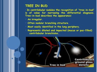
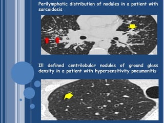
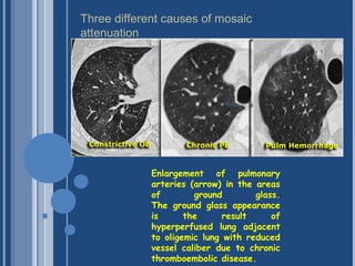
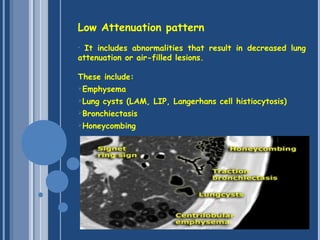
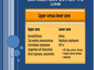
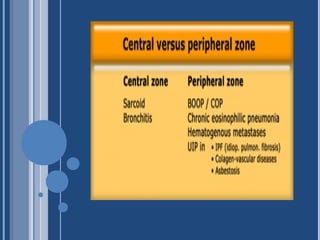
THE BASIC INTERPRETATION
OF HRCT
SECONDARY LOBULE
• Knowledge of the lung anatomy is essential for
understanding HRCT.
• The secondary lobule is the basic anatomic unit of
pulmonary structure and function.
• It measures about 1-2 cm and is made up of 5-15
pulmonary acini.
• The secondary lobule is supplied by a small bronchiole
(terminal bronchiole) in the center, that is paralleled by the
centrilobular artery.
• Pulmonary veins and lymphatics run in the periphery of the
lobule within the interlobular septa.
 
Centrilobular area
 
• The central part of the secondary lobule.
• Site of diseases, that enter the lung through the
airways
• Hypersensitivity pneumonitis, respiratory bronchiolitis,
centrilobular emphysema.
Perilymphatic area
• The peripheral part of the secondary lobule.
• Site of diseases, that are located in the lymphatics of
the interlobular septa.
• Sarcoid, lymphangitic carcinomatosis, pulmonary edema).
Basic Interpretation
• A structured approach to interpretation of HRCT involves
the following questions:
What is the dominant HR-pattern:
 Reticular
 Nodular
 High attenuation (ground-glass, consolidation)
 Low attenuation (emphysema, cystic)
Where is it located within the secondary lobule
(centrilobular, perilymphatic or random)?
Is there an upper versus lower zone or a central versus
peripheral predominance?
Are there additional findings (pleural fluid,
lymphadenopathy, traction bronchiectasis)?
RETICULAR PATTERN
• In the reticular pattern there are too many lines,
either as a result of thickening of the interlobular septa
or as a result of fibrosis as in honeycombing.
Septal Thickening
• Thickening of the lung interstitium by fluid, fibrous
tissue, or infiltration by cells results in a pattern of
reticular opacities due to thickening of the interlobular
septa.
Focal Septal Thickening in Lymphangitic
Carcinomatosis
Septal thickening and ground-glass
opacity with a gravitational distribution
in a patient with cardiogenic pulmonary
edema.
NODULAR PATTERN
TREE IN BUD
• In centrilobular nodules the recognition of 'tree-in-bud'
is of value for narrowing the differential diagnosis.
Tree-in-bud describes the appearance:
 An irregular.
 Often nodular branching structure.
 Most easily identified in the lung periphery. 
 Represents dilated and impacted (mucus or pus-filled)
centrilobular bronchioles.
Perilymphatic distribution of nodules in a patient with
sarcoidosis
Ill defined centrilobular nodules of ground glass
density in a patient with hypersensitivity pneumonitis
Random Distribution of Nodules in Miliary Tuberculosis
HIGH ATTENUATION PATTERN
• Increased lung attenuation is called ground-glass-
opacity (GGO) if there is a hazy increase in lung opacity
without obscuration of underlying vessels and is called
consolidation if the increase in lung opacity obscures the
vessels.
•In consolidation, there is exclusively air left intrabronchial. 
This is called the 'air bronchogram'.
BRONCHOALVEOLAR CARCINOMA WITH
GROUNG GLASS OPACITIES AND
CONSOLIDATION
MOSAIC ATTENUATION
Patchwork of regions of variable (black and grey)lung
density seen in
 Obliterative airway disease.
 Vascular disease
 Infilterative disease
The role of the radiologist is to determine which part is
abnormal: the black or the grey lung.
A disparity between the number/calibre of vessels in black
and grey lungs suggest that the black lung is abnormal.
In which case the likely causes are
Obliterative airway disease
or
Vascular disease
Diagnostic hint for further differentiation:
• Look at expiratory scans for air trapping( Attenuation
differences are accentuated in obliterative airway diseases)
In patients with infilterative disease, there is no obvious
discrepancy in the number/calibre of pulmonary vessels.
Enlargement of pulmonary
arteries (arrow) in the areas
of ground glass.
The ground glass appearance
is the result of
hyperperfused lung adjacent
to oligemic lung with reduced
vessel caliber due to chronic
thromboembolic disease.
Three different causes of mosaic
attenuation
CRAZY PAVING
• Crazy Paving is a combination of ground glass opacity with
superimposed septal thickening (5).
• It was first thought to be specific for alveolar
proteinosis, but later was also seen in other diseases.
Crazy Pavin can also be seen in:
 Alveolar proteinosis
 Sarcoid
 NSIP
 Organizing pneumonia
 Edema (heart failure, ARDS)
Low Attenuation pattern
• It includes abnormalities that result in decreased lung
attenuation or air-filled lesions.
These include:
Emphysema
Lung cysts (LAM, LIP, Langerhans cell histiocytosis)
Bronchiectasis
Honeycombing
Emphysema typically presents as areas of low attenuation
without visible walls as a result of parenchymal destruction.
Lung cysts are defined as radiolucent areas with a wall
thickness of less than 4mm. 
Cavities are defined as radiolucent areas with a wall
thickness of more than 4mm and are seen in infection
Bronchiectasis is defined as localized bronchial dilatation.
A signet-ring sign represents an axial cut of a dilated
bronchus (ring) with its accompanying small artery (signet).
Honeycombing is defined by the presence of small cystic
spaces with irregularly thickened walls composed of fibrous
tissue.
Honeycomb cysts often predominate in the peripheral and
subpleural lung regions regardless of their cause
DISTRIBUTION WITHIN THE
LUNG
The basic interpretation of hrct

The basic interpretation of hrct

  • 2.
  • 3.
    SECONDARY LOBULE • Knowledgeof the lung anatomy is essential for understanding HRCT. • The secondary lobule is the basic anatomic unit of pulmonary structure and function. • It measures about 1-2 cm and is made up of 5-15 pulmonary acini. • The secondary lobule is supplied by a small bronchiole (terminal bronchiole) in the center, that is paralleled by the centrilobular artery. • Pulmonary veins and lymphatics run in the periphery of the lobule within the interlobular septa.  
  • 4.
    Centrilobular area   • Thecentral part of the secondary lobule. • Site of diseases, that enter the lung through the airways • Hypersensitivity pneumonitis, respiratory bronchiolitis, centrilobular emphysema. Perilymphatic area • The peripheral part of the secondary lobule. • Site of diseases, that are located in the lymphatics of the interlobular septa. • Sarcoid, lymphangitic carcinomatosis, pulmonary edema).
  • 5.
    Basic Interpretation • Astructured approach to interpretation of HRCT involves the following questions: What is the dominant HR-pattern:  Reticular  Nodular  High attenuation (ground-glass, consolidation)  Low attenuation (emphysema, cystic) Where is it located within the secondary lobule (centrilobular, perilymphatic or random)? Is there an upper versus lower zone or a central versus peripheral predominance? Are there additional findings (pleural fluid, lymphadenopathy, traction bronchiectasis)?
  • 6.
    RETICULAR PATTERN • Inthe reticular pattern there are too many lines, either as a result of thickening of the interlobular septa or as a result of fibrosis as in honeycombing. Septal Thickening • Thickening of the lung interstitium by fluid, fibrous tissue, or infiltration by cells results in a pattern of reticular opacities due to thickening of the interlobular septa.
  • 7.
    Focal Septal Thickeningin Lymphangitic Carcinomatosis
  • 8.
    Septal thickening andground-glass opacity with a gravitational distribution in a patient with cardiogenic pulmonary edema.
  • 10.
  • 11.
    TREE IN BUD •In centrilobular nodules the recognition of 'tree-in-bud' is of value for narrowing the differential diagnosis. Tree-in-bud describes the appearance:  An irregular.  Often nodular branching structure.  Most easily identified in the lung periphery.   Represents dilated and impacted (mucus or pus-filled) centrilobular bronchioles.
  • 12.
    Perilymphatic distribution ofnodules in a patient with sarcoidosis Ill defined centrilobular nodules of ground glass density in a patient with hypersensitivity pneumonitis
  • 13.
    Random Distribution ofNodules in Miliary Tuberculosis
  • 14.
    HIGH ATTENUATION PATTERN •Increased lung attenuation is called ground-glass- opacity (GGO) if there is a hazy increase in lung opacity without obscuration of underlying vessels and is called consolidation if the increase in lung opacity obscures the vessels. •In consolidation, there is exclusively air left intrabronchial.  This is called the 'air bronchogram'.
  • 15.
    BRONCHOALVEOLAR CARCINOMA WITH GROUNGGLASS OPACITIES AND CONSOLIDATION
  • 16.
    MOSAIC ATTENUATION Patchwork ofregions of variable (black and grey)lung density seen in  Obliterative airway disease.  Vascular disease  Infilterative disease The role of the radiologist is to determine which part is abnormal: the black or the grey lung. A disparity between the number/calibre of vessels in black and grey lungs suggest that the black lung is abnormal. In which case the likely causes are Obliterative airway disease or Vascular disease Diagnostic hint for further differentiation: • Look at expiratory scans for air trapping( Attenuation differences are accentuated in obliterative airway diseases) In patients with infilterative disease, there is no obvious discrepancy in the number/calibre of pulmonary vessels.
  • 17.
    Enlargement of pulmonary arteries(arrow) in the areas of ground glass. The ground glass appearance is the result of hyperperfused lung adjacent to oligemic lung with reduced vessel caliber due to chronic thromboembolic disease. Three different causes of mosaic attenuation
  • 18.
    CRAZY PAVING • CrazyPaving is a combination of ground glass opacity with superimposed septal thickening (5). • It was first thought to be specific for alveolar proteinosis, but later was also seen in other diseases. Crazy Pavin can also be seen in:  Alveolar proteinosis  Sarcoid  NSIP  Organizing pneumonia  Edema (heart failure, ARDS)
  • 19.
    Low Attenuation pattern •It includes abnormalities that result in decreased lung attenuation or air-filled lesions. These include: Emphysema Lung cysts (LAM, LIP, Langerhans cell histiocytosis) Bronchiectasis Honeycombing
  • 20.
    Emphysema typically presentsas areas of low attenuation without visible walls as a result of parenchymal destruction. Lung cysts are defined as radiolucent areas with a wall thickness of less than 4mm.  Cavities are defined as radiolucent areas with a wall thickness of more than 4mm and are seen in infection Bronchiectasis is defined as localized bronchial dilatation. A signet-ring sign represents an axial cut of a dilated bronchus (ring) with its accompanying small artery (signet). Honeycombing is defined by the presence of small cystic spaces with irregularly thickened walls composed of fibrous tissue. Honeycomb cysts often predominate in the peripheral and subpleural lung regions regardless of their cause
  • 21.