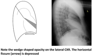
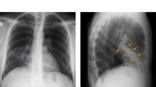
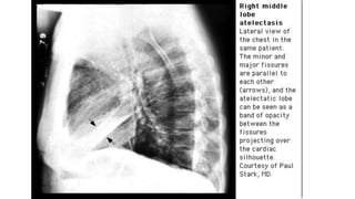
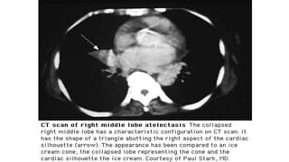
RIGHT MIDDLE LOBE COLLAPSE
Dr. Haseeb Manzoor
Department of Radiology
SMCH Lahore
• Right middle lobe is only 10% of total lung volume.
• Right middle lobe (RML) atelectasis is one form of the lobar collapse.
• of all the lobes, the right middle lobe is the most likely to be
chronically collapsed.
• RML atelectasis varies greatly from case to case in its radiographic
appearances.
Chronic RML Atelactasis
• often called "right middle lobe syndrome“
• Frequently nonobstructive
• Accompanied by scarring and bronchiectasis
• Often found in elderly women
PA view:
• Radiographic findings can be subtle
• the normal horizontal fissure is no longer visible (as it rotates down)
• Small triangular opacity pointing laterally
• blurring of the right heart border (loss of silhouette sign)
Lateral view:
• relatively easy to identify
• obliquely oriented triangular opacity with apex pointed toward hilum
and its base more peripheral in the lung
• Right horizontal and oblique fissure move towards each other
• The horizontal fissure is displaced inferiorly and the inferior part of
the oblique fissure, displaced anteriosuperiorly
Non-specific signs:
may be present but due to the small size of the right middle lobe they
are subtle or absent
• elevation of the hemidiaphragm
• crowding of the right sided ribs
• shift of the mediastinum to the right
Presence of linear opacities in the collapsed right middle lobe should
suggest that the collapse is chronic (right middle lobe syndrome), with
associated bronchiectasis
The right heart border is blurred
Note the wedge shaped opacity on the lateral CXR. The horizontal
fissure (arrow) is depressed
CT scan:
• the atelectatic right middle lobe presents as a triangular opacity with
its apex pointing laterally and with its medial contour apposed against
the right heart border.
• This has been called the "tilted ice cream cone" appearance
THANK YOU !

right middle lobe collapse

  • 1.
    RIGHT MIDDLE LOBECOLLAPSE Dr. Haseeb Manzoor Department of Radiology SMCH Lahore
  • 3.
    • Right middlelobe is only 10% of total lung volume. • Right middle lobe (RML) atelectasis is one form of the lobar collapse. • of all the lobes, the right middle lobe is the most likely to be chronically collapsed. • RML atelectasis varies greatly from case to case in its radiographic appearances. Chronic RML Atelactasis • often called "right middle lobe syndrome“ • Frequently nonobstructive • Accompanied by scarring and bronchiectasis • Often found in elderly women
  • 4.
    PA view: • Radiographicfindings can be subtle • the normal horizontal fissure is no longer visible (as it rotates down) • Small triangular opacity pointing laterally • blurring of the right heart border (loss of silhouette sign) Lateral view: • relatively easy to identify • obliquely oriented triangular opacity with apex pointed toward hilum and its base more peripheral in the lung • Right horizontal and oblique fissure move towards each other • The horizontal fissure is displaced inferiorly and the inferior part of the oblique fissure, displaced anteriosuperiorly
  • 5.
    Non-specific signs: may bepresent but due to the small size of the right middle lobe they are subtle or absent • elevation of the hemidiaphragm • crowding of the right sided ribs • shift of the mediastinum to the right Presence of linear opacities in the collapsed right middle lobe should suggest that the collapse is chronic (right middle lobe syndrome), with associated bronchiectasis
  • 6.
    The right heartborder is blurred
  • 7.
    Note the wedgeshaped opacity on the lateral CXR. The horizontal fissure (arrow) is depressed
  • 11.
    CT scan: • theatelectatic right middle lobe presents as a triangular opacity with its apex pointing laterally and with its medial contour apposed against the right heart border. • This has been called the "tilted ice cream cone" appearance
  • 13.