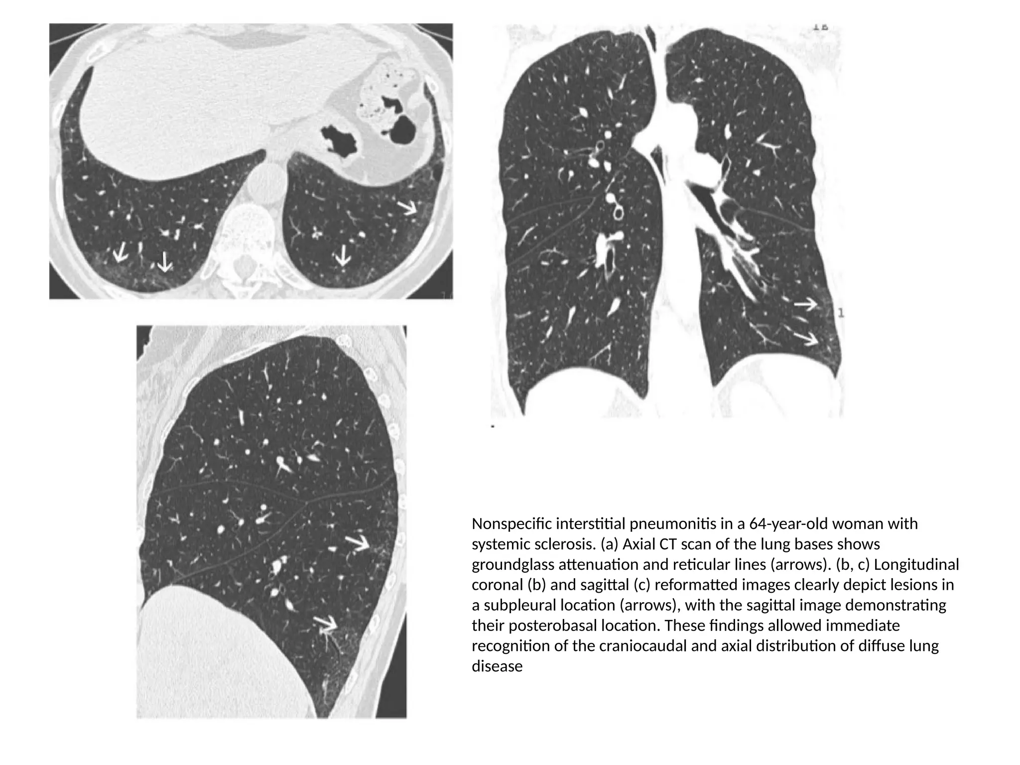
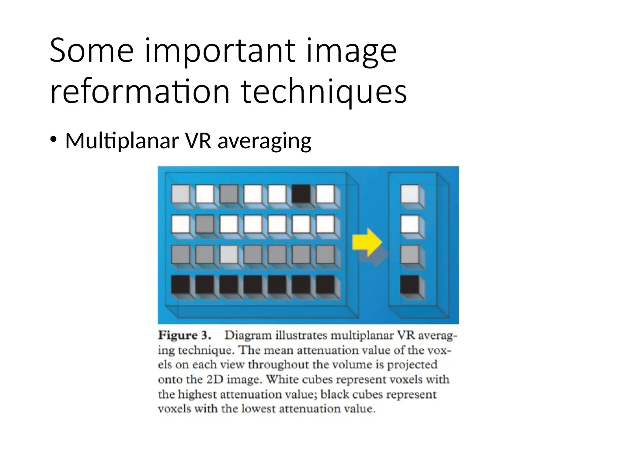
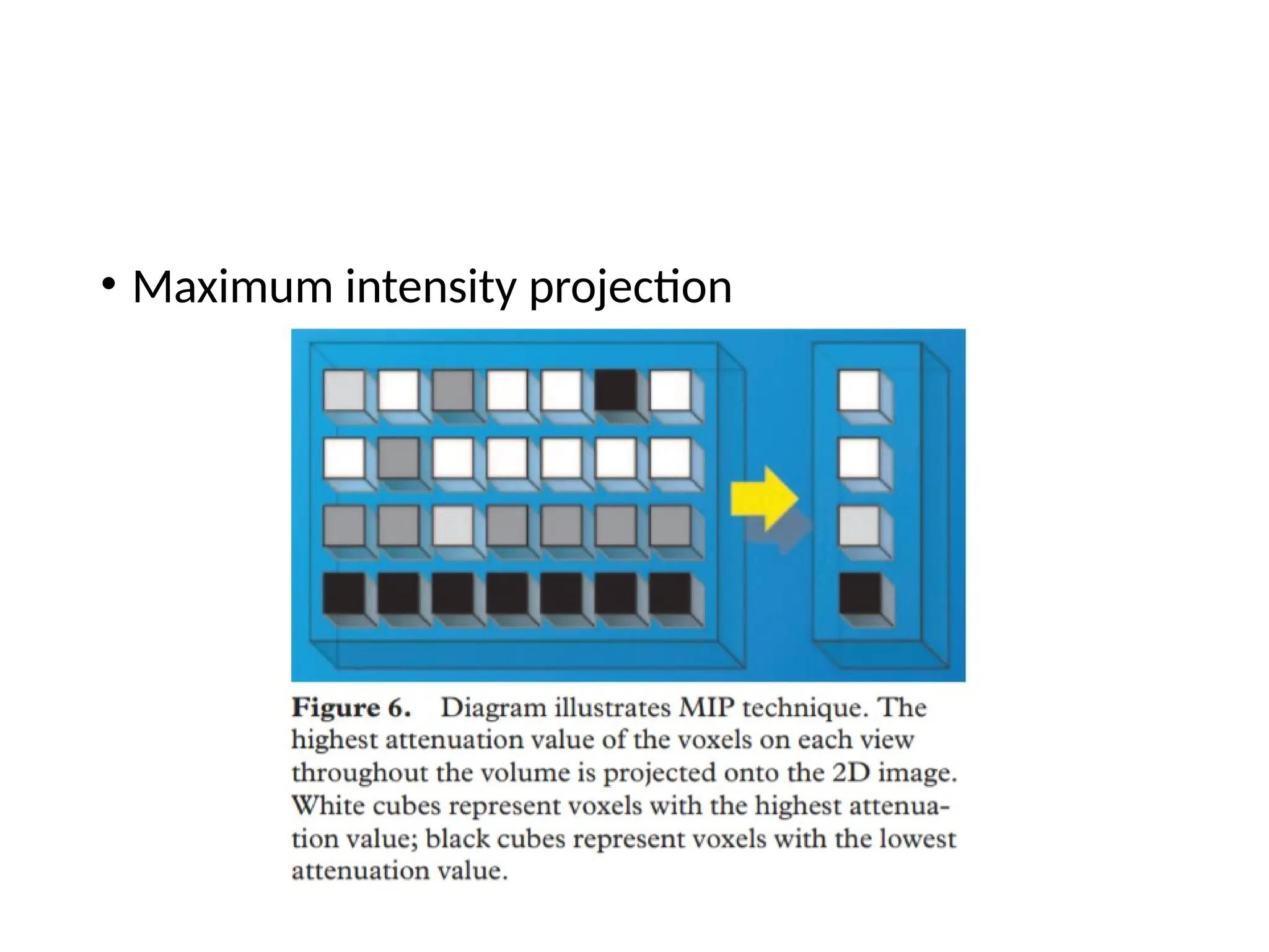
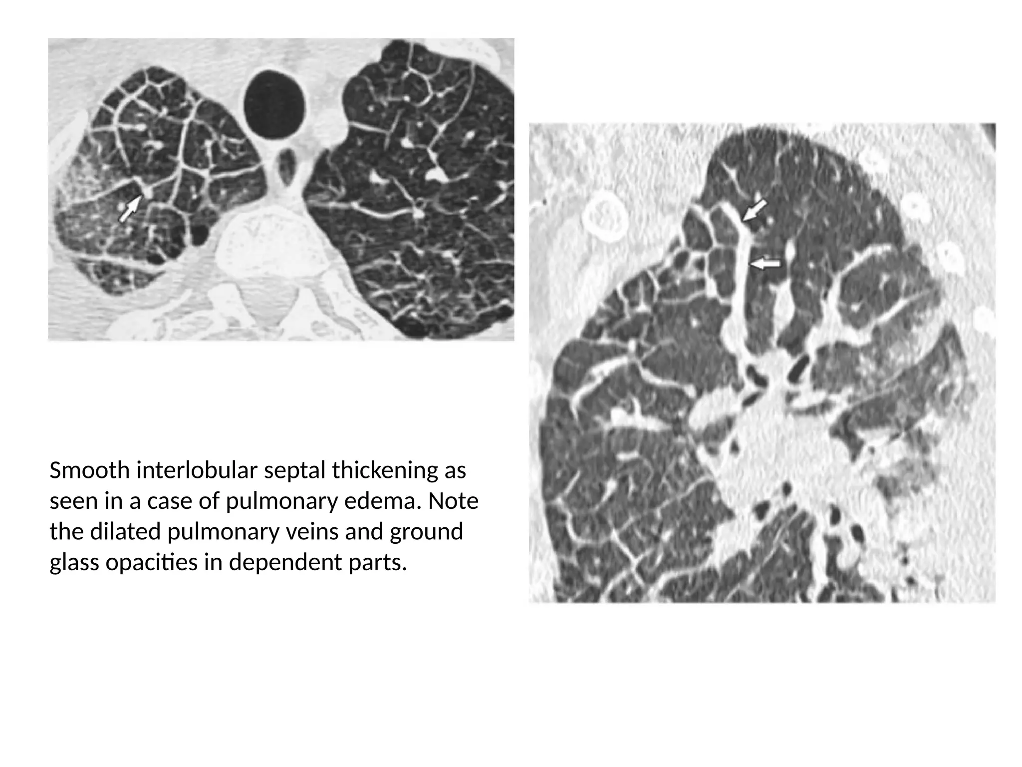
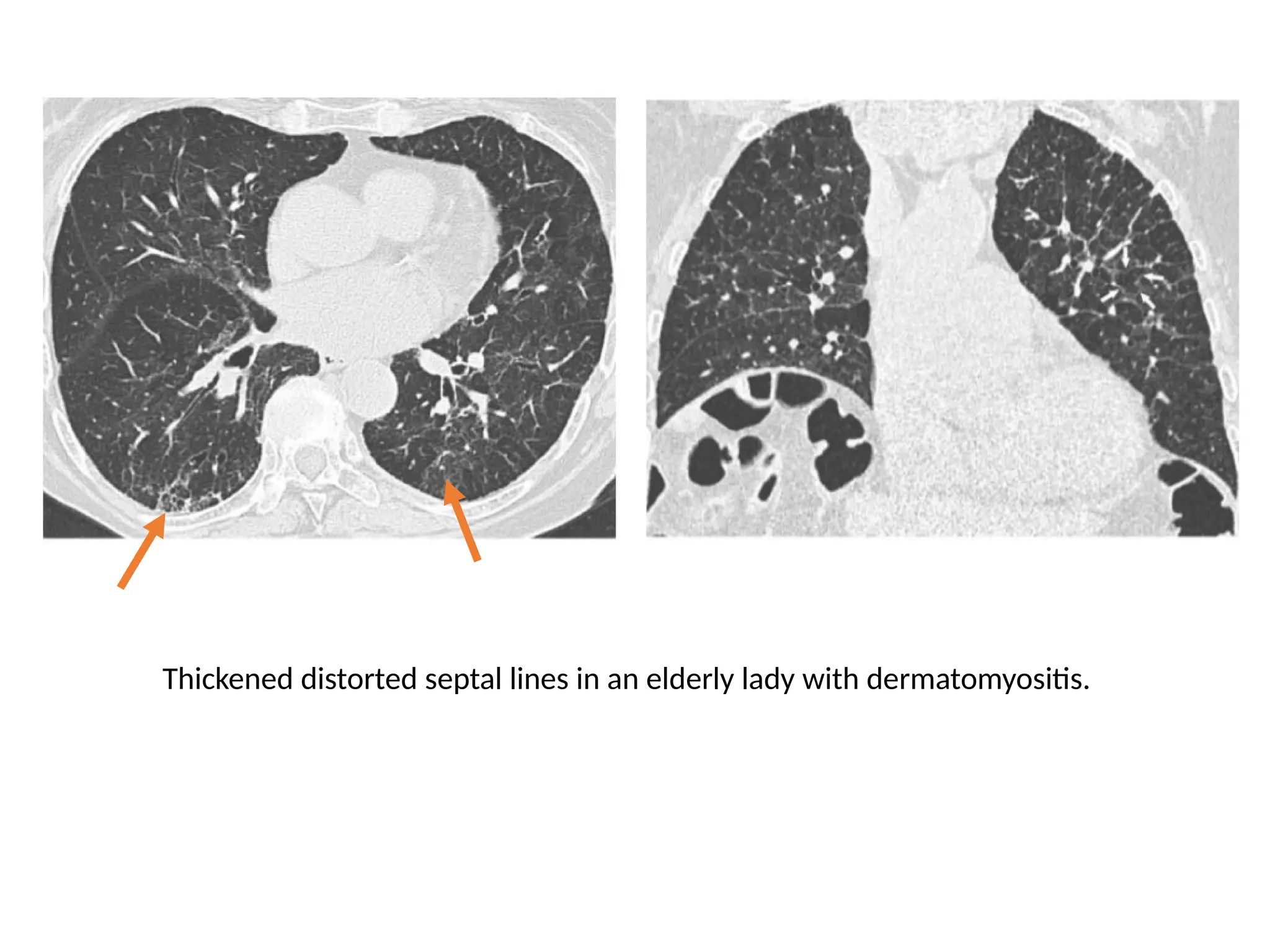
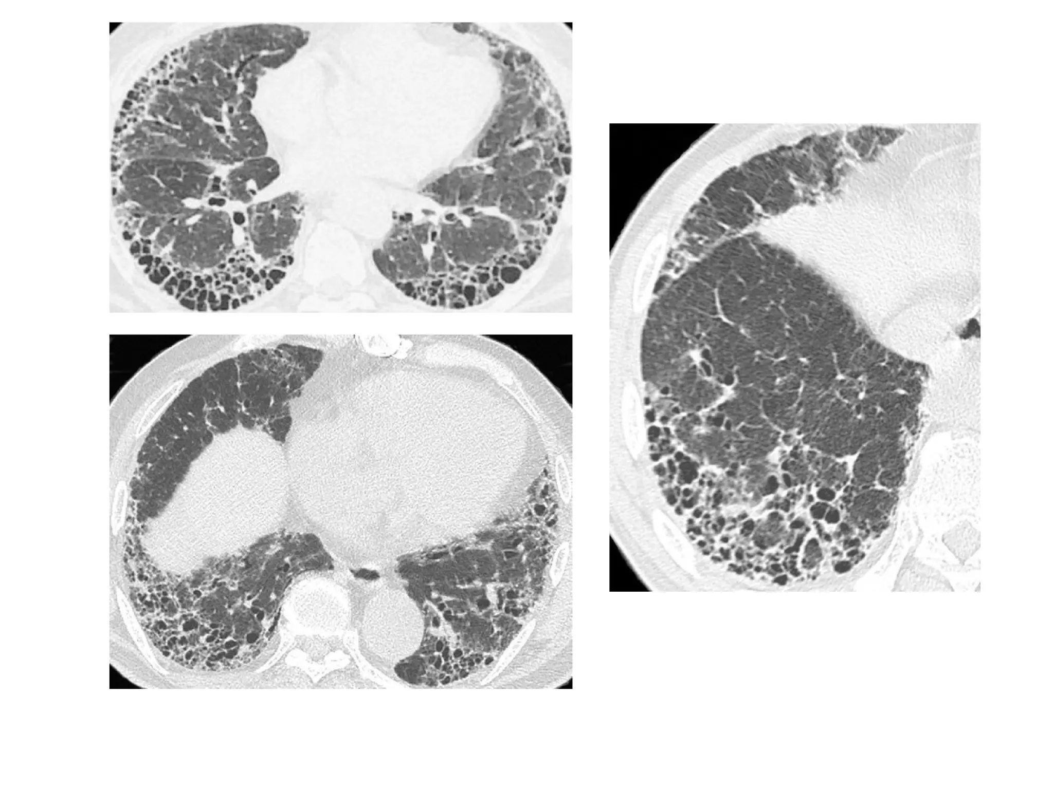
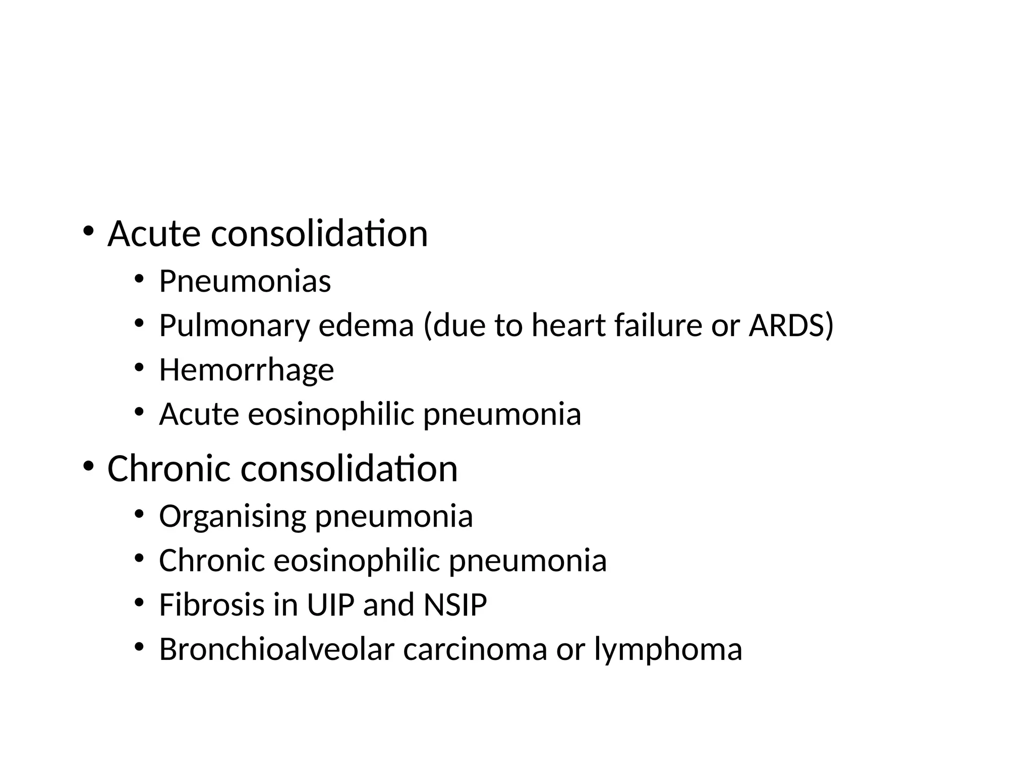
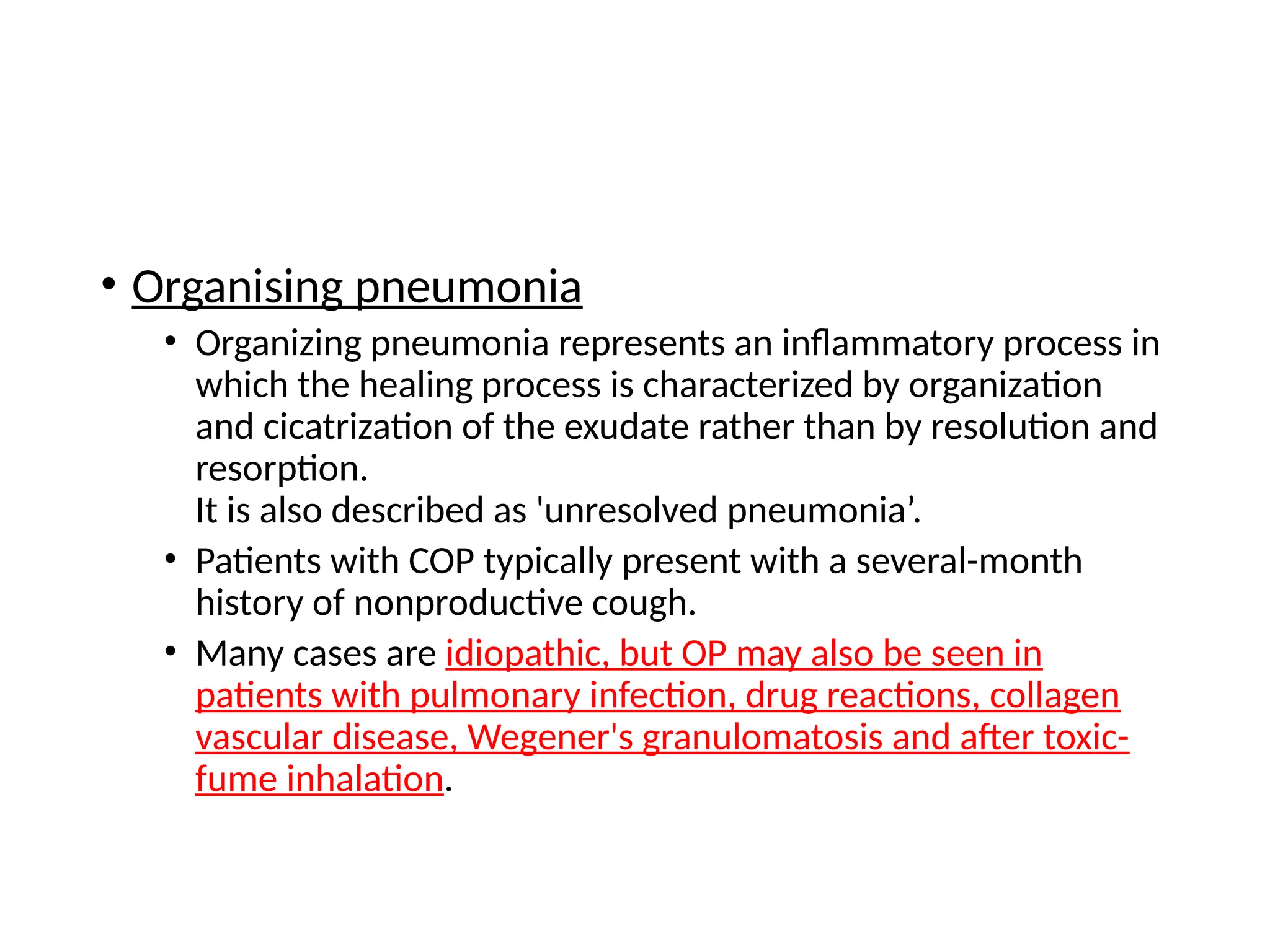
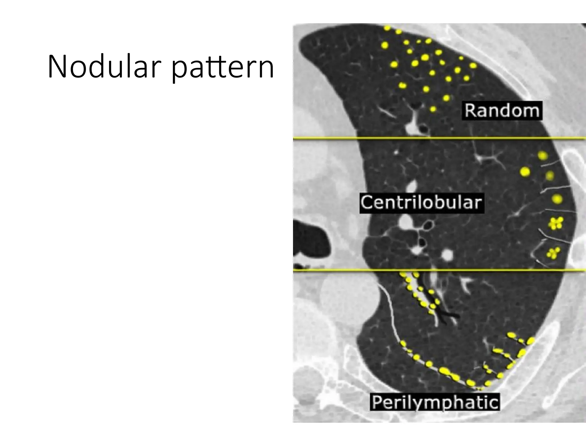
HRCT
Patterns in Lung Disease
Presented by Dr. Utkarsh Yadav
3rd
year Junior Resident
Department of Radiodiagnosis
S.N. Medical College, Agra
• HRCT with 1 mm thick sections obtained at 10 mm
intervals has widely been accepted as the imaging
standard for diffuse lung disease.
• However since only about 10% of lung parenchyma
is scanned, characteristic foci of disease may be
missed.
• Multidetector row CT generates isotropic
volumetric high resolution data and allows
contiguous visualisation of lung parenchyma.
Advantages of MDCT over
traditional HRCT
• MDCT allows
• Visualisation of three dimensions by providing an
isotropic volumetric data set.
• The ability to create 2D and 3D images of excellent
quality and significance
Langerhans cell histiocytosis in a 51-year-old man with chronic dyspnea. (a) Axial CT
scan demonstrates multiple irregular cysts in both upper lung lobes. (b) Coronal MPR
image clearly shows that the cysts are located predominantly in the upper and middle
lung zones. Note the normal size of the bronchi. MPR helped confirm the diagnosis in
less time than it took to review the more numerous axial images
Nonspecific interstitial pneumonitis in a 64-year-old woman with
systemic sclerosis. (a) Axial CT scan of the lung bases shows
groundglass attenuation and reticular lines (arrows). (b, c) Longitudinal
coronal (b) and sagittal (c) reformatted images clearly depict lesions in
a subpleural location (arrows), with the sagittal image demonstrating
their posterobasal location. These findings allowed immediate
recognition of the craniocaudal and axial distribution of diffuse lung
disease
Scan parameters
kVp 100 (≤ 80kg)
120 (80-113 kg)
140 (>113 kg)
mA 35
Tube current 130-200
Collimation Lowest possible on scanner (eg. 0.6)
Pitch 1.2
Rotation time 0.5
Slice thickness 1.2 – 1.5 mm
Matrix 512 x 512
Expected radiation dose 3 – 8 mSv
Bhalla, et al.: Recommendations for imaging protocols for CT chest
Some important image
reformation techniques
• Multiplanar VR averaging
• Minimum intensity projection (mIP)
• The subtle difference in density between the
endobronchial (pure) air and the lung parenchyma,
corresponding to a difference in attenuation of 50 –
150 HU, permits visualization of the bronchi below
the subsubsegmental level. mIP is the optimal tool
for the detection, localization, and quantification of
ground-glass and linear attenuation patterns.
• Maximum intensity projection
• The primary clinical application of MIP is to improve
the detection of pulmonary nodules and assess
their profusion.
• In addition, MIP sections of variable thickness are
excellent for assessing the size and location of
vessels, including the pulmonary arteries and veins.
Relevant HRCT lung
anatomy
Secondary lobule
• It is the basic anatomical unit of pulmonary
structure and function.
• It measures about 1-2 cm and is made up of 5-15
pulmonary acini.
• It is supplied by a terminal bronchiole at its center
paralleled by centrilobular artery.
• Pulmonary vein and lymphatics run in the periphery
of the lobule, within the interlobular septa.
Approach to HRCT of
lungs
• A structured approach to interpretation of HRCT involves the following
questions:
• What is the dominant HR-pattern:
• reticular
• nodular
• high attenuation (ground-glass, consolidation)
• low attenuation (emphysema, cystic)
• Where is it located within the secondary lobule HR-pattern:
• centrilobular
• perilymphatic
• random
• Is there an upper versus lower zone or a central versus peripheral
predominance
• Are there additional findings HR-pattern:
• pleural fluid
• lymphadenopathy
• traction bronchiectasis
Linear pattern
• It may be due to
• Thickening of the interstitial fiber network owing to fluid
• Fibrous tissue
• Interstitial infiltration by cells or other material
• Reticular patterns, which usually reflect intralobular
interstitial thickening, are most commonly seen in
patients with pulmonary fibrosis.
• Interlobular septal can be of two types based on
etiology
Smooth interlobular septal thickening as
seen in a case of pulmonary edema. Note
the dilated pulmonary veins and ground
glass opacities in dependent parts.
Irregular nodular interlobular septal
thickening due to lymphangitis
carcinomatosis
Thickened distorted septal lines in an elderly lady with dermatomyositis.
Honeycombing
• It represents the second type of reticular opacity
• Fleischner Society definition
• “On CT scans, the appearance is clustered cystic air spaces, typically
of comparable diameters on the order of 3–10 mm but occasionally
as large as 2.5 cm. Honeycombing is usually subpleural and is
characterized by well-defined walls. It is a CT feature of established
pulmonary fibrosis and is an important criterion in the diagnosis of
usual interstitial pneumonia.”
Is it honeycombing or not?
High attenuation pattern
• Increased lung attenuation is called ground-glass-opacity
(GGO) if there is a hazy increase in lung opacity without
obscuration of underlying vessels and is called consolidation
if the increase in lung opacity obscures the vessels.
• In both ground glass and consolidation the increase in lung
density is the result of replacement of air in the alveoli by
fluid, cells or fibrosis.
• Ground glass opacity – dark bronchus sign
• Consolidation – air bronchogram sign
Ground glass opacity
• Ground-glass opacity (GGO) represents:
• Filling of the alveolar spaces with pus, edema, hemorrhage,
inflammation or tumor cells.
• Thickening of the interstitium or alveolar walls below the spatial
resolution of the HRCT as seen in fibrosis.
• So ground-glass opacification may either be the result of air space
disease (filling of the alveoli) or interstitial lung disease (i.e. fibrosis).
• The location of the abnormalities in ground glass pattern can be
helpful:
• Upper zone predominance: Respiratory bronchiolitis, Pneumocystis
pneumonia.
• Lower zone predominance: UIP, NSIP, DIP.
• Centrilobular distribution: Hypersensitivity pneumonitis, Respiratory
bronchiolitis
consolidation and ground-glass opacity in a patient with persistent
chest abnormalities and weight loss without signs of infection.
• Ground-glass opacity is nonspecific, but a highly significant
finding since 60-80% of patients with ground-glass opacity
on HRCT have an active and potentially treatable lung
disease.
• In the other 20-40% of the cases the lung disease is not
treatable and the ground-glass pattern is the result of
fibrosis.
• In those cases there are usually associated HRCT findings of
fibrosis, such as traction bronchiectasis and honeycombing.
Mosaic attenuation
• Density differences between affected and non-affected lung
areas.
• When ground glass opacity presents as mosaic attenuation
consider:
• Infiltrative process adjacent to normal lung.
• Normal lung appearing relatively dense adjacent to lung with air-
trapping.
• Hyperperfused lung adjacent to hypoperfused lung due to chronic
thromboembolic disease.
How to differentiate???
• There are two diagnostic hints for further differentiation:
• Look at expiratory scans for air trapping
• Look at the vessels
• If the vessels are difficult to see in the 'black' lung as compared
to the 'white' lung, than it is likely that the 'black' lung is
abnormal.
Then there are two possibilities: obstructive bronchiolitis or
chronic pulmonary embolism.
Sometimes these can be differentiated with an expiratory scan.
If the vessels are the same in the 'black' lung and 'white' lung,
then you are looking at a patient with infiltrative lung disease,
like the one on the right with the pulmonary hemorrhage.
Inspiratory scan
Expiratory scan
Inspiratory scan
Expiratory scan
Inspiratory scan
Expiratory scan
Normal or pathologic ????
Small airway disease or chronic thromboembolism???
54-year-old man with chronic cough and poorly controlled
asthma.
41-year-old man with diffuse alveolar
hemorrhage
Crazy Paving
• The “crazy-paving” pattern at thin-section computed
tomography (CT) of the lungs is characterized by scattered
or diffuse ground-glass attenuation with superimposed
interlobular septal thickening and intralobular lines.
32-year-old man with acquired immunodeficiency
syndrome
Diffuse mucinous bronchioloalveolar
carcinoma in a 78-year-old man.
Replacement of normal epithelial alveolar cells
by neoplastic cells containing abundant mucin
Alveolar proteinosis in a 37-year-old woman
with a nonproductive cough and dyspnea.
Acute diffuse pulmonary hemorrhage in a
53-year-old woman with systemic lupus
erythematosus and massive hemoptysis.
Consolidation
• Consolidation is synonymous with airspace disease.
• What is replacing the air in the alveoli?
• Pus, edema, blood or tumor cells.
• Acute consolidation
• Pneumonias
• Pulmonary edema (due to heart failure or ARDS)
• Hemorrhage
• Acute eosinophilic pneumonia
• Chronic consolidation
• Organising pneumonia
• Chronic eosinophilic pneumonia
• Fibrosis in UIP and NSIP
• Bronchioalveolar carcinoma or lymphoma
• Organising pneumonia
• Organizing pneumonia represents an inflammatory process in
which the healing process is characterized by organization
and cicatrization of the exudate rather than by resolution and
resorption.
It is also described as 'unresolved pneumonia’.
• Patients with COP typically present with a several-month
history of nonproductive cough.
• Many cases are idiopathic, but OP may also be seen in
patients with pulmonary infection, drug reactions, collagen
vascular disease, Wegener's granulomatosis and after toxic-
fume inhalation.
Initial scan
After 10 years follow-up scan
• Chronic eosinophilic pneumonia
• Chronic eosinophilic pneumonia is an idiopathic
condition characterized by filling of the alveoli with
eosinophils.
• Often affects patients with a history of atopy, asthma
and allergic rhinitis.
• Symptoms usually present for >1 month and include cough,
high fever, night sweats, dyspnea, malaise, and marked
weight loss
• Blood eosinophilia present in 90% of patients and sputum
eosinophilia in 50%.
• Imaging Findings
• Ground glass and consolidation in a peripheral and
upper lobe distribution (photographic negative of
central pulmonary edema)
• Not usually migratory (differentiates from organising
pneumonia)
• Responds dramatically to steroid treatment.
• After treatment residual fibrosis seen in some patients
Low attenuation pattern
• It is comprised of abnormalities that result in
decreased lung attenuation.
• Emphysema
• Lung cysts
• Bronchiectasis
• Honeycombing
Emphysema
• It is characterised by permanently enlarged airspaces
distal to terminal bronchioles with destruction of alveolar
walls
• “without visible walls”
• 4 types
• Centrilobular - proximal respiratory bronchioles dilated, leaving
normal distal alveolar ducts and sacs
• Para-septal - enlarged airspace at the periphery of acini
• Panlobular - uniform dilatation of the air space from the
respiratory bronchioles to the alveoli
• Para-cicatricial – air space enlargement adjacent to lung
scarring
Centrilobular
emphysema
Asso. with cigarette smoking
Panlobular
emphysema
Asso. with alpha 1
antitrypsin deficiency
Basal predominance
Para-septal
emphysema
Paracicatricial emphysema from progressive massive fibrosis caused by silicosis
Bullous emphysematous lung
disease
• Also called “Vanishing lung syndrome”
• Characterised by giant emphysematous bullae
commonly developing in upper lobe and occupy at
least one-third of a hemithorax.
• Idiopathic condition
• Associated with
• Marfan syndrome
• Ehlers-Danlos syndrome
• Alpha 1 antitrypsin deficiency
Giant bullae or pneumothorax ?
• The double wall sign
• This sign occurs when one sees air outlining both sides of
the bulla wall parallel to the chest wall
• Absence of this sign provides further evidence and
increased confidence against the diagnosis of
pneumothorax, which can prevent unnecessary chest tube
placement.
???
• One potential pitfall in the appreciation of the double-wall
sign of pneumothorax occurs when two large bullae are
adjacent to one another.
• This situation can produce an apparent double-wall sign,
mimicking pneumothorax.
• However, careful scrutiny of multiple images will show the
absence of air in the pleural space and that the bulla wall is
not parallel to the chest wall or parietal pleura
Congenital lobar emphysema
• Characterised by progressive lobar overexpansion, usually
with compression of the remaining (ipsilateral) lung.
• Underlying cause can be secondary to an intrinsic
cartilaginous abnormality with resultant weak or absent
bronchial cartilage or extrinsic compression of an airway
(eg, by a large pulmonary artery or a bronchogenic cyst).
• Although the alveoli expand, the alveolar walls remain
intact; therefore, the term emphysema is technically
inaccurate
Cystic lung disease
• Pulmonary cyst (Fleischner Society definition)
“A cyst appears as a round parenchymal lucency or low
attenuating area with a well defined interface with normal
lung”
• Most commonly associated with following;
• Langerhans cell histiocytosis (LCH)
• Lymphangioleiomyomatosis (LAM)
• Lymphocytic interstitial pneumonia (LIP)
• Birt Hogge Dube syndrome (BHD)
• Pneumatoceles
• Honeycombing
Langerhans cell histiocytosis
• Smokers, Females = Males
• Nodules and cysts
• Cysts have wall of variable thickness
• “bizarre shaped cysts”
• Mid to upper lung predominance
• Spares CP angles
• Ill defined nodules → Ill defined nodules + bizarre
shaped cysts → Bizarre shaped cysts
• May be associated with other smoking related
diseases – RB-ILD, Emphysema.
High-resolution CT image shows thin-walled cysts. Cysts may
be round but are frequently irregular, bilobed (arrow),
cloverleaf-shaped, or bizarre shapes. Note striking upper lobe
predominance.
High-resolution CT image shows sparing of lung bases.
High-resolution CT images show thin-walled cysts
and nodules. Again, note striking upper lobe
predominance and, in D, sparing of costophrenic
angles, one feature that can be used to distinguish
LCH from lymphangioleiomyomatosis.
Lymphangioleiomyomatosis
• Females >> Males, women of child bearing age
• Smooth rounded cysts with thin walls
• Diffusely distributed cysts.
• Normal intervening lung.
• Involves CP angles.
• Associated findings
• Chlyous effusions
• Pneumothorax
• Renal angiomyolipomas
High-resolution CT images show
characteristic appearance of
lymphangioleiomyomatosis: diffuse
thin-walled cysts surrounded by
normal lung. Cysts are usually 2–5
mm but can be as large as 25–30
mm. Cysts are typically round or
ovoid but may become polygonal
(arrow, A) when parenchymal
involvement is severe
Coronal reformatted
image shows diffuse
involvement with lack
of regional sparing and
involvement of
costophrenic sulci.
41 Y lady with
shortness of breath
Lymphocytic Interstitial
Pneumonia
• Females >> Males, 5th
decade of life
• Thin walled cysts in lower lobe predominance.
• The cysts are perivascular in location.
• Associated with:
• Autoimmune diseases, AIDS
• GGO, septal and bronchovascular bundle thickening
40 Y lady with lupus
52 Y lady with Sjogren’s syndrome
38 Y lady with RA
Birt Hogge Dube Syndrome
• Associated with folliculin gene (FLCN-S)
• Autosomal dominant multiorgan disorder
• Facial papules – fibrofolliculomas
• Renal tumours – often multifocal chromophobe RCCs
and oncocytomas.
• Thin walled pulmonary cysts
• LL predominate, paramediastinal, subpleural
• Oval or septated
• Cyst rupture → pneumothorax (33 – 38 % pts)
• Present in 3rd
to 4th
decade of life
Bronchiectasis
• Irreversible localised or diffuse bronchial dilatation
usually resulting from chronic infection, proximal
airway obstruction or congenital bronchial
abnormality.
• Detected on HRCT by
• bronchial dilatation (signet-ring sign)
• bronchial wall thickening
• lack of normal tapering with visibility of airways in the
peripheral lung
• mucus retention in the broncial lumen
• associated atelectasis and sometimes air trapping
• 3 types
• Cylindrical
• Varicoid
• Cystic
Normal lung Cylindrical bronchiectasis
Cystic bronchiectasis
Varicoid bronchiectasis
Cystic fibrosis. Transverse and
coronal images show upper lobe
predominance of cystic
bronchiectasis (arrows) and volume
loss, enlarged lung volumes
Sarcoidosis. A and B,
Transverse images show
fibrosis and traction
bronchiectasis (arrows,
B) that predominantly
involve upper lobes.
Mycobacterium avium-intracellulare infection.
Bronchiectasis (arrows) predominantly involves right
middle lobe and lingula.
Usual interstitial pneumonia. Bibasilar and subpleural
reticulation and traction bronchiectasis are seen in areas of
fibrosis (arrows).
Allergic bronchopulmonary aspergillosis. A,
Chest radiograph shows central
bronchiectasis and mucoid impaction, so-
called finger-in-glove appearance (arrows).
B, Transverse CT image shows central
bronchiectasis, mucoid impaction (large
arrow), and distal bronchiolitis (small arrow).
Nodular pattern
Perilymphatic distribution
• Nodules are seen in relation to pleural surfaces,
interlobular septa and the peribronchovascular
interstitium.
• Nodules are almost always visible in a subpleural
location, particularly in relation to the fissures.
• Causes:
• Sarcoidosis
• Lymphangitic spread of carcinoma
• Silicosis
• Coal workers pneumoconiosis
Centrilobular distribution
• Nodules spare the pleural surfaces.
• Seen in diseases which enter the lung through the
airways.
• Causes:
• Hypersensitivity pneumonitis
• Respiratory bronchiolitis in smokers
• Infectious airways diseases (endobronchial spread of TB
or NTB, bronchopneumonia)
• In many cases nodules may be of ground glass
density (acinar nodules)
Subacute hypersensitivity pneumonitis in a bird fancier
Tree in bud nodules
• It describes the appearance of an irregular and
often nodular branching structure, most easily
identified in the lung periphery.
• It represents the dilated and impacted (mucus or
pus filled) centrilobular bronchioles.
Typical Tree-in-bud appearance in a patient with active TB.
Random distribution
• The random distribution is a result of
hematogenous spread of infection.
• Causes:
• Hematogenous metastases
• Miliary TB
• Miliary fungal infections
• Sarcoidosis
• Langerhans cell histiocytosis (early nodular stage)
Random distribution of nodules in miliary tuberculosis
Langerhans cell histiocytosis: early nodular stage before the
typical cysts appear.
Summary
THANK YOU

HRCT patterns in interstitial lung disease

  • 1.
    HRCT Patterns in LungDisease Presented by Dr. Utkarsh Yadav 3rd year Junior Resident Department of Radiodiagnosis S.N. Medical College, Agra
  • 2.
    • HRCT with1 mm thick sections obtained at 10 mm intervals has widely been accepted as the imaging standard for diffuse lung disease. • However since only about 10% of lung parenchyma is scanned, characteristic foci of disease may be missed. • Multidetector row CT generates isotropic volumetric high resolution data and allows contiguous visualisation of lung parenchyma.
  • 3.
    Advantages of MDCTover traditional HRCT • MDCT allows • Visualisation of three dimensions by providing an isotropic volumetric data set. • The ability to create 2D and 3D images of excellent quality and significance
  • 4.
    Langerhans cell histiocytosisin a 51-year-old man with chronic dyspnea. (a) Axial CT scan demonstrates multiple irregular cysts in both upper lung lobes. (b) Coronal MPR image clearly shows that the cysts are located predominantly in the upper and middle lung zones. Note the normal size of the bronchi. MPR helped confirm the diagnosis in less time than it took to review the more numerous axial images
  • 5.
    Nonspecific interstitial pneumonitisin a 64-year-old woman with systemic sclerosis. (a) Axial CT scan of the lung bases shows groundglass attenuation and reticular lines (arrows). (b, c) Longitudinal coronal (b) and sagittal (c) reformatted images clearly depict lesions in a subpleural location (arrows), with the sagittal image demonstrating their posterobasal location. These findings allowed immediate recognition of the craniocaudal and axial distribution of diffuse lung disease
  • 6.
    Scan parameters kVp 100(≤ 80kg) 120 (80-113 kg) 140 (>113 kg) mA 35 Tube current 130-200 Collimation Lowest possible on scanner (eg. 0.6) Pitch 1.2 Rotation time 0.5 Slice thickness 1.2 – 1.5 mm Matrix 512 x 512 Expected radiation dose 3 – 8 mSv Bhalla, et al.: Recommendations for imaging protocols for CT chest
  • 7.
    Some important image reformationtechniques • Multiplanar VR averaging
  • 8.
    • Minimum intensityprojection (mIP)
  • 9.
    • The subtledifference in density between the endobronchial (pure) air and the lung parenchyma, corresponding to a difference in attenuation of 50 – 150 HU, permits visualization of the bronchi below the subsubsegmental level. mIP is the optimal tool for the detection, localization, and quantification of ground-glass and linear attenuation patterns.
  • 11.
  • 12.
    • The primaryclinical application of MIP is to improve the detection of pulmonary nodules and assess their profusion. • In addition, MIP sections of variable thickness are excellent for assessing the size and location of vessels, including the pulmonary arteries and veins.
  • 14.
  • 15.
    Secondary lobule • Itis the basic anatomical unit of pulmonary structure and function. • It measures about 1-2 cm and is made up of 5-15 pulmonary acini. • It is supplied by a terminal bronchiole at its center paralleled by centrilobular artery. • Pulmonary vein and lymphatics run in the periphery of the lobule, within the interlobular septa.
  • 18.
  • 19.
    • A structuredapproach to interpretation of HRCT involves the following questions: • What is the dominant HR-pattern: • reticular • nodular • high attenuation (ground-glass, consolidation) • low attenuation (emphysema, cystic) • Where is it located within the secondary lobule HR-pattern: • centrilobular • perilymphatic • random • Is there an upper versus lower zone or a central versus peripheral predominance • Are there additional findings HR-pattern: • pleural fluid • lymphadenopathy • traction bronchiectasis
  • 20.
    Linear pattern • Itmay be due to • Thickening of the interstitial fiber network owing to fluid • Fibrous tissue • Interstitial infiltration by cells or other material • Reticular patterns, which usually reflect intralobular interstitial thickening, are most commonly seen in patients with pulmonary fibrosis.
  • 21.
    • Interlobular septalcan be of two types based on etiology
  • 22.
    Smooth interlobular septalthickening as seen in a case of pulmonary edema. Note the dilated pulmonary veins and ground glass opacities in dependent parts.
  • 23.
    Irregular nodular interlobularseptal thickening due to lymphangitis carcinomatosis
  • 24.
    Thickened distorted septallines in an elderly lady with dermatomyositis.
  • 25.
    Honeycombing • It representsthe second type of reticular opacity • Fleischner Society definition • “On CT scans, the appearance is clustered cystic air spaces, typically of comparable diameters on the order of 3–10 mm but occasionally as large as 2.5 cm. Honeycombing is usually subpleural and is characterized by well-defined walls. It is a CT feature of established pulmonary fibrosis and is an important criterion in the diagnosis of usual interstitial pneumonia.”
  • 27.
  • 30.
    High attenuation pattern •Increased lung attenuation is called ground-glass-opacity (GGO) if there is a hazy increase in lung opacity without obscuration of underlying vessels and is called consolidation if the increase in lung opacity obscures the vessels. • In both ground glass and consolidation the increase in lung density is the result of replacement of air in the alveoli by fluid, cells or fibrosis. • Ground glass opacity – dark bronchus sign • Consolidation – air bronchogram sign
  • 31.
    Ground glass opacity •Ground-glass opacity (GGO) represents: • Filling of the alveolar spaces with pus, edema, hemorrhage, inflammation or tumor cells. • Thickening of the interstitium or alveolar walls below the spatial resolution of the HRCT as seen in fibrosis. • So ground-glass opacification may either be the result of air space disease (filling of the alveoli) or interstitial lung disease (i.e. fibrosis). • The location of the abnormalities in ground glass pattern can be helpful: • Upper zone predominance: Respiratory bronchiolitis, Pneumocystis pneumonia. • Lower zone predominance: UIP, NSIP, DIP. • Centrilobular distribution: Hypersensitivity pneumonitis, Respiratory bronchiolitis
  • 33.
    consolidation and ground-glassopacity in a patient with persistent chest abnormalities and weight loss without signs of infection.
  • 34.
    • Ground-glass opacityis nonspecific, but a highly significant finding since 60-80% of patients with ground-glass opacity on HRCT have an active and potentially treatable lung disease. • In the other 20-40% of the cases the lung disease is not treatable and the ground-glass pattern is the result of fibrosis. • In those cases there are usually associated HRCT findings of fibrosis, such as traction bronchiectasis and honeycombing.
  • 36.
    Mosaic attenuation • Densitydifferences between affected and non-affected lung areas. • When ground glass opacity presents as mosaic attenuation consider: • Infiltrative process adjacent to normal lung. • Normal lung appearing relatively dense adjacent to lung with air- trapping. • Hyperperfused lung adjacent to hypoperfused lung due to chronic thromboembolic disease.
  • 37.
    How to differentiate??? •There are two diagnostic hints for further differentiation: • Look at expiratory scans for air trapping • Look at the vessels • If the vessels are difficult to see in the 'black' lung as compared to the 'white' lung, than it is likely that the 'black' lung is abnormal. Then there are two possibilities: obstructive bronchiolitis or chronic pulmonary embolism. Sometimes these can be differentiated with an expiratory scan. If the vessels are the same in the 'black' lung and 'white' lung, then you are looking at a patient with infiltrative lung disease, like the one on the right with the pulmonary hemorrhage.
  • 38.
  • 39.
  • 40.
  • 41.
    Small airway diseaseor chronic thromboembolism???
  • 42.
    54-year-old man withchronic cough and poorly controlled asthma.
  • 43.
    41-year-old man withdiffuse alveolar hemorrhage
  • 44.
    Crazy Paving • The“crazy-paving” pattern at thin-section computed tomography (CT) of the lungs is characterized by scattered or diffuse ground-glass attenuation with superimposed interlobular septal thickening and intralobular lines.
  • 45.
    32-year-old man withacquired immunodeficiency syndrome
  • 46.
    Diffuse mucinous bronchioloalveolar carcinomain a 78-year-old man. Replacement of normal epithelial alveolar cells by neoplastic cells containing abundant mucin
  • 47.
    Alveolar proteinosis ina 37-year-old woman with a nonproductive cough and dyspnea.
  • 48.
    Acute diffuse pulmonaryhemorrhage in a 53-year-old woman with systemic lupus erythematosus and massive hemoptysis.
  • 49.
    Consolidation • Consolidation issynonymous with airspace disease. • What is replacing the air in the alveoli? • Pus, edema, blood or tumor cells.
  • 50.
    • Acute consolidation •Pneumonias • Pulmonary edema (due to heart failure or ARDS) • Hemorrhage • Acute eosinophilic pneumonia • Chronic consolidation • Organising pneumonia • Chronic eosinophilic pneumonia • Fibrosis in UIP and NSIP • Bronchioalveolar carcinoma or lymphoma
  • 51.
    • Organising pneumonia •Organizing pneumonia represents an inflammatory process in which the healing process is characterized by organization and cicatrization of the exudate rather than by resolution and resorption. It is also described as 'unresolved pneumonia’. • Patients with COP typically present with a several-month history of nonproductive cough. • Many cases are idiopathic, but OP may also be seen in patients with pulmonary infection, drug reactions, collagen vascular disease, Wegener's granulomatosis and after toxic- fume inhalation.
  • 55.
    Initial scan After 10years follow-up scan
  • 56.
    • Chronic eosinophilicpneumonia • Chronic eosinophilic pneumonia is an idiopathic condition characterized by filling of the alveoli with eosinophils. • Often affects patients with a history of atopy, asthma and allergic rhinitis. • Symptoms usually present for >1 month and include cough, high fever, night sweats, dyspnea, malaise, and marked weight loss • Blood eosinophilia present in 90% of patients and sputum eosinophilia in 50%.
  • 57.
    • Imaging Findings •Ground glass and consolidation in a peripheral and upper lobe distribution (photographic negative of central pulmonary edema) • Not usually migratory (differentiates from organising pneumonia) • Responds dramatically to steroid treatment. • After treatment residual fibrosis seen in some patients
  • 60.
  • 61.
    • It iscomprised of abnormalities that result in decreased lung attenuation. • Emphysema • Lung cysts • Bronchiectasis • Honeycombing
  • 62.
    Emphysema • It ischaracterised by permanently enlarged airspaces distal to terminal bronchioles with destruction of alveolar walls • “without visible walls” • 4 types • Centrilobular - proximal respiratory bronchioles dilated, leaving normal distal alveolar ducts and sacs • Para-septal - enlarged airspace at the periphery of acini • Panlobular - uniform dilatation of the air space from the respiratory bronchioles to the alveoli • Para-cicatricial – air space enlargement adjacent to lung scarring
  • 63.
  • 64.
    Panlobular emphysema Asso. with alpha1 antitrypsin deficiency Basal predominance
  • 65.
  • 66.
    Paracicatricial emphysema fromprogressive massive fibrosis caused by silicosis
  • 67.
    Bullous emphysematous lung disease •Also called “Vanishing lung syndrome” • Characterised by giant emphysematous bullae commonly developing in upper lobe and occupy at least one-third of a hemithorax. • Idiopathic condition • Associated with • Marfan syndrome • Ehlers-Danlos syndrome • Alpha 1 antitrypsin deficiency
  • 69.
    Giant bullae orpneumothorax ?
  • 70.
    • The doublewall sign • This sign occurs when one sees air outlining both sides of the bulla wall parallel to the chest wall • Absence of this sign provides further evidence and increased confidence against the diagnosis of pneumothorax, which can prevent unnecessary chest tube placement.
  • 72.
  • 73.
    • One potentialpitfall in the appreciation of the double-wall sign of pneumothorax occurs when two large bullae are adjacent to one another. • This situation can produce an apparent double-wall sign, mimicking pneumothorax. • However, careful scrutiny of multiple images will show the absence of air in the pleural space and that the bulla wall is not parallel to the chest wall or parietal pleura
  • 75.
    Congenital lobar emphysema •Characterised by progressive lobar overexpansion, usually with compression of the remaining (ipsilateral) lung. • Underlying cause can be secondary to an intrinsic cartilaginous abnormality with resultant weak or absent bronchial cartilage or extrinsic compression of an airway (eg, by a large pulmonary artery or a bronchogenic cyst). • Although the alveoli expand, the alveolar walls remain intact; therefore, the term emphysema is technically inaccurate
  • 77.
    Cystic lung disease •Pulmonary cyst (Fleischner Society definition) “A cyst appears as a round parenchymal lucency or low attenuating area with a well defined interface with normal lung” • Most commonly associated with following; • Langerhans cell histiocytosis (LCH) • Lymphangioleiomyomatosis (LAM) • Lymphocytic interstitial pneumonia (LIP) • Birt Hogge Dube syndrome (BHD) • Pneumatoceles • Honeycombing
  • 78.
    Langerhans cell histiocytosis •Smokers, Females = Males • Nodules and cysts • Cysts have wall of variable thickness • “bizarre shaped cysts” • Mid to upper lung predominance • Spares CP angles • Ill defined nodules → Ill defined nodules + bizarre shaped cysts → Bizarre shaped cysts • May be associated with other smoking related diseases – RB-ILD, Emphysema.
  • 79.
    High-resolution CT imageshows thin-walled cysts. Cysts may be round but are frequently irregular, bilobed (arrow), cloverleaf-shaped, or bizarre shapes. Note striking upper lobe predominance.
  • 80.
    High-resolution CT imageshows sparing of lung bases.
  • 81.
    High-resolution CT imagesshow thin-walled cysts and nodules. Again, note striking upper lobe predominance and, in D, sparing of costophrenic angles, one feature that can be used to distinguish LCH from lymphangioleiomyomatosis.
  • 84.
    Lymphangioleiomyomatosis • Females >>Males, women of child bearing age • Smooth rounded cysts with thin walls • Diffusely distributed cysts. • Normal intervening lung. • Involves CP angles. • Associated findings • Chlyous effusions • Pneumothorax • Renal angiomyolipomas
  • 85.
    High-resolution CT imagesshow characteristic appearance of lymphangioleiomyomatosis: diffuse thin-walled cysts surrounded by normal lung. Cysts are usually 2–5 mm but can be as large as 25–30 mm. Cysts are typically round or ovoid but may become polygonal (arrow, A) when parenchymal involvement is severe
  • 86.
    Coronal reformatted image showsdiffuse involvement with lack of regional sparing and involvement of costophrenic sulci.
  • 87.
    41 Y ladywith shortness of breath
  • 88.
    Lymphocytic Interstitial Pneumonia • Females>> Males, 5th decade of life • Thin walled cysts in lower lobe predominance. • The cysts are perivascular in location. • Associated with: • Autoimmune diseases, AIDS • GGO, septal and bronchovascular bundle thickening
  • 89.
    40 Y ladywith lupus
  • 90.
    52 Y ladywith Sjogren’s syndrome
  • 91.
    38 Y ladywith RA
  • 92.
    Birt Hogge DubeSyndrome • Associated with folliculin gene (FLCN-S) • Autosomal dominant multiorgan disorder • Facial papules – fibrofolliculomas • Renal tumours – often multifocal chromophobe RCCs and oncocytomas. • Thin walled pulmonary cysts • LL predominate, paramediastinal, subpleural • Oval or septated • Cyst rupture → pneumothorax (33 – 38 % pts) • Present in 3rd to 4th decade of life
  • 95.
    Bronchiectasis • Irreversible localisedor diffuse bronchial dilatation usually resulting from chronic infection, proximal airway obstruction or congenital bronchial abnormality. • Detected on HRCT by • bronchial dilatation (signet-ring sign) • bronchial wall thickening • lack of normal tapering with visibility of airways in the peripheral lung • mucus retention in the broncial lumen • associated atelectasis and sometimes air trapping
  • 96.
    • 3 types •Cylindrical • Varicoid • Cystic
  • 97.
  • 98.
  • 100.
    Cystic fibrosis. Transverseand coronal images show upper lobe predominance of cystic bronchiectasis (arrows) and volume loss, enlarged lung volumes
  • 101.
    Sarcoidosis. A andB, Transverse images show fibrosis and traction bronchiectasis (arrows, B) that predominantly involve upper lobes.
  • 102.
    Mycobacterium avium-intracellulare infection. Bronchiectasis(arrows) predominantly involves right middle lobe and lingula.
  • 103.
    Usual interstitial pneumonia.Bibasilar and subpleural reticulation and traction bronchiectasis are seen in areas of fibrosis (arrows).
  • 104.
    Allergic bronchopulmonary aspergillosis.A, Chest radiograph shows central bronchiectasis and mucoid impaction, so- called finger-in-glove appearance (arrows). B, Transverse CT image shows central bronchiectasis, mucoid impaction (large arrow), and distal bronchiolitis (small arrow).
  • 105.
  • 107.
    Perilymphatic distribution • Nodulesare seen in relation to pleural surfaces, interlobular septa and the peribronchovascular interstitium. • Nodules are almost always visible in a subpleural location, particularly in relation to the fissures. • Causes: • Sarcoidosis • Lymphangitic spread of carcinoma • Silicosis • Coal workers pneumoconiosis
  • 110.
    Centrilobular distribution • Nodulesspare the pleural surfaces. • Seen in diseases which enter the lung through the airways. • Causes: • Hypersensitivity pneumonitis • Respiratory bronchiolitis in smokers • Infectious airways diseases (endobronchial spread of TB or NTB, bronchopneumonia) • In many cases nodules may be of ground glass density (acinar nodules)
  • 111.
  • 112.
    Tree in budnodules • It describes the appearance of an irregular and often nodular branching structure, most easily identified in the lung periphery. • It represents the dilated and impacted (mucus or pus filled) centrilobular bronchioles.
  • 114.
    Typical Tree-in-bud appearancein a patient with active TB.
  • 115.
    Random distribution • Therandom distribution is a result of hematogenous spread of infection. • Causes: • Hematogenous metastases • Miliary TB • Miliary fungal infections • Sarcoidosis • Langerhans cell histiocytosis (early nodular stage)
  • 116.
    Random distribution ofnodules in miliary tuberculosis
  • 117.
    Langerhans cell histiocytosis:early nodular stage before the typical cysts appear.
  • 118.
  • 122.

Editor's Notes

  • #38 Obliterative bronchiolitis
  • #39 Organising pneumonia