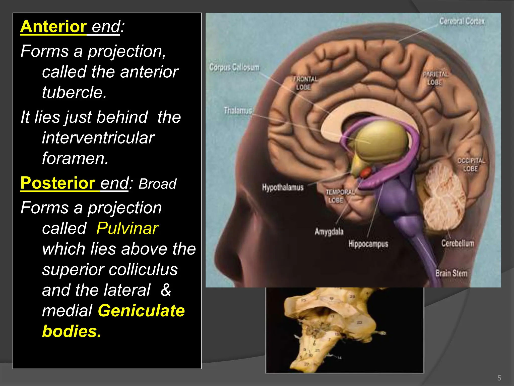
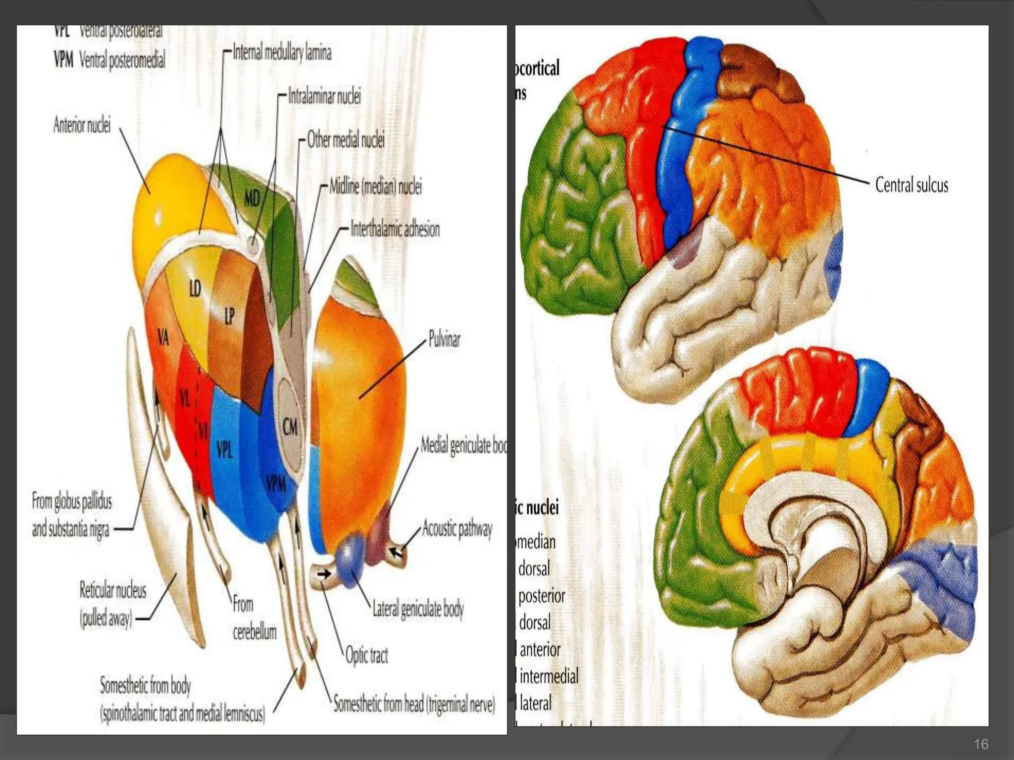
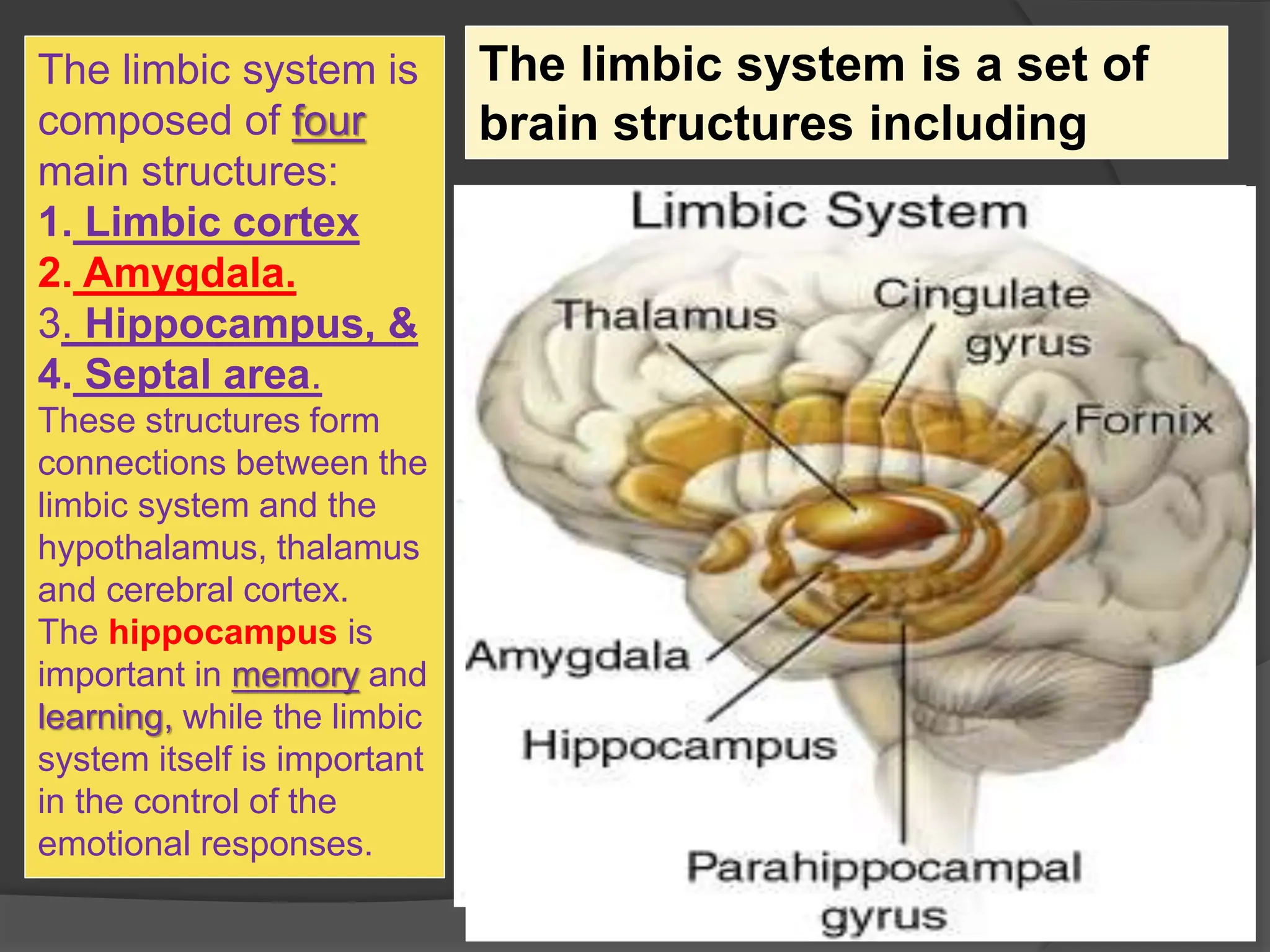
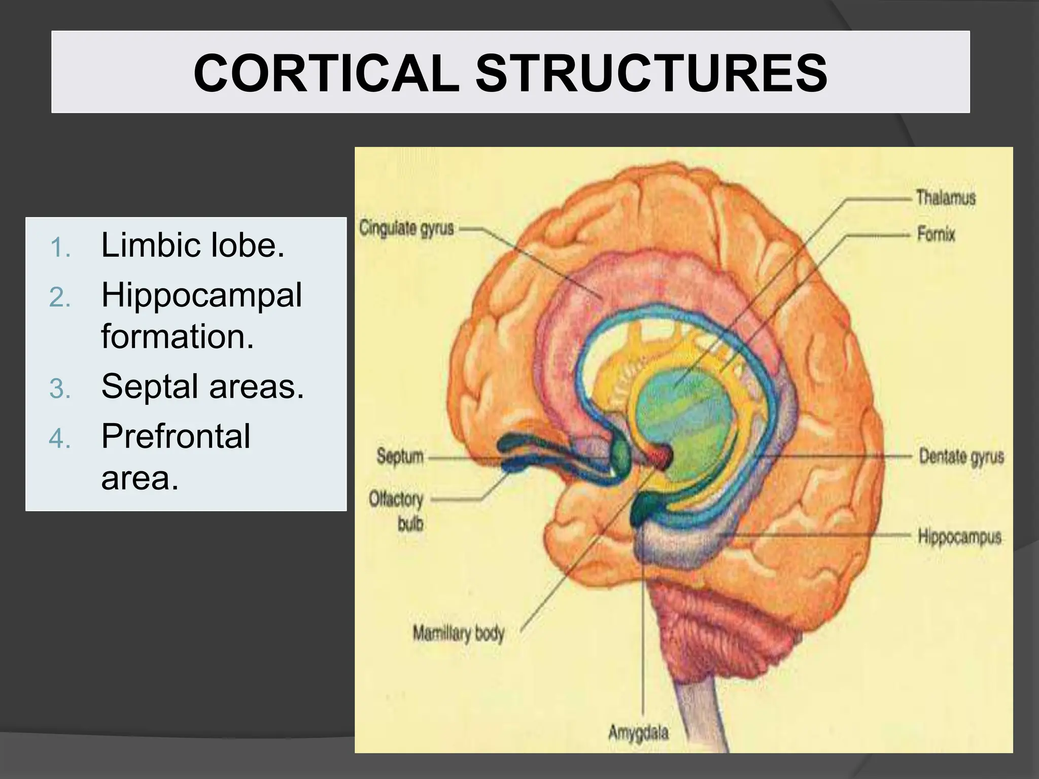
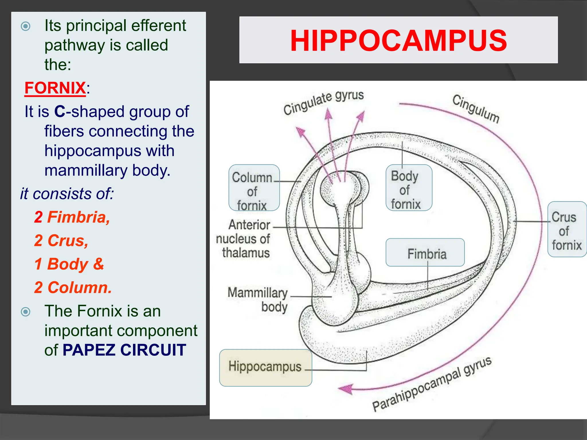
1
Prof. Saeed Abuel Makarem
2
THALAMUS
It is the largest
nuclear mass of the
whole body.
It is the largest
part of the
diencephalon
It is formed of
two oval masses of
grey matter.
It is the
gateway to the
cortex.
Resemble a small
hen.
Together with the
hypothalamus they
form the lateral wall
of the 3rd ventricle.
PONS
Thalamus
Corpus callosum
Midbrain
3
It sends received
information from different
brain regions to the
cerebral cortex.
Axons from every sensory
system (except olfaction)
synapse in the thalamus
as the last relay site 'last
pit stop' before the
information reaches the
cerebral cortex.
There are some thalamic
nuclei that receive input
from:
1. Cerebellar nuclei,
2. Basal ganglia- and
3. Limbic-related brain
regions.
Thalamus
It has 4 surfaces & 2 ends.
Surfaces
Lateral:(L)
Posterior limb of the internal
capsule
Medial: (3)
The 3rd ventricle
In some people it is connected
to the thalamus of the
opposite side by the
interthalamic connexus,
(adhesion) or Massa
intermedia.
Superior: (s)
Lateral ventricle and fornix.
Inferior: Hypothalamus,
anteriorly & Subthalamus
posteriorly.
4
Relations
I
S
L
3
T T
Anterior end:
Forms a projection,
called the anterior
tubercle.
It lies just behind the
interventricular
foramen.
Posterior end: Broad
Forms a projection
called Pulvinar
which lies above the
superior colliculus
and the lateral &
medial Geniculate
bodies.
5
Internal Structure
 White matter:
 External medullary
lamina:
 Covers the lateral surface.
 It consists of
thalamocortical &
corticothalamic fibers.
 Internal medullary lamina:
 Bundle of Y- shaped
myelinated (afferent &
efferent) fibers.
 It divides the thalamus into:
anterior , medial, lateral
nuclear groups.
 Each of these group is
subdivided into a number of
named nuclei.
6
Internal Structure
 Embedded
within the
internal
medullary
lamina lie
intralaminar
nuclei.
 The external
medullary
lamina covers
the lateral
surface, in
which lies thin
reticular
nucleus.
7
8
 It is divided into: Dorsal
& Ventral tiers.
 Dorsal tier:
 which contains:
1. Lateral Dorsal (LD).
2. Lateral Posterior (LP).
3. Pulvinar.
 Ventral tier,
 Which contains :
1. Ventral Anterior (VA).
2. Ventral Lateral (VL).
3. Ventral Intermediate (VI).
4. Ventral Posterior (VP).
(VPL, VPM).
5. Medial & Lateral geniculate
nuclei.
Lateral Nuclear Group
9
Projection of thalamic nuclei
Anterior Thalamic
Nucleus
 Afferent: Mammillary
body.
 Efferent: Cingulate
gyrus, (limbic system)
 -------------------------------
Medial Nucleus
 Afferent: Hypothalamus.
 Efferent: Prefrontal
cortex.
10
Projection of thalamic nuclei
Ventral Anterior Nucleus
 Afferent: Globus
pallidus and substantial
nigra.
 Efferent: Premotor
cortex.
 -------------------------------
Ventral Lateral Nucleus
 Afferent: Dentate
Nucleus.
 Efferent: Primary Motor
Cortex.
11
Projection of thalamic nuclei
Ventral Posterior Lateral
Nucleus
 Afferent: Medial and
Spinal Leminsci.
 Efferent: Sensory
Cortex.
 -------------------------------
Ventral Posterior Medial
Nucleus
 Afferent: Trigeminal
Leminiscus.
 Efferent: Sensory
Cortex.
12
Projection of thalamic nuclei
Lateral Geniculate
Nucleus
 Afferent: Optic tract.
 Efferent: Visual Cortex.
 -------------------------------
Medial Geniculate
Nucleus
 Afferent: Lateral
Leminiscus.
 Efferent: Auditory
Cortex.
13
14
15
16
17
Input & Output of
thalamic nuclei
Mammillary
Body
Optic
Tract
Lateral
Leminiscus
Medial &
Spinal
Leminsci
Trigeminal
Leminiscus
Globus
pallidus and
Substantia
Nigra
Dentate
Nucleus
Hypothalamus
 The term "limbic" is
from the Latin word
Limbus, for
"border" or "edge".
 It separates the
medial surface of
the cerebral cortex
from the
diencephalon
 It consists of a
number of cortical
& subcortical
structures with
looped connections
that all project to
the hypothalamus.
LIMBIC SYSTEM
It control a variety of
functions including:
 Emotions,
 Emotional responses
 Behaviour & Mood
(happy, cry, laugh, sad,
afraid, aggression,
depression)
 Motivation.
 Memory.
 Visceral & Motor
responses involved in
(sex, pleasure, hunger,
and reproduction).
 Olfaction.
WHAT IS THE FUNCTION OF THE LIMBIC
SYSTEM?
MEMORY
OLFACTION
Pleasure
sensation
The limbic system is
composed of four
main structures:
1. Limbic cortex
2. Amygdala.
3. Hippocampus, &
4. Septal area.
These structures form
connections between the
limbic system and the
hypothalamus, thalamus
and cerebral cortex.
The hippocampus is
important in memory and
learning, while the limbic
system itself is important
in the control of the
emotional responses.
The limbic system is a set of
brain structures including
CORTICAL STRUCTURES
1. Limbic lobe.
2. Hippocampal
formation.
3. Septal areas.
4. Prefrontal
area.
LIMBIC LOBE
 C-shaped ring of grey
matter on the medial
surface of each
cerebral hemisphere,
surrounding the
corpus callosum.
 It includes:
1. Subcallosal area
2. Cingulate gyrus
3. Isthmus
4. Parahippocampal
gyrus and the
5. Uncus.
It is a limbic system structure that is
involved in:
Formation,
Organization, and
Storage of memories.
It is important in forming new memories
and connecting emotions and senses,
such as smell and sound, to memories.
It is a horseshoe paired structure, one
in each cerebral hemisphere.
It acts as a memory indexer by sending
memories to the appropriate part of the
cerebral hemisphere for long-term
storage and retrieving them when
necessary.
HIPPOCAMPUS
HIPPOCAMPUS
 Site:
 It is a scrolled
structure in the
inferomedial part
of the temporal
lobe.
 Function:
 Memory (file new
memories as they
occur).
 The hippocampus
& its connections
are necessary for
consolidation of
new short-term
memories.
HIPPOCAMPUS
 Its principal efferent
pathway is called
the:
FORNIX:
It is C-shaped group of
fibers connecting the
hippocampus with
mammillary body.
it consists of:
2 Fimbria,
2 Crus,
1 Body &
2 Column.
 The Fornix is an
important component
of PAPEZ CIRCUIT
AMYGDALA
 Site:
 almond shaped
mass of nuclei
that
lies near the
temporal pole,
close to the tail
of the caudate
nucleus.
 Function:
 It is involved in
1. FEAR ,
2. Emotions
3. Anger, &
4. Hormonal
secretions.
CONNECTIONS OF
AMYGDALA
 Inputs:
 Association areas
of visual, auditory &
somatosensory
cortices.
 Outputs:
 Hypothalamus &
 Autonomic nuclei in
the brain stem,
 Lesion:
 Lack of emotional
responses &
docility.
Site:
Located anterior to the
interventricular
septum
Main connections:
1. To Hypothalamus
2. To Habenular nuclei
Function:
It is the pleasure zone.
Septal nuclei
 Korsakoff’s psychosis
(Retrograde = loss of new
memories at the time of lesion
with retained old memories &
anterograde amnesia= inability
to gain new memories)
 Temporal lobe epilepsy
 The hippocampus is a common
focus site in epilepsy, and can be
damaged through chronic
seizures.
 It is sometimes damaged in
diseases such as herpes
encephalitis,
 Alzheimer’s disease: The
hippocampus is one of the first
brain areas to show damage in
Alzheimer's disease
 Schizophrenia.
Lesions associated with
limbic lobe disorders
THANK YOU
AND
GOOD LUCK
30

17- Thalamus Limbic System.ppt

  • 1.
  • 2.
    2 THALAMUS It is thelargest nuclear mass of the whole body. It is the largest part of the diencephalon It is formed of two oval masses of grey matter. It is the gateway to the cortex. Resemble a small hen. Together with the hypothalamus they form the lateral wall of the 3rd ventricle. PONS Thalamus Corpus callosum Midbrain
  • 3.
    3 It sends received informationfrom different brain regions to the cerebral cortex. Axons from every sensory system (except olfaction) synapse in the thalamus as the last relay site 'last pit stop' before the information reaches the cerebral cortex. There are some thalamic nuclei that receive input from: 1. Cerebellar nuclei, 2. Basal ganglia- and 3. Limbic-related brain regions. Thalamus
  • 4.
    It has 4surfaces & 2 ends. Surfaces Lateral:(L) Posterior limb of the internal capsule Medial: (3) The 3rd ventricle In some people it is connected to the thalamus of the opposite side by the interthalamic connexus, (adhesion) or Massa intermedia. Superior: (s) Lateral ventricle and fornix. Inferior: Hypothalamus, anteriorly & Subthalamus posteriorly. 4 Relations I S L 3 T T
  • 5.
    Anterior end: Forms aprojection, called the anterior tubercle. It lies just behind the interventricular foramen. Posterior end: Broad Forms a projection called Pulvinar which lies above the superior colliculus and the lateral & medial Geniculate bodies. 5
  • 6.
    Internal Structure  Whitematter:  External medullary lamina:  Covers the lateral surface.  It consists of thalamocortical & corticothalamic fibers.  Internal medullary lamina:  Bundle of Y- shaped myelinated (afferent & efferent) fibers.  It divides the thalamus into: anterior , medial, lateral nuclear groups.  Each of these group is subdivided into a number of named nuclei. 6
  • 7.
    Internal Structure  Embedded withinthe internal medullary lamina lie intralaminar nuclei.  The external medullary lamina covers the lateral surface, in which lies thin reticular nucleus. 7
  • 8.
    8  It isdivided into: Dorsal & Ventral tiers.  Dorsal tier:  which contains: 1. Lateral Dorsal (LD). 2. Lateral Posterior (LP). 3. Pulvinar.  Ventral tier,  Which contains : 1. Ventral Anterior (VA). 2. Ventral Lateral (VL). 3. Ventral Intermediate (VI). 4. Ventral Posterior (VP). (VPL, VPM). 5. Medial & Lateral geniculate nuclei. Lateral Nuclear Group
  • 9.
  • 10.
    Projection of thalamicnuclei Anterior Thalamic Nucleus  Afferent: Mammillary body.  Efferent: Cingulate gyrus, (limbic system)  ------------------------------- Medial Nucleus  Afferent: Hypothalamus.  Efferent: Prefrontal cortex. 10
  • 11.
    Projection of thalamicnuclei Ventral Anterior Nucleus  Afferent: Globus pallidus and substantial nigra.  Efferent: Premotor cortex.  ------------------------------- Ventral Lateral Nucleus  Afferent: Dentate Nucleus.  Efferent: Primary Motor Cortex. 11
  • 12.
    Projection of thalamicnuclei Ventral Posterior Lateral Nucleus  Afferent: Medial and Spinal Leminsci.  Efferent: Sensory Cortex.  ------------------------------- Ventral Posterior Medial Nucleus  Afferent: Trigeminal Leminiscus.  Efferent: Sensory Cortex. 12
  • 13.
    Projection of thalamicnuclei Lateral Geniculate Nucleus  Afferent: Optic tract.  Efferent: Visual Cortex.  ------------------------------- Medial Geniculate Nucleus  Afferent: Lateral Leminiscus.  Efferent: Auditory Cortex. 13
  • 14.
  • 15.
  • 16.
  • 17.
    17 Input & Outputof thalamic nuclei Mammillary Body Optic Tract Lateral Leminiscus Medial & Spinal Leminsci Trigeminal Leminiscus Globus pallidus and Substantia Nigra Dentate Nucleus Hypothalamus
  • 18.
     The term"limbic" is from the Latin word Limbus, for "border" or "edge".  It separates the medial surface of the cerebral cortex from the diencephalon  It consists of a number of cortical & subcortical structures with looped connections that all project to the hypothalamus. LIMBIC SYSTEM
  • 19.
    It control avariety of functions including:  Emotions,  Emotional responses  Behaviour & Mood (happy, cry, laugh, sad, afraid, aggression, depression)  Motivation.  Memory.  Visceral & Motor responses involved in (sex, pleasure, hunger, and reproduction).  Olfaction. WHAT IS THE FUNCTION OF THE LIMBIC SYSTEM? MEMORY OLFACTION Pleasure sensation
  • 20.
    The limbic systemis composed of four main structures: 1. Limbic cortex 2. Amygdala. 3. Hippocampus, & 4. Septal area. These structures form connections between the limbic system and the hypothalamus, thalamus and cerebral cortex. The hippocampus is important in memory and learning, while the limbic system itself is important in the control of the emotional responses. The limbic system is a set of brain structures including
  • 21.
    CORTICAL STRUCTURES 1. Limbiclobe. 2. Hippocampal formation. 3. Septal areas. 4. Prefrontal area.
  • 22.
    LIMBIC LOBE  C-shapedring of grey matter on the medial surface of each cerebral hemisphere, surrounding the corpus callosum.  It includes: 1. Subcallosal area 2. Cingulate gyrus 3. Isthmus 4. Parahippocampal gyrus and the 5. Uncus.
  • 23.
    It is alimbic system structure that is involved in: Formation, Organization, and Storage of memories. It is important in forming new memories and connecting emotions and senses, such as smell and sound, to memories. It is a horseshoe paired structure, one in each cerebral hemisphere. It acts as a memory indexer by sending memories to the appropriate part of the cerebral hemisphere for long-term storage and retrieving them when necessary. HIPPOCAMPUS
  • 24.
    HIPPOCAMPUS  Site:  Itis a scrolled structure in the inferomedial part of the temporal lobe.  Function:  Memory (file new memories as they occur).  The hippocampus & its connections are necessary for consolidation of new short-term memories.
  • 25.
    HIPPOCAMPUS  Its principalefferent pathway is called the: FORNIX: It is C-shaped group of fibers connecting the hippocampus with mammillary body. it consists of: 2 Fimbria, 2 Crus, 1 Body & 2 Column.  The Fornix is an important component of PAPEZ CIRCUIT
  • 26.
    AMYGDALA  Site:  almondshaped mass of nuclei that lies near the temporal pole, close to the tail of the caudate nucleus.  Function:  It is involved in 1. FEAR , 2. Emotions 3. Anger, & 4. Hormonal secretions.
  • 27.
    CONNECTIONS OF AMYGDALA  Inputs: Association areas of visual, auditory & somatosensory cortices.  Outputs:  Hypothalamus &  Autonomic nuclei in the brain stem,  Lesion:  Lack of emotional responses & docility.
  • 28.
    Site: Located anterior tothe interventricular septum Main connections: 1. To Hypothalamus 2. To Habenular nuclei Function: It is the pleasure zone. Septal nuclei
  • 29.
     Korsakoff’s psychosis (Retrograde= loss of new memories at the time of lesion with retained old memories & anterograde amnesia= inability to gain new memories)  Temporal lobe epilepsy  The hippocampus is a common focus site in epilepsy, and can be damaged through chronic seizures.  It is sometimes damaged in diseases such as herpes encephalitis,  Alzheimer’s disease: The hippocampus is one of the first brain areas to show damage in Alzheimer's disease  Schizophrenia. Lesions associated with limbic lobe disorders
  • 30.