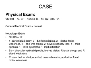
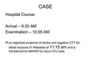
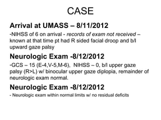
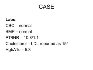
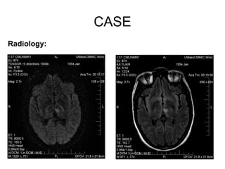
THALAMIC VASCULAR
   SYNDROMES
     Chris Robinson, DO
    Deparment of Neurology
 Loyola University Medical Center
              2012.
CASE
• 58 y/o female driving w/ husband @ 9:30
  AM w/ sudden onset blurred vision and
  difficulty focusing.

• 5 min later w/ R facial droop, somnolence,
  aphasia and R hemiparesis

• Present to OSH at 10:05 AM
CASE
PMH/PSH:
1.   Infiltrating DCIS s/p lumpectomy/radiation – 2006
2.   Insomnia – 2006
3.   Reported incidental heart murmur – 2007

Social Hx:
Tob – negative
ETOH – socially
Illicits – negative
Married
Speech/Language Pathologist

Family Hx:
CAD, Breast Cancer, Brother w/ brain aneurysm
CASE
Medications:
Lorazepam prn insomnia


Allergies:
NKDA
CASE
Physical Exam:
VS: HR – 73 BP – 154/83 R – 14 O2- 98% RA

General Medical Exam – normal

Neurologic Exam
- NIHSS – 12
- 1- partial gaze palsy, 3 – b/l hemianopsia, 2 – partial facial
  weakness, 1 – one limb ataxia, 2- severe sensory loss, 1 – mild
  aphasia, 1 – mild dysarthria, 1- mild extinction
- Sx – binocular vertical diplopia, blurred vision, R facial droop, and R
  sided weakness
- Pt recorded as alert, oriented, comprehensive, and w/out focal
  motor weakness
CASE
Hospital Course:

Arrival – 9:30 AM
Examination – 10:05 AM

Pt w/ objective evidence of stroke and negative CTT for
  bleed recieves IV Alteplase at 11:15 am and is
  transferred to UMASS for neuro ICU care.
CASE
Arrival at UMASS – 8/11/2012
-NIHSS of 6 on arrival - records of exam not received –
known at that time pt had R sided facial droop and b/l
upward gaze palsy
Neurologic Exam -8/12/2012
-GCS – 15 (E-4,V-5,M-6), NIHSS – 0, b/l upper gaze
palsy (R>L) w/ binocular upper gaze diplopia, remainder of
neurologic exam normal.
Neurologic Exam -8/12/2012
- Neurologic exam within normal limits w/ no residual deficits
CASE

Labs:
CBC – normal
BMP – normal
PT/INR – 10.8/1.1
Cholesterol – LDL reported as 154
HgbA1c – 5.3
CASE
Radiology:
CASE

Radiology Cont:
MRI Brain – acute L thalamic infarction

CTA – No stenosis in the anterior or the posterior
circulation, small nodule L lobe of thyroid

TTE – evidence of ? ASD
Thalamic Blood Supply
•   4 Major Vacular Territories
    1. Tuberothalamic or Polar Artery
       (PCOM)
    2. Paramedian
       (Basilar Communicating Artery
    3. Inferolateral or Thalamogeniculate
       (PCA)
    4. Posterior Choroidal
       (PCA)
Artery of Percheron
• Paramedian A.
Thalamic Infarction Localization
Posterolateral Inarction
• Occlusion of thalamogeniculate a.
• P2 segment of PCA
• 3 common clinical syndromes
  1. Pure Sensory
  2. Sensorimotor
  3. Dejerine-Roussy - contralateral sensory, thalamic pain
     of involved side, vasomotor disturbance, transient contralateral
     hemiparesis, and mild hyperkinetic disturbances
Anterior Infarction
• Occlusion of polar or turberothalamic a.
• Branch of PCOM

• Clinical Manifestations
  - Alteration in consciousness, abulia,
    disorientation, personality disturbances, visual
    field deficits
  L sided – thalamic aphasia
  R sided – hemineglect, alien hand
Paramedian Infarction
• Paramedian a. -
• Branch of basilar communication a.
  – Basilar a. prior to ostium of PCOM


• Clinical Manifestations – somnolence, memory
  loss, mood disturbances, vertical gaze
  abnormalities
• Can be b/l – artery of percheron –
  hypersomnolence and marked memory
  impairment
Dorsal Infarction
• Posterior Choroidal a.
• Branch of P2 segment of PCA

• Clinical Manifestations – homonymous
  quadrantanopia or homonymous
  horizontal sectoranopias
  – If pulvinar affected can have aphasia or
    cardiac dysthymia's
Thalamic Infarction Localization
Case

Plan:
-Pt d/c on zocor 40 mg daily and ASA 325
mg
-Presented to Neurology Clinic 8/22/2012 pt
continues to be asymptomatic

-Cont ASA and statin for now, 30 day ECAT
monitor, TEE
Thalamic infarction
Thalamic infarction

Thalamic infarction

  • 1.
    THALAMIC VASCULAR SYNDROMES Chris Robinson, DO Deparment of Neurology Loyola University Medical Center 2012.
  • 2.
    CASE • 58 y/ofemale driving w/ husband @ 9:30 AM w/ sudden onset blurred vision and difficulty focusing. • 5 min later w/ R facial droop, somnolence, aphasia and R hemiparesis • Present to OSH at 10:05 AM
  • 3.
    CASE PMH/PSH: 1. Infiltrating DCIS s/p lumpectomy/radiation – 2006 2. Insomnia – 2006 3. Reported incidental heart murmur – 2007 Social Hx: Tob – negative ETOH – socially Illicits – negative Married Speech/Language Pathologist Family Hx: CAD, Breast Cancer, Brother w/ brain aneurysm
  • 4.
  • 5.
    CASE Physical Exam: VS: HR– 73 BP – 154/83 R – 14 O2- 98% RA General Medical Exam – normal Neurologic Exam - NIHSS – 12 - 1- partial gaze palsy, 3 – b/l hemianopsia, 2 – partial facial weakness, 1 – one limb ataxia, 2- severe sensory loss, 1 – mild aphasia, 1 – mild dysarthria, 1- mild extinction - Sx – binocular vertical diplopia, blurred vision, R facial droop, and R sided weakness - Pt recorded as alert, oriented, comprehensive, and w/out focal motor weakness
  • 6.
    CASE Hospital Course: Arrival –9:30 AM Examination – 10:05 AM Pt w/ objective evidence of stroke and negative CTT for bleed recieves IV Alteplase at 11:15 am and is transferred to UMASS for neuro ICU care.
  • 7.
    CASE Arrival at UMASS– 8/11/2012 -NIHSS of 6 on arrival - records of exam not received – known at that time pt had R sided facial droop and b/l upward gaze palsy Neurologic Exam -8/12/2012 -GCS – 15 (E-4,V-5,M-6), NIHSS – 0, b/l upper gaze palsy (R>L) w/ binocular upper gaze diplopia, remainder of neurologic exam normal. Neurologic Exam -8/12/2012 - Neurologic exam within normal limits w/ no residual deficits
  • 8.
    CASE Labs: CBC – normal BMP– normal PT/INR – 10.8/1.1 Cholesterol – LDL reported as 154 HgbA1c – 5.3
  • 9.
  • 10.
    CASE Radiology Cont: MRI Brain– acute L thalamic infarction CTA – No stenosis in the anterior or the posterior circulation, small nodule L lobe of thyroid TTE – evidence of ? ASD
  • 11.
    Thalamic Blood Supply • 4 Major Vacular Territories 1. Tuberothalamic or Polar Artery (PCOM) 2. Paramedian (Basilar Communicating Artery 3. Inferolateral or Thalamogeniculate (PCA) 4. Posterior Choroidal (PCA)
  • 13.
  • 14.
  • 15.
    Posterolateral Inarction • Occlusionof thalamogeniculate a. • P2 segment of PCA • 3 common clinical syndromes 1. Pure Sensory 2. Sensorimotor 3. Dejerine-Roussy - contralateral sensory, thalamic pain of involved side, vasomotor disturbance, transient contralateral hemiparesis, and mild hyperkinetic disturbances
  • 16.
    Anterior Infarction • Occlusionof polar or turberothalamic a. • Branch of PCOM • Clinical Manifestations - Alteration in consciousness, abulia, disorientation, personality disturbances, visual field deficits L sided – thalamic aphasia R sided – hemineglect, alien hand
  • 17.
    Paramedian Infarction • Paramediana. - • Branch of basilar communication a. – Basilar a. prior to ostium of PCOM • Clinical Manifestations – somnolence, memory loss, mood disturbances, vertical gaze abnormalities • Can be b/l – artery of percheron – hypersomnolence and marked memory impairment
  • 18.
    Dorsal Infarction • PosteriorChoroidal a. • Branch of P2 segment of PCA • Clinical Manifestations – homonymous quadrantanopia or homonymous horizontal sectoranopias – If pulvinar affected can have aphasia or cardiac dysthymia's
  • 19.
  • 20.
    Case Plan: -Pt d/c onzocor 40 mg daily and ASA 325 mg -Presented to Neurology Clinic 8/22/2012 pt continues to be asymptomatic -Cont ASA and statin for now, 30 day ECAT monitor, TEE