- CHAITRA CHINMAYEE M K
CORPUS CALLOSUM
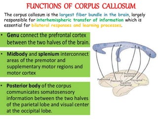
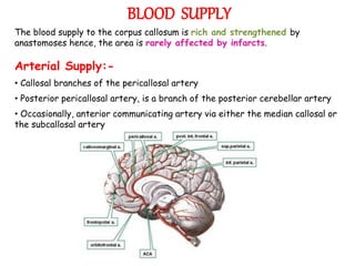
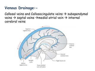
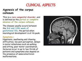
• The corpus callosum is the
largest commissure of the
brain.
• It is a thick band of nerve
fibres that connects the left
and right sides of the
cerebral cortex, allowing
communication between them.
• It transfers motor, sensory,
and cognitive information
between the cerebral
hemispheres.
CORPUS CALLOSUM
EXTERNAL FEATURES
• Corpus callosum forms a massive
arched interhemispheric bridge
in the floor of the median
longitudinal cerebral fissure
connecting the medial surfaces
of the two cerebral
hemispheres.
• In sagittal section, it is seen as
a C-shaped mass of white
fibres on the medial surface of
the hemisphere forming the
roof of the lateral ventricle.
• The concave inferior aspect of
corpus callosum is attached with
the convex superior aspect of
the fornix by septum
pellucidum.
PARTS OF CORPUS CALLOSUM
The Corpus Callosum has four parts:-
• Genu - It is thick curved anterior
extremity of corpus callosum which
lies 4 cm behind the frontal pole.
• Rostrum: The genu extends
downwards and backwards as a
thin prolongation to join the lamina
terminalis forming the rostrum.
• Trunk/Body: The trunk is the main
part of the corpus callosum
between its anterior and posterior
extremities.
• Splenium: The splenium is the
massive posterior extremity of the
corpus callosum, lying 6 cm in front
of the occipital pole whose fibres
connect the parietal, temporal, and
occipital lobes of the two
hemispheres.
FUNCTIONS OF CORPUS CALLOSUM
The corpus callosum is the largest fiber bundle in the brain, largely
responsible for interhemispheric transfer of information which is
essential for bilateral responses and learning processes.
BLOOD SUPPLY
The blood supply to the corpus callosum is rich and strengthened by
anastomoses hence, the area is rarely affected by infarcts.
Arterial Supply:-
• Callosal branches of the pericallosal artery
• Posterior pericallosal artery, is a branch of the posterior cerebellar artery
• Occasionally, anterior communicating artery via either the median callosal or
the subcallosal artery
Venous Drainage:-
Callosal veins and Callosocingulate veins  subependymal
veins  septal veins medial atrial vein  internal
cerebral veins
CLINICAL ASPECTS
Agenesis of the corpus
callosum
This is a rare congenital disorder, and
is defined as the partial or complete
absence of the corpus callosum.
The disease usually occurs between
the 3rd and 12th week of
gestational life, the period when
neurological development is at its peak.
Symptoms –
Hypotonia, swallowing and chewing
difficulties, low pain perception, delays
in motor milestones such as walking
and sitting, poor motor coordination.
Seizures occur in up to two thirds of
patients and symptoms also depend on
the presence of associated brain
malformations.
Stereopsis /binocular vision
This is our ability to perceive depth, and is dependent
on our corpus callosum, allowing us to interpret visual
information from one eye in conjunction with the visual
information from our other eye.
Corpus callosum impingement syndrome
Impingement of the corpus callosum fibres on the inferior
free margin of the falx cerebri. It usually results from
longstanding hydrocephalus. Ischaemia results and
eventually causes atrophy. It does not usually produce
symptoms.
Disconnection syndrome
This is a syndrome that occurs when the connection
between the two hemispheres is disrupted, either as a
result of brain surgery, stroke or trauma. The patient
may be completely normal to their family and friends, but
specific tests elicit the abnormalities.
CLINICAL SIGNIFICANCE
Corpus Callosotomy
The corpus callosum is cut through in an effort to limit the spread of
epileptic activity between the two halves of the brain.
It is a pallative surgical procedure for the treatment of medically
refractory epilepsy.
After the operation the brain has much more difficulty sending messages
between the hemispheres. Although the corpus callosum is the largest
white matter tract connecting the hemispheres, some limited
interhemispheric communication is still possible via the anterior
commissure and posterior commissure.
VARIATION IN SEXES
Advanced analytical techniques of computational
Neuro-anatomy, showed that sex differences
were clear but confined to certain parts of the
corpus callosum, and that they correlated with
cognitive performance in certain tests.
Recent study using MRI found that the mid-
sagittal corpus callosum cross-sectional area is, on
average, proportionately larger in females.
Using diffusion tensor techniques on MRI machines
anisotropy can be measured and used as an indirect
measurement of anatomical connection strength.
These sequences have found consistent sex
differences in human corpus callosal shape and
microstructure.
Front portion of the human corpus callosum was found
to be 11% larger in left handed and ambidextrous
people than right-handed people. This difference was
evident in the anterior and posterior regions of the
corpus callosum, but not in the splenium.
THANK YOU

Corpus callosum

  • 1.
    - CHAITRA CHINMAYEEM K CORPUS CALLOSUM
  • 2.
    • The corpuscallosum is the largest commissure of the brain. • It is a thick band of nerve fibres that connects the left and right sides of the cerebral cortex, allowing communication between them. • It transfers motor, sensory, and cognitive information between the cerebral hemispheres. CORPUS CALLOSUM
  • 3.
    EXTERNAL FEATURES • Corpuscallosum forms a massive arched interhemispheric bridge in the floor of the median longitudinal cerebral fissure connecting the medial surfaces of the two cerebral hemispheres. • In sagittal section, it is seen as a C-shaped mass of white fibres on the medial surface of the hemisphere forming the roof of the lateral ventricle. • The concave inferior aspect of corpus callosum is attached with the convex superior aspect of the fornix by septum pellucidum.
  • 4.
    PARTS OF CORPUSCALLOSUM The Corpus Callosum has four parts:- • Genu - It is thick curved anterior extremity of corpus callosum which lies 4 cm behind the frontal pole. • Rostrum: The genu extends downwards and backwards as a thin prolongation to join the lamina terminalis forming the rostrum. • Trunk/Body: The trunk is the main part of the corpus callosum between its anterior and posterior extremities. • Splenium: The splenium is the massive posterior extremity of the corpus callosum, lying 6 cm in front of the occipital pole whose fibres connect the parietal, temporal, and occipital lobes of the two hemispheres.
  • 5.
    FUNCTIONS OF CORPUSCALLOSUM The corpus callosum is the largest fiber bundle in the brain, largely responsible for interhemispheric transfer of information which is essential for bilateral responses and learning processes.
  • 6.
    BLOOD SUPPLY The bloodsupply to the corpus callosum is rich and strengthened by anastomoses hence, the area is rarely affected by infarcts. Arterial Supply:- • Callosal branches of the pericallosal artery • Posterior pericallosal artery, is a branch of the posterior cerebellar artery • Occasionally, anterior communicating artery via either the median callosal or the subcallosal artery
  • 7.
    Venous Drainage:- Callosal veinsand Callosocingulate veins  subependymal veins  septal veins medial atrial vein  internal cerebral veins
  • 8.
    CLINICAL ASPECTS Agenesis ofthe corpus callosum This is a rare congenital disorder, and is defined as the partial or complete absence of the corpus callosum. The disease usually occurs between the 3rd and 12th week of gestational life, the period when neurological development is at its peak. Symptoms – Hypotonia, swallowing and chewing difficulties, low pain perception, delays in motor milestones such as walking and sitting, poor motor coordination. Seizures occur in up to two thirds of patients and symptoms also depend on the presence of associated brain malformations.
  • 9.
    Stereopsis /binocular vision Thisis our ability to perceive depth, and is dependent on our corpus callosum, allowing us to interpret visual information from one eye in conjunction with the visual information from our other eye.
  • 10.
    Corpus callosum impingementsyndrome Impingement of the corpus callosum fibres on the inferior free margin of the falx cerebri. It usually results from longstanding hydrocephalus. Ischaemia results and eventually causes atrophy. It does not usually produce symptoms. Disconnection syndrome This is a syndrome that occurs when the connection between the two hemispheres is disrupted, either as a result of brain surgery, stroke or trauma. The patient may be completely normal to their family and friends, but specific tests elicit the abnormalities.
  • 11.
    CLINICAL SIGNIFICANCE Corpus Callosotomy Thecorpus callosum is cut through in an effort to limit the spread of epileptic activity between the two halves of the brain. It is a pallative surgical procedure for the treatment of medically refractory epilepsy. After the operation the brain has much more difficulty sending messages between the hemispheres. Although the corpus callosum is the largest white matter tract connecting the hemispheres, some limited interhemispheric communication is still possible via the anterior commissure and posterior commissure.
  • 12.
    VARIATION IN SEXES Advancedanalytical techniques of computational Neuro-anatomy, showed that sex differences were clear but confined to certain parts of the corpus callosum, and that they correlated with cognitive performance in certain tests. Recent study using MRI found that the mid- sagittal corpus callosum cross-sectional area is, on average, proportionately larger in females. Using diffusion tensor techniques on MRI machines anisotropy can be measured and used as an indirect measurement of anatomical connection strength. These sequences have found consistent sex differences in human corpus callosal shape and microstructure. Front portion of the human corpus callosum was found to be 11% larger in left handed and ambidextrous people than right-handed people. This difference was evident in the anterior and posterior regions of the corpus callosum, but not in the splenium.
  • 13.