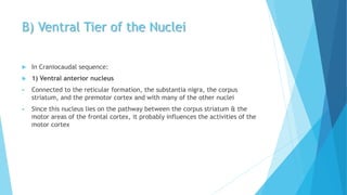
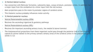
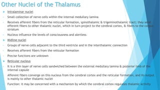
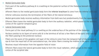
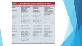
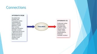
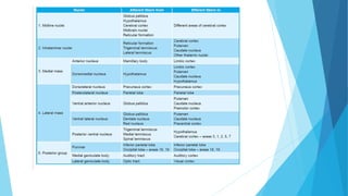
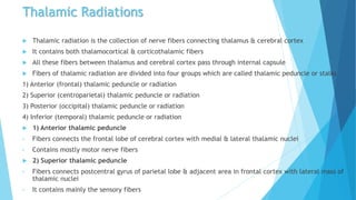
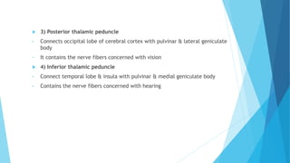
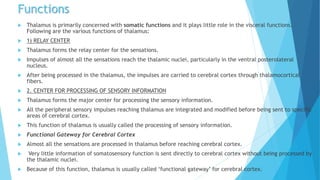
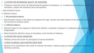
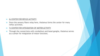
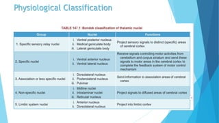
Thalamus which is the Relay center in our Body.
Anatomy & Physiology of Thalamus
Book references:- Snell's Anatomy and K. and prema Sembuligum
Medical
-Yash Bhandari (Physiotherapist)
The thalamus acts as a relay center and plays a key role in sensory and motor functions. It is divided into nuclear groups including anterior, medial, lateral, and intralaminar. Specific nuclei relay information from particular sensory pathways to corresponding cortical areas, while nonspecific nuclei have broader connections. The thalamus receives subcortical inputs and sends outputs to widespread areas of cortex, regulating cortical activity and integrating sensory, motor, and limbic functions. Damage to different thalamic regions can cause sensory deficits, involuntary movements, or pain syndromes on the opposite side of the body.
The thalamus is a large mass of gray matter located in the diencephalon that acts as a relay center for sensory and motor signals sent to the cerebral cortex. It has several nuclei that receive input from various sensory systems and subcortical structures and projects this information to different areas of the cortex via thalamic radiations. Damage to the thalamus can result in thalamic syndrome, where a person experiences hypersensitivity to pain throughout the body due to disruptions in sensory processing.
The thalamus is a structure located in the middle of the brain between the cerebral cortex and midbrain. It is the largest component of the diencephalon. The thalamus acts as a relay station for sensory information (except smell) sending signals to the appropriate areas of the cerebral cortex. It is divided into nuclei that each have distinct functions and connections related to motor control, sensory processing and integration, arousal, memory and cognition. Damage to different thalamic nuclei can disrupt various functions and result in sensory deficits, movement problems or changes to consciousness.
The thalamus is a midline paired structure in the brain that relays sensory and motor signals to the cerebral cortex. It contains several nuclei that subserve different functions. The ventral posterior nuclei relay sensory information from the body and face. The ventral anterior and ventral lateral nuclei are major motor relay nuclei. The medial geniculate and lateral geniculate bodies relay auditory and visual information respectively. Damage to specific nuclei causes localized neurological deficits, while more widespread damage can impact functions like arousal, memory and behavior.
The basal ganglia are large masses of gray matter located in the cerebral hemispheres. They are comprised of the caudate nucleus, lentiform nucleus (putamen and globus pallidus), amygdaloid nuclear complex, and claustrum. The basal ganglia receive input from the cerebral cortex and thalamus and output mainly to the globus pallidus and substantia nigra. They are involved in motor control and planning through direct and indirect pathways that facilitate or inhibit motor activity. Disorders like Parkinson's and Huntington's result from disruptions to these circuits.
The document discusses the limbic system, which is a group of brain structures involved in emotion, behavior, motivation, long-term memory, and olfaction. It describes the key structures that make up the limbic system, including the hippocampus, amygdala, hypothalamus, cingulate gyrus, and others. It also discusses the connections between limbic structures and their roles in functions like memory, emotional behavior, and endocrine regulation. Damage to limbic structures can cause issues like loss of recent memory and changes in emotional behavior.
There are 12 pairs of cranial nerves in the brain with motor and/or sensory nuclei in the brain stem. Each cranial nerve has its own nucleus of origin or termination. These nuclei are arranged medio-laterally and include somatic, visceral, special, and general fibers. The medio-lateral arrangement includes sensory, special visceral efferent, general visceral efferent, general visceral afferent, special visceral afferent, general somatic afferent, and special somatic afferent. Cranial nerve nuclei are located in the midbrain, pons, and medulla.
The three meninges (membranous layers) that cover the brain and spinal cord are the dura mater, arachnoid mater, and pia mater. The dura mater is the outermost layer and consists of two layers. The arachnoid mater lies interior to the dura mater and the subarachnoid space contains cerebrospinal fluid. The pia mater is the innermost layer and closely adheres to the brain and spinal cord. Several folds of dura mater form septa that partition brain regions including the falx cerebri, tentorium cerebelli, and falx cerebelli. Several venous sinuses are contained within the dura mater layers. The
The pons lies between the medulla oblongata and midbrain, connecting them. It contains motor and sensory nuclei for cranial nerves 5-8 and helps transmit signals between the cerebellum and cerebral cortex. The pons has anterior and posterior surfaces and contains fibers, nuclei, and tracts that process sensory information and coordinate motor functions. Damage to different areas can cause deficits like hemiplegia, hearing loss, or facial paralysis.
The medulla oblongata connects the pons superiorly to the spinal cord inferiorly. It has important cranial nerve nuclei and tracts that control vital functions like breathing and heart rate. A key feature is the decussation of the pyramids, where most corticospinal fibers cross to the opposite side. The medulla also contains nuclei that relay proprioceptive and fine touch sensations from the body to the brain.
The document summarizes key aspects of the brainstem and its functions. It discusses three regions of the brainstem - medulla oblongata, pons, and midbrain. Each region contains cranial nerves and nuclei that control autonomic functions. The reticular formation is a network of neurons running through the core of the brainstem that is involved in arousal, alertness, sleep, and motor control. Sensory information is received by the reticular formation and it projects both upwards and downwards. The brainstem modulates consciousness, sensory and motor functions, and plays an important role in emotion, memory, and learning.
The reticular formation is a network of neurons located in the brainstem that serves several important functions. It is involved in regulating states of consciousness like sleep and wakefulness via the ascending reticular activating system. The reticular formation also controls posture and balance through reticulospinal tracts that project to motor neurons in the spinal cord. It is comprised of several nuclei that contain different neurotransmitters and integrate sensory information from various modalities to coordinate motor functions. Lesions in specific areas of the reticular formation can impact muscle tone and result in conditions like decorticate posturing.
The thalamus is a paired, symmetrical structure located in the center of the brain near the brainstem. Each half is bulb-shaped and around the size of a walnut. The thalamus contains several nuclei that receive and relay sensory information to the cerebral cortex. It plays an important role in regulating states of consciousness and sleep/wake cycles. Damage to the thalamus can cause sensory deficits, involuntary movements, and even permanent coma. Common pathologies include lesions from blocked blood vessels and thalamic syndromes that cause loss of sensation.
The limbic system is a group of brain structures located on the edge of the brain involved in emotion, behavior, motivation, long-term memory, and olfaction. It includes structures like the hippocampus, amygdala, cingulate gyrus, parahippocampal gyrus, and others. The limbic system is interconnected and influences functions like emotional responses, memory formation, instinctual behaviors, and the autonomic nervous system. It is also involved in regulating behaviors important for survival like fear, sex, and memory through connections with the hypothalamus and brainstem.
Love you guys with my whole Limbic system...
Limbic System from where the main feelings comes... Here we go..
It got the main structures seen here along with some other related areas...
CONNECTIONS (Papez circuit simplified)
Different functions of Limbic system... Fear vs. Rage, sham rage, reward and punisment, hippocampus and memory related functions...
Some applied aspects are also covered...
THANK YOU
The document provides information about the anatomy and functions of the cerebellum. It can be summarized as follows:
1. The cerebellum is located in the posterior cranial fossa and is separated into two hemispheres and a median vermis. It has three lobes and three functional subdivisions that control balance, coordination of movements, and muscle tone.
2. The cerebellum connects to the brainstem via three cerebellar peduncles - superior, middle, and inferior - which carry afferent and efferent fibers. Deep within the cerebellum are four nuclei - dentate, globose, emboliformis, and fastigius.
3. The cerebell
The limbic system can be divided into a rostral and caudal region. The rostral region including the amygdala, septum, orbitofrontal cortex, anterior insula, and anterior cingulate is important for emotion. The caudal region including the hippocampus, posterior parahippocampal cortex and posterior cingulate is important for memory and visual-spatial functions. The limbic system is involved in various functions including emotional responses, reward and punishment, memory, social cognition, and more. Key structures in the limbic system that support these functions are the amygdala, hippocampus, hypothalamus, and cingulate gyrus.
The document discusses the limbic system, which is a set of brain structures involved in emotion, learning, and memory. It includes the hippocampus, amygdala, hypothalamus, and connections between cortical and subcortical regions. The limbic system regulates many functions including emotion, learning, memory formation, and autonomic nervous system activity. Key structures like the hippocampus and amygdala each have distinct roles - the hippocampus is important for memory formation while the amygdala processes emotions. Lesions in different limbic system areas can impact neurophysiological functions and lead to effects on behaviors regulated by that region.
Anatomy of the cerebrum; Anatomy - January 2015Kareem Alnakeeb
The document provides detailed information about the structure and functions of the cerebrum. It describes the lobes, sulci, gyri, poles and borders of each cerebral hemisphere. It then outlines the primary motor, sensory and association cortices and their functions. Specifically, it discusses the primary motor cortex, premotor cortex, supplementary motor cortex, frontal eye field, Broca's area, primary somatosensory cortex, primary auditory cortex, primary visual cortex, Wernicke's area and their roles in movement, speech, senses and language.
The brainstem is located between the cerebrum and spinal cord. It consists of the midbrain, pons, and medulla oblongata. The midbrain connects the pons and cerebrum and contains the superior and inferior colliculi. The pons connects to the cerebellum via peduncles and contains pontine nuclei and cranial nerve nuclei. The medulla oblongata connects to the spinal cord and contains cranial nerve nuclei, the inferior olives, and tracts such as the gracile and cuneate fasciculi.
The thalamus is a paired symmetrical structure located in the center of the brain that relays sensory and motor signals between the brainstem and cerebral cortex. It is divided into several nuclei that have distinct connections and functions. The document provides detailed information on the anatomy, physiology, functional organization and clinical syndromes associated with lesions of different thalamic nuclei. Key points include a description of the gross anatomy and location of the thalamus, its blood supply, the nuclei and their connections, and syndromes associated with infarcts in the posterolateral and medial thalamic territories.
The document discusses the thalamus and its functions. It begins with objectives and physiological anatomy of the thalamus. It then discusses the internal structure of the thalamus including its white and grey matter. Next, it covers the connections of the thalamus including its sensory, motor, visceral, and integrative nuclei. It lists the functions of the thalamus such as acting as a sensory relay center and in arousal, perception, emotions, and motor integration. Finally, it discusses some applied aspects including thalamic syndrome, Korsakoff's syndrome, and frontal lobotomy.
The thalamus is a structure located in the middle of the brain between the cerebral cortex and midbrain. It is the largest component of the diencephalon. The thalamus acts as a relay station for sensory information (except smell) sending signals to the appropriate areas of the cerebral cortex. It is divided into nuclei that each have distinct functions and connections related to motor control, sensory processing and integration, arousal, memory and cognition. Damage to specific thalamic nuclei can disrupt functions like sensory perception, movement, consciousness and memory formation.
The thalamus is a paired structure located in the diencephalon that relays sensory and motor information to and from the cerebral cortex. It contains several nuclei that can be categorized as specific relay nuclei, association nuclei, and nonspecific nuclei. The thalamus plays a key role in sensory relay, feedback loops between cortex and subcortical structures, and regulating arousal and attention. Lesions of the thalamus can cause motor, sensory, cognitive and arousal disturbances depending on the location within the thalamus.
The thalamus is a large egg-shaped structure located in the brain that serves as a relay center for sensory and motor signals to and from the cerebral cortex. It has multiple nuclei that receive input from various sensory systems and project to different regions of the cortex. The thalamus plays an important role in sensory perception and motor control through its connections with the cortex, cerebellum, and basal ganglia.
The diencephalon includes structures like the thalamus, hypothalamus, epithalamus, and subthalamus. The thalamus relays sensory and motor signals to the cerebral cortex. It contains nuclei that relay specific sensations like vision, hearing, and somatosensation. The hypothalamus controls autonomic functions and regulates behaviors related to hunger, thirst, temperature, sleep, and reproduction. It also controls the pituitary gland. The epithalamus includes the pineal gland and habenular nucleus. The subthalamus contains the subthalamic nucleus and is involved in motor control.
The document discusses the thalamus and hypothalamus. It provides details on the anatomy, internal structure, nuclei, connections and blood supply of the thalamus. It describes the relations, boundaries, nuclei and connections of the hypothalamus. The thalamus acts as a relay station and integrative center for sensory information to the cortex. The hypothalamus regulates the autonomic nervous system, endocrine system and limbic system through its nuclei and connections. Lesions of the thalamus can cause sensory loss, involuntary movements and effects on motor control through disruption of thalamic circuits.
The thalamus is a paired, oval structure located in the diencephalon that serves as a relay center for sensory and motor signals to and from the cerebral cortex. It is divided into several nuclei that process different sensory modalities. The thalamus receives input from various areas and projects to specific regions of the cortex. Damage to certain thalamic nuclei can disrupt motor control, sensory processing, and cause syndromes like thalamic pain. Surgical procedures targeting thalamic nuclei have been used to treat chronic pain conditions.
The thalamus is a paired, oval structure located in the diencephalon that serves as a relay center for sensory and motor signals to and from the cerebral cortex. It is divided into several nuclei that process different sensory modalities. The thalamus receives input from various areas and projects to specific regions of the cortex. Damage to certain thalamic nuclei can disrupt sensory and motor functions and cause syndromes like thalamic pain. Surgical procedures targeting thalamic nuclei have been used to treat intractable pain.
The document discusses the diencephalon region of the forebrain, focusing on the thalamus and hypothalamus. It describes the thalamus as the major part of the diencephalon, located on each side of the third ventricle. The thalamus is divided into anterior, medial, and lateral parts containing different thalamic nuclei. The hypothalamus lies ventral to the thalamus and is divided into preoptic, supraoptic, tuberal, and mamillary regions containing important nuclei that regulate functions like autonomic control, endocrine control, temperature, hunger and thirst, emotions, and circadian rhythms.
The thalamus is an egg-shaped structure atop the brain stem that acts as a sensory relay station. It is composed of several discrete nuclei that are classified anatomically and physiologically. Anatomically, the thalamus is divided into anterior, medial, and lateral groups of nuclei. Physiologically, the nuclei are grouped into specific relay nuclei, association nuclei, nonspecific nuclei, and motor nuclei. The thalamus receives ascending sensory inputs and projects them to sensory cortical areas, functioning as a relay for somatic sensations and special senses. It also plays roles in arousal, memory, emotion, motor functions, and sleep-wake cycles.
The document summarizes key structures and functions of the forebrain and brainstem. It discusses the major components of the forebrain - the telencephalon including the cerebral hemispheres, limbic system, and basal ganglia. It also describes the diencephalon including the thalamus, hypothalamus, and epithalamus. The brainstem is formed of the medulla, pons, and midbrain. Key structures in the midbrain include the tectum, tegmentum, red nucleus, and substantia nigra. The document outlines functions of sensory processing, motor control, arousal, autonomic functions, and other roles of different brain regions.
The thalamus is a paired structure located in the brain that serves as a relay center and integrator for sensory and motor signals. It receives input from various areas of the body and brain and relays this information to the appropriate regions of the cerebral cortex. The thalamus is divided into several nuclei that serve functions like relaying sensory information, regulating states of consciousness, and participating in memory and emotion. Damage to certain thalamic nuclei can cause syndromes with loss of sensory abilities and motor impairments on the opposite side of the body.
The central nervous system consists of the brain and spinal cord. The brain is divided into the forebrain, midbrain, and hindbrain. The forebrain contains structures like the cerebral cortex that control functions like thinking and producing language. The midbrain regulates sensory processes and body movement. The hindbrain includes the cerebellum, pons, and medulla, which are involved in motor control, balance, and vital functions. The spinal cord connects the brain to the rest of the body and transmits motor and sensory signals.
The interhemispheric transcallosal transventricular approach provides access to thalamic tumors that protrude into the lateral ventricle. It involves a U-shaped skin incision and craniotomy exposing the sagittal suture and superior temporal line. The dura is opened in a C-shape along the superior sagittal sinus. This allows visualization of the frontal and parietal cortex, cingulate gyrus, and corpus callosum for tumor resection through the ventricle.
The thalamus is a large gray mass located in the diencephalon that contains several nuclei with different functions. It acts as a relay station for sensory information sent to the cerebral cortex. Specific nuclei relay information for different senses like vision, hearing, and somatosensation. Diffuse nuclei connect with broad areas of the cortex and are involved in attention, motor control, and limbic functions. Damage to thalamic nuclei can cause syndromes like contralateral sensory loss, ataxia, hemianopia, and pain.
The basal ganglia are clusters of grey matter in the brain that are involved in motor control and regulation. They consist of the striatum, globus pallidus, substantia nigra, and subthalamic nucleus. The basal ganglia have direct and indirect pathways that facilitate or inhibit movement through connections with the cortex and thalamus. Dopamine levels modulate these pathways. Diseases that impact the basal ganglia can cause either excessive or reduced movement. Parkinson's disease results from dopamine deficiency in the substantia nigra, leading to hypokinetic movements.
EXTERNAL FEATURES OF MIDBRAIN, ANATOMY OF INTERNAL FEATURES OF MIDBRAIN, CRUS CEREBRI, SUBSTANTIA NIGRA, CEREBRAL PEDUNCLE,INFERIOR COLLICULUS,LEMNISCI
Function of Thalamus & thalamic syndromeDr Sara Sadiq
The thalamus is located in the middle of the brain between the cerebral cortex and midbrain. It acts as a relay station for all sensory pathways and regulates states of sleep and wakefulness. The thalamus is divided into sensory, motor, limbic, and association nuclei that receive and project different types of sensory and motor information to various parts of the cerebral cortex. Damage to the thalamus can lead to a condition called thalamic syndrome, also known as Dejerine-Roussy syndrome, which involves loss of fine sensation, exaggerated pain sensation, and motor problems due to disturbances in thalamocortical connections.
This document provides an overview of the trigeminal nerve (cranial nerve V) in 12 sections. It discusses the elementary structure of neurons, classification of cranial nerves, embryology and nuclei of the trigeminal nerve, the trigeminal ganglion, and the three divisions (ophthalmic, maxillary, mandibular) and their branches. Diagrams are included to illustrate the course and branches of the trigeminal nerve. The presentation provides detailed anatomical information about the trigeminal nerve and related structures.
A Visual Guide to 1 Samuel | A Tale of Two HeartsSteve Thomason
These slides walk through the story of 1 Samuel. Samuel is the last judge of Israel. The people reject God and want a king. Saul is anointed as the first king, but he is not a good king. David, the shepherd boy is anointed and Saul is envious of him. David shows honor while Saul continues to self destruct.
This presentation was provided by Rebecca Benner, Ph.D., of the American Society of Anesthesiologists, for the second session of NISO's 2024 Training Series "DEIA in the Scholarly Landscape." Session Two: 'Expanding Pathways to Publishing Careers,' was held June 13, 2024.
Walmart Business+ and Spark Good for Nonprofits.pdfTechSoup
"Learn about all the ways Walmart supports nonprofit organizations.
You will hear from Liz Willett, the Head of Nonprofits, and hear about what Walmart is doing to help nonprofits, including Walmart Business and Spark Good. Walmart Business+ is a new offer for nonprofits that offers discounts and also streamlines nonprofits order and expense tracking, saving time and money.
The webinar may also give some examples on how nonprofits can best leverage Walmart Business+.
The event will cover the following::
Walmart Business + (https://business.walmart.com/plus) is a new shopping experience for nonprofits, schools, and local business customers that connects an exclusive online shopping experience to stores. Benefits include free delivery and shipping, a 'Spend Analytics” feature, special discounts, deals and tax-exempt shopping.
Special TechSoup offer for a free 180 days membership, and up to $150 in discounts on eligible orders.
Spark Good (walmart.com/sparkgood) is a charitable platform that enables nonprofits to receive donations directly from customers and associates.
Answers about how you can do more with Walmart!"
This document provides an overview of wound healing, its functions, stages, mechanisms, factors affecting it, and complications.
A wound is a break in the integrity of the skin or tissues, which may be associated with disruption of the structure and function.
Healing is the body’s response to injury in an attempt to restore normal structure and functions.
Healing can occur in two ways: Regeneration and Repair
There are 4 phases of wound healing: hemostasis, inflammation, proliferation, and remodeling. This document also describes the mechanism of wound healing. Factors that affect healing include infection, uncontrolled diabetes, poor nutrition, age, anemia, the presence of foreign bodies, etc.
Complications of wound healing like infection, hyperpigmentation of scar, contractures, and keloid formation.
ISO/IEC 27001, ISO/IEC 42001, and GDPR: Best Practices for Implementation and...PECB
Denis is a dynamic and results-driven Chief Information Officer (CIO) with a distinguished career spanning information systems analysis and technical project management. With a proven track record of spearheading the design and delivery of cutting-edge Information Management solutions, he has consistently elevated business operations, streamlined reporting functions, and maximized process efficiency.
Certified as an ISO/IEC 27001: Information Security Management Systems (ISMS) Lead Implementer, Data Protection Officer, and Cyber Risks Analyst, Denis brings a heightened focus on data security, privacy, and cyber resilience to every endeavor.
His expertise extends across a diverse spectrum of reporting, database, and web development applications, underpinned by an exceptional grasp of data storage and virtualization technologies. His proficiency in application testing, database administration, and data cleansing ensures seamless execution of complex projects.
What sets Denis apart is his comprehensive understanding of Business and Systems Analysis technologies, honed through involvement in all phases of the Software Development Lifecycle (SDLC). From meticulous requirements gathering to precise analysis, innovative design, rigorous development, thorough testing, and successful implementation, he has consistently delivered exceptional results.
Throughout his career, he has taken on multifaceted roles, from leading technical project management teams to owning solutions that drive operational excellence. His conscientious and proactive approach is unwavering, whether he is working independently or collaboratively within a team. His ability to connect with colleagues on a personal level underscores his commitment to fostering a harmonious and productive workplace environment.
Date: May 29, 2024
Tags: Information Security, ISO/IEC 27001, ISO/IEC 42001, Artificial Intelligence, GDPR
-------------------------------------------------------------------------------
Find out more about ISO training and certification services
Training: ISO/IEC 27001 Information Security Management System - EN | PECB
ISO/IEC 42001 Artificial Intelligence Management System - EN | PECB
General Data Protection Regulation (GDPR) - Training Courses - EN | PECB
Webinars: https://pecb.com/webinars
Article: https://pecb.com/article
-------------------------------------------------------------------------------
For more information about PECB:
Website: https://pecb.com/
LinkedIn: https://www.linkedin.com/company/pecb/
Facebook: https://www.facebook.com/PECBInternational/
Slideshare: http://www.slideshare.net/PECBCERTIFICATION
Leveraging Generative AI to Drive Nonprofit InnovationTechSoup
In this webinar, participants learned how to utilize Generative AI to streamline operations and elevate member engagement. Amazon Web Service experts provided a customer specific use cases and dived into low/no-code tools that are quick and easy to deploy through Amazon Web Service (AWS.)
2. Introduction
Situated at the rostral end of the brainstem
Function : Important relay & Integrative station for information passing to all
areas of the cerebral cortex, the basal ganglia, the hypothalamus, and the
brainstem
3. General Appearances
Large, egg shaped mass of grey matter
Forms major part of the diencephalon
2 thalami situated on each side of the third ventricle
ANTERIOR END- is narrow and rounder
- forms the posterior boundary of the interventricular foramen
POSTERIOR END- expanded to form Pulvinar, which overhangs the Superior colliculus
INFERIOR SURFACE- continuous with the tegmentum of the midbrain
MEDIAL SURFACE- forms part of lateral wall of the third ventricle & it usually connected to the
opposite thalamus by a band of grey matter called the Interthalamic connection
4.
5. Subdivision
Covered on its- superior surface by a thin layer of white matter, called the Stratum zonale &
lateral surface by another layer, the External medullary lamina
Grey matter of thalamus is divided by a vertical sheet of white matter, the Internal medullary lamina,
into medial & lateral halves
Internal medullary lamina consists of nerve fibers that pass from one thalamic nucleus to another
Anterosuperiorly, the internal medullary lamina splits, resembling a Y shape
Anterior part lies between the limbs of the Y
Medial & Lateral lie on the sides of the stem of the Y
Each of the three parts of the thalamus contains a group of thalamic nuclei
Smaller nuclear groups are situated within the internal medullary lamina & some are on medial & lateral
side
Thalamus
Anterior
part
Medial
Part
Lateral
Part
6. Anterior Part
Contains the anterior thalamic nuclei
Receives mammillothalamic tract from the mammillary nuclei
Also receives reciprocal connections with the cingulate gyrus & hypothalamus
Function: it is closely associated with that of the limbic system & is concerned with emotional tone &
the mechanism of recent memory
7. Medial Part
Contains the large Dorsomedial nucleus & several smaller nuclei
It has two connections with the whole prefrontal cortex of frontal lobe of cerebral hemisphere
It also has connections with the hypothalamic nuclei
It is interconnected with all other groups of thalamic nuclei
Function: Integration of a large variety of sensory information, including somatic, visceral, and olfactory
information, including somatic, visceral, and olfactory information, and the relation of this information
to one’s emotional feelings & subjective states
8. Lateral Part
Lateral Part
Dorsal tier of
Nuclei
Ventral Tier of
Nuclei
1) Ventral
anterior
nucleus
2) Ventral
lateral nucleus
3) Ventral
posterior
nucleus
Ventral
Posteromedial
nucleus (VPM)
Ventral
Posterolateral
nucleus (VPL)
9. A) Dorsal Tier of the Nuclei
Includes the Lateral dorsal nucleus, the lateral posterior nucleus and the
pulvinar
Nuclei detail is not clear
They are know, However, to have interconnections with other thalamus nuclei
and with the parietal lobe, cingulate gyrus, and occipital & temporal lobes
10. B) Ventral Tier of the Nuclei
In Craniocaudal sequence:
1) Ventral anterior nucleus
Connected to the reticular formation, the substantia nigra, the corpus
striatum, and the premotor cortex and with many of the other nuclei
Since this nucleus lies on the pathway between the corpus striatum & the
motor areas of the frontal cortex, it probably influences the activities of the
motor cortex
11. 2) Ventral lateral nucleus
Has connection with Reticular formation, substantia nigra, corpus striatum, premotor cortex, in addition, has
a major input from the cerebellum & a minor input from the red nucleus
Main projections pass to the motor & premotor regions of cerebral cortex
This thalamic nucleus probably influences motor activity
3) Ventral posterior nucleus
Ventral Posteromedial nucleus (VPM)
Receives the ascending trigeminal & gustatory pathways
Ventral Posterolateral nucleus (VPL)
Receives the important ascending sensory tracts, the medial & lateral lemnisci
The thalamocortical projections from these important nuclei pass through the posterior limb of the internal
capsule & corona radiate to the primary somatic sensory areas of the cerebral cortex in the postcentral gyrus
(areas 3,1,2)
12. Other Nuclei of the Thalamus
Intralaminar nuclei
• Small collection of nerve cells within the internal medullary lamina
• Receives afferent fibers from the reticular formation, spinothalamic & trigeminothalamic tract; they send
efferent fibers to other thalamic nuclei, which in turn project to the cerebral cortex, & fibers to the corpus
striatum
• Nucleus influence the levels of consciousness and alertless
Midline nuclei
• Groups of nerve cells adjacent to the third ventricle and in the interthalamic connection
• Receives afferent fibers from the reticular formation
• Precise functions are unknown
Reticular nucleus
• It is a thin layer of nerve cells sandwiched between the external medullary lamina & posterior limb of the
internal capsule
• Afferent fibers converge on this nucleus from the cerebral cortex and the reticular formation, and its output
is mainly to other thalamic nuclei
• Function: it may be concerned with a mechanism by which the cerebral cortex regulates thalamic activity
13. Medial Geniculate body
• Form part of the auditory pathway & is a swelling on the posterior surface of the thalamus beneath the
pulvinar
• Afferent fibers to the medial geniculate body form the inferior brachium & come from the inferior colliculus
• Inferior colliculus receives the termination of the fibers of the lateral lemniscus
• Medial geniculate body receives auditory information from both ears but predominantly from the opposite ear
• Efferent fibers leave the medial geniculate body to form the auditory radiation, which passes to the auditory
cortex of the superior temporal gyrus
Lateral Geniculate body
• Forms part of the visual pathway & is a swelling on the undersurface of the pulvinar of the thalamus
• Nucleus consists to six layers of nerve cells & is the terminus of all but a few fibers of the optic tract (except
the fibers passing to the pretectal nucleus)
• Fibers are the axons of the ganglion cell layer of the retina & come from the temporal half of the ipsilateral
eye & from the nasal half of the contralateral eye, the latter fibers crossing the midline in the optic chiasma
• Receives visual information from the opposite field of vision
• Efferent fibers leave the lateral geniculate body to form the visual radiation, which passes to the visual
cortex of the occipital lobe
17. Thalamic Radiations
Thalamic radiation is the collection of nerve fibers connecting thalamus & cerebral cortex
It contains both thalamocortical & corticothalamic fibers
All these fibers between thalamus and cerebral cortex pass through internal capsule
Fibers of thalamic radiation are divided into four groups which are called thalamic peduncle or stalks
1) Anterior (frontal) thalamic peduncle or radiation
2) Superior (centroparietal) thalamic peduncle or radiation
3) Posterior (occipital) thalamic peduncle or radiation
4) Inferior (temporal) thalamic peduncle or radiation
1) Anterior thalamic peduncle
• Fibers connects the frontal lobe of cerebral cortex with medial & lateral thalamic nuclei
• Contains mostly motor nerve fibers
2) Superior thalamic peduncle
• Fibers connects postcentral gyrus of parietal lobe & adjacent area in frontal cortex with lateral mass of
thalamic nuclei
• It contains mainly the sensory fibers
18. 3) Posterior thalamic peduncle
• Connects occipital lobe of cerebral cortex with pulvinar & lateral geniculate
body
• It contains the nerve fibers concerned with vision
4) Inferior thalamic peduncle
• Connect temporal lobe & insula with pulvinar & medial geniculate body
• Contains the nerve fibers concerned with hearing
19. Functions
Thalamus is primarily concerned with somatic functions and it plays little role in the visceral functions.
Following are the various functions of thalamus:
1) RELAY CENTER
Thalamus forms the relay center for the sensations.
Impulses of almost all the sensations reach the thalamic nuclei, particularly in the ventral posterolateral
nucleus.
After being processed in the thalamus, the impulses are carried to cerebral cortex through thalamocortical
fibers.
2. CENTER FOR PROCESSING OF SENSORY INFORMATION
Thalamus forms the major center for processing the sensory information.
All the peripheral sensory impulses reaching thalamus are integrated and modified before being sent to specific
areas of cerebral cortex.
This function of thalamus is usually called the processing of sensory information.
Functional Gateway for Cerebral Cortex
Almost all the sensations are processed in thalamus before reaching cerebral cortex.
Very little information of somatosensory function is sent directly to cerebral cortex without being processed by
the thalamic nuclei.
Because of this function, thalamus is usually called ‘functional gateway’ for cerebral cortex.
20. 3) CENTER FOR DETERMINING QUALITY OF SENSATIONS
Thalamus is also the center for determining the quality of sensations, i.e. to determine the affective nature of
sensations. Usually the sensations have two qualities:
i. Discriminative nature
ii. Affective nature.
i. Discriminative Nature
Discriminative nature is the ability to recognize the type, location and other details of the sensations and it is
the function of cerebral cortex.
ii. Affective Nature
Affective nature is the capacity to determine whether a sensation is pleasant or unpleasant and agreeable or
disagreeable.
Determining the affective nature of sensations is the function of thalamus.
4) CENTER FOR SEXUAL SENSATIONS
Thalamus forms the center for perception of sexual sensations.
5) ROLE IN AROUSAL AND ALERTNESS REACTIONS
Because of its connections with nuclei of reticular formation, thalamus plays an important role in arousal and
alertness reactions.
21. 6) CENTER FOR REFLEX ACTIVITY
Since the sensory fibers relay here, thalamus forms the center for many
reflex activities.
7) CENTER FOR INTEGRATION OF MOTOR ACTIVITY
Through the connections with cerebellum and basal ganglia, thalamus serves
as a center for integration of motor functions.
23. Applied
THALAMIC LESION
Thalamic lesion occurs mainly because of blockage (due to thrombosis) in thalamogeniculate branch of posterior
cerebral artery.
Mostly, posteroventral nuclei of thalamus are affected because the thalamogeniculate branch of posterior cerebral
artery supply this part of thalamus.
Lesion of thalamus leads to a condition called thalamic syndrome.
THALAMIC SYNDROME
Thalamic syndrome is the neurological disease caused by infarction of posteroventral part of thalamus.
It is a rare disease and it has many names
Synonyms of thalamic syndrome
1. Dejerine-Roussy syndrome
2. Thalamic hyperesthetic anesthesia
3. Thalamic pain syndrome
4. Central pain syndrome
5. Central poststroke pain syndrome
6. Posterior thalamic syndrome
7. Retrolenticular syndrome
24. In thalamic syndrome, whole body becomes hypersensitive to pain.
Effects of thalamic lesion occur in the contralateral (opposite) side
1. Loss of Sensations
Loss of all sensations (anesthesia) occurs as the sensory relay system in thalamus is affected.
2. Astereognosis
Astereognosis is the loss of ability to recognize a known object by touch with closed eyes.
It is due to the loss of tactile and kinesthetic sensations in thalamic syndrome.
3. Ataxia
Ataxia refers to incoordination of voluntary movements.
It occurs due to loss of kinesthetic sensation.
This type of ataxia due to loss of sensation is called sensory ataxia.
It is very common in thalamic syndrome.
4. Thalamic Phantom Limb
The patient is unable to locate the position of a limb with closed eyes.
The patient may search for the limb in air or may have the illusion that the limb is lost.
This is called thalamic phantom limb.
25. 5. Anosognosia
Anosognosia is the lack of awareness or denial of existance of a neurological defect or general illness or any
disability.
6. Spontaneous Pain and Thalamic Over-reaction
Spontaneous pain occurs often. Pain stimulus is felt more acutely than in normal conditions (hyperalgesia).
Pain may be so intense, that it even resists the action of powerful sedatives like morphine.
Threshold for pain is very much reduced.
Even the light touch may be unpleasant.
Sometimes, the patient feels pain even in the absence of pain stimulus.
It becomes worst in conditions such as emotional disturbance and exposure to cold or heat.
Pain is due to over activity of medial mass of nuclei of thalamus, which escape the lesion.
Abnormal reaction to various stimuli is called thalamic over-reaction.
7. Involuntary Movements
Thalamic syndrome is always associated with some involuntary motor movements.
Athetosis
Athetosis means slow writhing and twisting movements.
Chorea
Chorea means quick, jerky, involuntary movements.
26. Intention tremor
Tremor is defined as rapid alternate rhythmic and involuntary movement of
flexion and extension in the joints of fingers and wrist or elbow.
Intention tremor is the tremor that develops while attempting to do any
voluntary act.
Intention tremor is the common feature of thalamic syndrome.
8. Thalamic Hand or Athetoid Hand
Athetoid hand is the abnormal attitude of hand in thalamic lesion.
It is characterized by moderate flexion at wrist and hyperextension of all
fingers.