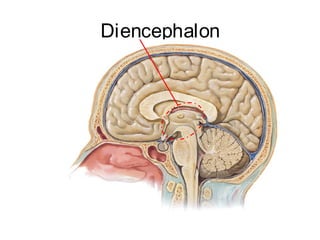
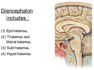
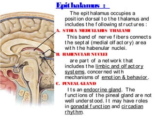
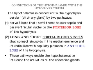
The document provides an overview of the diencephalon, which is the region of the brain between the cerebral hemispheres and below the thalamus. It describes the gross topography and borders of the diencephalon. The diencephalon includes the epithalamus, thalamus, metathalamus, subthalamus, and hypothalamus. Details are given on the structures and functions of these regions, including the thalamic nuclei and their connections. Clinical notes discuss lesions of the thalamus and how they can cause sensory loss or pain.
the ddep structure of brain, diencephalon, third ventricle, thalamus, hypothalamus, epithalamus, meta thalamus, boudaries of diencephalon, extent of diencephalon, boundaries of thalamus, boundaries of hypothalamus, functions of meta thalamus, functions of sub thalamus.components of epithalamus, functions of epithalamus, fornix, third ventricle, optic chiasma,
The thalamus is the large mass of gray matter in the dorsal part of the diencephalon of the brain with several functions such as relaying of sensory signals, including motor signals, to the cerebral cortex and the regulation of consciousness, sleep, and alertness.
The somatosensory system is the part of the sensory system concerned with the conscious perception of touch, pressure, pain, temperature, position, movement, and vibration, which arise from the muscles, joints, skin, and fascia.
The somatosensory system is a 3-neuron system that relays sensations detected in the periphery and conveys them via pathways through the spinal cord, brainstem, and thalamic relay nuclei to the sensory cortex in the parietal lobe
Impulses are carried from receptors via sensory afferents to the dorsal root ganglia, where the cell bodies of the first-order neurons are located.
Here the fibers split into 2 functional groups: a lateral group (or anterolateral system) and a medial group (or dorsal column-medial lemniscal system).
The lateral group carries mainly unmyelinated fibers that subserve pain and temperature sensations, whereas the medial group carries mainly myelinated fibers that convey proprioceptive impulses
Their axons then travel through the spinal cord either in an ipsilateral or a contralateral fashion. Note that second-order neuron cell bodies are located in different anatomical areas depending on the sensation they carry.
Broadly, the spinal cord contains the second-order neurons for the fibers carrying pain, touch, and temperature sensations.
The lateral group of fibers enters the spinal cord, then ascend to terminate on the substantia gelatinosa and the nucleus proprius, where the second-order neurons are housed
Fibers then ascend via the brainstem to the thalamus in the spinothalamic tracts (or STT).
The medulla contains the second-order neurons for fibers carrying touch, position, and vibratory sensations. The fibers are then either conveyed to the thalamus (where the third-order neurons are located)
The medial group also sends its fibers into the posterior spinal cord; however, upon reaching it, most fibers ascend to the dorsal column nuclei in the medulla and synapse there
These tracts synapse on a second-order neuron in the nucleus gracilis and cuneatus, which are located in the medulla.
Their axons then decussate form a bundle known as the medial lemniscus.
Fibers of the posterior columns and medial lemniscus are concerned primarily with position sense and fine discriminative touch
These fibers travel to the midbrain on their way to the thalamus. Once in the thalamus, they synapse on third-order neurons in the ventral posterior lateral (VPL) nucleus.
The third-order neurons then project to the primary somatosensory cortex, which is located in the postcentral gyrus (also known as Brodmann areas 1, 2, and 3) of the parietal lobe
Primary somatosensory cortex subserves general and proprioceptive sensations and serves to integrate sensory information
Somesthetic cortex is organized in a sensory homunculus
Body areas particularly important to the sensory system (for example the face, lips, and hand) are given larger representation than other areas
Sensory receptorsperipheral nerve dorsal
A simple basic professional content, which is suitable for representation by medical students, physicians and surgeons.
Your comments are more than welcomed.
A simple basic professional content, which is suitable for representation by medical students, physicians and surgeons.
Your comments are more than welcomed.
A simple basic professional content, which is suitable for representation by medical students, physicians and surgeons.
Your comments are more than welcomed.
A simple basic professional content, which is suitable for representation by medical students, physicians and surgeons.
Your comments are more than welcomed.
Prix Galien International 2024 Forum ProgramLevi Shapiro
June 20, 2024, Prix Galien International and Jerusalem Ethics Forum in ROME. Detailed agenda including panels:
- ADVANCES IN CARDIOLOGY: A NEW PARADIGM IS COMING
- WOMEN’S HEALTH: FERTILITY PRESERVATION
- WHAT’S NEW IN THE TREATMENT OF INFECTIOUS,
ONCOLOGICAL AND INFLAMMATORY SKIN DISEASES?
- ARTIFICIAL INTELLIGENCE AND ETHICS
- GENE THERAPY
- BEYOND BORDERS: GLOBAL INITIATIVES FOR DEMOCRATIZING LIFE SCIENCE TECHNOLOGIES AND PROMOTING ACCESS TO HEALTHCARE
- ETHICAL CHALLENGES IN LIFE SCIENCES
- Prix Galien International Awards Ceremony
Report Back from SGO 2024: What’s the Latest in Cervical Cancer?bkling
Are you curious about what’s new in cervical cancer research or unsure what the findings mean? Join Dr. Emily Ko, a gynecologic oncologist at Penn Medicine, to learn about the latest updates from the Society of Gynecologic Oncology (SGO) 2024 Annual Meeting on Women’s Cancer. Dr. Ko will discuss what the research presented at the conference means for you and answer your questions about the new developments.
New Directions in Targeted Therapeutic Approaches for Older Adults With Mantl...i3 Health
i3 Health is pleased to make the speaker slides from this activity available for use as a non-accredited self-study or teaching resource.
This slide deck presented by Dr. Kami Maddocks, Professor-Clinical in the Division of Hematology and
Associate Division Director for Ambulatory Operations
The Ohio State University Comprehensive Cancer Center, will provide insight into new directions in targeted therapeutic approaches for older adults with mantle cell lymphoma.
STATEMENT OF NEED
Mantle cell lymphoma (MCL) is a rare, aggressive B-cell non-Hodgkin lymphoma (NHL) accounting for 5% to 7% of all lymphomas. Its prognosis ranges from indolent disease that does not require treatment for years to very aggressive disease, which is associated with poor survival (Silkenstedt et al, 2021). Typically, MCL is diagnosed at advanced stage and in older patients who cannot tolerate intensive therapy (NCCN, 2022). Although recent advances have slightly increased remission rates, recurrence and relapse remain very common, leading to a median overall survival between 3 and 6 years (LLS, 2021). Though there are several effective options, progress is still needed towards establishing an accepted frontline approach for MCL (Castellino et al, 2022). Treatment selection and management of MCL are complicated by the heterogeneity of prognosis, advanced age and comorbidities of patients, and lack of an established standard approach for treatment, making it vital that clinicians be familiar with the latest research and advances in this area. In this activity chaired by Michael Wang, MD, Professor in the Department of Lymphoma & Myeloma at MD Anderson Cancer Center, expert faculty will discuss prognostic factors informing treatment, the promising results of recent trials in new therapeutic approaches, and the implications of treatment resistance in therapeutic selection for MCL.
Target Audience
Hematology/oncology fellows, attending faculty, and other health care professionals involved in the treatment of patients with mantle cell lymphoma (MCL).
Learning Objectives
1.) Identify clinical and biological prognostic factors that can guide treatment decision making for older adults with MCL
2.) Evaluate emerging data on targeted therapeutic approaches for treatment-naive and relapsed/refractory MCL and their applicability to older adults
3.) Assess mechanisms of resistance to targeted therapies for MCL and their implications for treatment selection
Title: Sense of Smell
Presenter: Dr. Faiza, Assistant Professor of Physiology
Qualifications:
MBBS (Best Graduate, AIMC Lahore)
FCPS Physiology
ICMT, CHPE, DHPE (STMU)
MPH (GC University, Faisalabad)
MBA (Virtual University of Pakistan)
Learning Objectives:
Describe the primary categories of smells and the concept of odor blindness.
Explain the structure and location of the olfactory membrane and mucosa, including the types and roles of cells involved in olfaction.
Describe the pathway and mechanisms of olfactory signal transmission from the olfactory receptors to the brain.
Illustrate the biochemical cascade triggered by odorant binding to olfactory receptors, including the role of G-proteins and second messengers in generating an action potential.
Identify different types of olfactory disorders such as anosmia, hyposmia, hyperosmia, and dysosmia, including their potential causes.
Key Topics:
Olfactory Genes:
3% of the human genome accounts for olfactory genes.
400 genes for odorant receptors.
Olfactory Membrane:
Located in the superior part of the nasal cavity.
Medially: Folds downward along the superior septum.
Laterally: Folds over the superior turbinate and upper surface of the middle turbinate.
Total surface area: 5-10 square centimeters.
Olfactory Mucosa:
Olfactory Cells: Bipolar nerve cells derived from the CNS (100 million), with 4-25 olfactory cilia per cell.
Sustentacular Cells: Produce mucus and maintain ionic and molecular environment.
Basal Cells: Replace worn-out olfactory cells with an average lifespan of 1-2 months.
Bowman’s Gland: Secretes mucus.
Stimulation of Olfactory Cells:
Odorant dissolves in mucus and attaches to receptors on olfactory cilia.
Involves a cascade effect through G-proteins and second messengers, leading to depolarization and action potential generation in the olfactory nerve.
Quality of a Good Odorant:
Small (3-20 Carbon atoms), volatile, water-soluble, and lipid-soluble.
Facilitated by odorant-binding proteins in mucus.
Membrane Potential and Action Potential:
Resting membrane potential: -55mV.
Action potential frequency in the olfactory nerve increases with odorant strength.
Adaptation Towards the Sense of Smell:
Rapid adaptation within the first second, with further slow adaptation.
Psychological adaptation greater than receptor adaptation, involving feedback inhibition from the central nervous system.
Primary Sensations of Smell:
Camphoraceous, Musky, Floral, Pepperminty, Ethereal, Pungent, Putrid.
Odor Detection Threshold:
Examples: Hydrogen sulfide (0.0005 ppm), Methyl-mercaptan (0.002 ppm).
Some toxic substances are odorless at lethal concentrations.
Characteristics of Smell:
Odor blindness for single substances due to lack of appropriate receptor protein.
Behavioral and emotional influences of smell.
Transmission of Olfactory Signals:
From olfactory cells to glomeruli in the olfactory bulb, involving lateral inhibition.
Primitive, less old, and new olfactory systems with different path
The prostate is an exocrine gland of the male mammalian reproductive system
It is a walnut-sized gland that forms part of the male reproductive system and is located in front of the rectum and just below the urinary bladder
Function is to store and secrete a clear, slightly alkaline fluid that constitutes 10-30% of the volume of the seminal fluid that along with the spermatozoa, constitutes semen
A healthy human prostate measures (4cm-vertical, by 3cm-horizontal, 2cm ant-post ).
It surrounds the urethra just below the urinary bladder. It has anterior, median, posterior and two lateral lobes
It’s work is regulated by androgens which are responsible for male sex characteristics
Generalised disease of the prostate due to hormonal derangement which leads to non malignant enlargement of the gland (increase in the number of epithelial cells and stromal tissue)to cause compression of the urethra leading to symptoms (LUTS
Factory Supply Best Quality Pmk Oil CAS 28578–16–7 PMK Powder in Stockrebeccabio
Factory Supply Best Quality Pmk Oil CAS 28578–16–7 PMK Powder in Stock
Telegram: bmksupplier
signal: +85264872720
threema: TUD4A6YC
You can contact me on Telegram or Threema
Communicate promptly and reply
Free of customs clearance, Double Clearance 100% pass delivery to USA, Canada, Spain, Germany, Netherland, Poland, Italy, Sweden, UK, Czech Republic, Australia, Mexico, Russia, Ukraine, Kazakhstan.Door to door service
Hot Selling Organic intermediates
Explore natural remedies for syphilis treatment in Singapore. Discover alternative therapies, herbal remedies, and lifestyle changes that may complement conventional treatments. Learn about holistic approaches to managing syphilis symptoms and supporting overall health.
These simplified slides by Dr. Sidra Arshad present an overview of the non-respiratory functions of the respiratory tract.
Learning objectives:
1. Enlist the non-respiratory functions of the respiratory tract
2. Briefly explain how these functions are carried out
3. Discuss the significance of dead space
4. Differentiate between minute ventilation and alveolar ventilation
5. Describe the cough and sneeze reflexes
Study Resources:
1. Chapter 39, Guyton and Hall Textbook of Medical Physiology, 14th edition
2. Chapter 34, Ganong’s Review of Medical Physiology, 26th edition
3. Chapter 17, Human Physiology by Lauralee Sherwood, 9th edition
4. Non-respiratory functions of the lungs https://academic.oup.com/bjaed/article/13/3/98/278874
micro teaching on communication m.sc nursing.pdfAnurag Sharma
Microteaching is a unique model of practice teaching. It is a viable instrument for the. desired change in the teaching behavior or the behavior potential which, in specified types of real. classroom situations, tends to facilitate the achievement of specified types of objectives.
Pulmonary Thromboembolism - etilogy, types, medical- Surgical and nursing man...VarunMahajani
Disruption of blood supply to lung alveoli due to blockage of one or more pulmonary blood vessels is called as Pulmonary thromboembolism. In this presentation we will discuss its causes, types and its management in depth.
Anti ulcer drugs and their Advance pharmacology ||
Anti-ulcer drugs are medications used to prevent and treat ulcers in the stomach and upper part of the small intestine (duodenal ulcers). These ulcers are often caused by an imbalance between stomach acid and the mucosal lining, which protects the stomach lining.
||Scope: Overview of various classes of anti-ulcer drugs, their mechanisms of action, indications, side effects, and clinical considerations.
Title: Sense of Taste
Presenter: Dr. Faiza, Assistant Professor of Physiology
Qualifications:
MBBS (Best Graduate, AIMC Lahore)
FCPS Physiology
ICMT, CHPE, DHPE (STMU)
MPH (GC University, Faisalabad)
MBA (Virtual University of Pakistan)
Learning Objectives:
Describe the structure and function of taste buds.
Describe the relationship between the taste threshold and taste index of common substances.
Explain the chemical basis and signal transduction of taste perception for each type of primary taste sensation.
Recognize different abnormalities of taste perception and their causes.
Key Topics:
Significance of Taste Sensation:
Differentiation between pleasant and harmful food
Influence on behavior
Selection of food based on metabolic needs
Receptors of Taste:
Taste buds on the tongue
Influence of sense of smell, texture of food, and pain stimulation (e.g., by pepper)
Primary and Secondary Taste Sensations:
Primary taste sensations: Sweet, Sour, Salty, Bitter, Umami
Chemical basis and signal transduction mechanisms for each taste
Taste Threshold and Index:
Taste threshold values for Sweet (sucrose), Salty (NaCl), Sour (HCl), and Bitter (Quinine)
Taste index relationship: Inversely proportional to taste threshold
Taste Blindness:
Inability to taste certain substances, particularly thiourea compounds
Example: Phenylthiocarbamide
Structure and Function of Taste Buds:
Composition: Epithelial cells, Sustentacular/Supporting cells, Taste cells, Basal cells
Features: Taste pores, Taste hairs/microvilli, and Taste nerve fibers
Location of Taste Buds:
Found in papillae of the tongue (Fungiform, Circumvallate, Foliate)
Also present on the palate, tonsillar pillars, epiglottis, and proximal esophagus
Mechanism of Taste Stimulation:
Interaction of taste substances with receptors on microvilli
Signal transduction pathways for Umami, Sweet, Bitter, Sour, and Salty tastes
Taste Sensitivity and Adaptation:
Decrease in sensitivity with age
Rapid adaptation of taste sensation
Role of Saliva in Taste:
Dissolution of tastants to reach receptors
Washing away the stimulus
Taste Preferences and Aversions:
Mechanisms behind taste preference and aversion
Influence of receptors and neural pathways
Impact of Sensory Nerve Damage:
Degeneration of taste buds if the sensory nerve fiber is cut
Abnormalities of Taste Detection:
Conditions: Ageusia, Hypogeusia, Dysgeusia (parageusia)
Causes: Nerve damage, neurological disorders, infections, poor oral hygiene, adverse drug effects, deficiencies, aging, tobacco use, altered neurotransmitter levels
Neurotransmitters and Taste Threshold:
Effects of serotonin (5-HT) and norepinephrine (NE) on taste sensitivity
Supertasters:
25% of the population with heightened sensitivity to taste, especially bitterness
Increased number of fungiform papillae
3. GROSS TOPOGRAPHY OF
DI ENCEPHALON,
“or in-bet ween br ain,”
• is complet ely sur rounded by
t he cer ebral hemispher es
except at it s vent ral
surf ace.
Borders :
• Ant er ior ly :
Foramen of Monro
Lamina t er minalis
• Post erior ly :
Post erior commissur e
4. • Super ior ly :
f or ms f loor of
lat er al vent ricle
• Medially :
f or ms lat eral wall
of
3rd
vent r icle.
• lat er ally :
Post er ior limb of
int er nal capsule
Lat er al and 3rd
vent r icles
6. • A groove ext ending
bet ween t he f oramen
of Monro and t he
aqueduct of Sylvius
(The Hypot halamic
Sulcus)
t hat divides
diencephalon int o
1. A vent ral port ion,
The
7. • The t wo t halami are
connect ed acr oss
t he midline in about
70% of humans
t hrough t he
(int ert halamic
adhesion)
10. Epithalamus :
The epit halamus occupies a
posit ion dorsal t o t he t halamus and
includes t he f ollowing st ruct ures :
A. STRIA MEDULLARIS THALAMI
This band of nerve f ibers connect s
t he sept al (medial olf act ory) area
wit h t he habenular nuclei.
B. HABENULAR NUCLEI
are part of a net work t hat
includes t he limbic and olf act ory
syst ems, concerned wit h
mechanisms of emot ion & behavior .
C. PINEAL GLAND
I t s an endocrine gland. The
f unct ions of t he pineal gland are not
well underst ood. I t may have roles
in gonadal f unct ion and circadian
rhyt hm.
12. Thalamus :
General Appearances
• The t halamus is a large,
egg-shaped mass of gr ay
mat t er t hat f or ms t he
maj or par t of t he
diencephalon.
• Ther e ar e t wo t halami, and
one is sit uat ed on each side
of t he t hir d vent r icle.
13. • Ant er ior end
of t he t halamus is
nar row and r ounded
• Post er ior end
is expanded t o f or m
t he pulvinar , which
overhangs t he
super ior colliculus .
• Usually connect ed t o
t he opposit e t halamus
by int er t halamic
adhesion
14. Relat ions :
• Ant erior ly: int ervent ricular
f oramen
• Post eriorly : expand as Pulvinar
• Superiorly : f loor of lat eral
vent ricle & Tela choroidae of
3rd
vent ricle
• I nf erior ly : I t rest s on
subt halamus & hypot halamus.
Midbrain.
• Medially : f orms part of lat eral
wall of t he 3rd
vent ricle. St ria
medullaris t halami
15. Subdivisions of t he Thalamus
The gr ay mat t er of t he
t halamus is divided by a
ver t ical sheet of whit e
mat t er,
int er nal medullar yint er nal medullar y
laminalamina,
int o medial and lat er al
halves. t halamus t hus is
subdivided int o t hree main
part s;
1. Ant er ior part : lies bet ween
t he limbs of t he Y.
2. Medial part : on medial side
of Y st em.
Anterior
Medial
Lateral
16. • Each of t he t hree par t s of t he t halamus cont ains
a gr oup of :
THALAMI C NUCLEI .
• Ot her smaller nuclear groups are :
1. Wit hin int ernal medullar y lamina ( int r alaminar ),
2. On medial surf ace of t he t halamus
3. On lat er al sur f ace of t he t halamus.
4. Ret icular nucleus.
17. 1. Ant er ior par t :
• Ant er ior t halamic nuclei
2. Medial par t :
• Dor somedial nucleus
• Sever al smaller nuclei
3. Lat er al par t :
A. Dorsal Tier of t he Nuclei :
• Lat eral dorsal nucleus
( LD ),
• Lat eral post erior nucleus (
LP ),
• Pulvinar
B. Vent ral Tier of t he Nuclei :
• Vent ral ant erior nucleus
( VA )
• Vent ral lat eral nucleus ( VL
)
• Vent ral post erior nuclei
( VP ):
1. Vent ral post eromedial ( VPM
Thalamic Nuclei :
19. Funct ion of t he Thalamus
Alt hough an enormous amount of research has
been devot ed t o t his area, we st ill know very
lit t le about t he f unct ional signif icance of many
of t he nuclei.
1. A vast amount of sensory inf ormat ion of all
t ypes (except smell t hat inegrat ed wit h t ast e
t hen) converges on t he t halamus and presumably
is int egrat ed t hrough t he int erconnect ions
bet ween t he nuclei. The result ing inf ormat ion
pat t ern is dist ribut ed t o ot her part s of t he
cent ral nervous syst em.
2.The t halamus possesses cert ain very import ant
nuclei whose connect ions have been clearly
est ablished. These include t he ventralventral
posteromedialposteromedial nucleus, t he ventralventral
posterolateralposterolateral nucleus, t he medial geniculatemedial geniculate
body, and t he lateral geniculatelateral geniculate body. Their
20. 3. Anat omically and f unct ionally, t he
t halamus and t he cerebral cort ex are
closely linked. The f iber connect ions
have been est ablished, and it is known
t hat f ollowing removal of t he cort ex,
t he t halamus can appreciat e crude
sensat ions. However, t he cerebral
cort ex is required f or t he
int erpret at ion of sensat ions based on
past experiences. For example, if t he
sensory cort ex is dest royed :
• one can st ill appreciat e t he presence
of a hot obj ect in t he hand;
• appreciat ion of t he shape, weight , and
exact t emperat ure of t he obj ect
22. Thalamic
Nucleus
Af f er ent
Neuronal Loop
Ef f er ent
Neur onal Loop
Funct ion
Ant er ior
Nucleus
Cingulat e gyr us,
Hypot halamus
• Emot ional
t one
• Recent
memor y
23. Thalamic
Nucleus
Af f er ent
Neur onal
Loop
Ef f er ent
Neur onal Loop
Funct ion
Medial
Nuclei:
Dor somedial
Pr ef r ont al cor t ex,
hypot halamus,
ot her t halamic nuclei
I nt egr at ion
of somat ic,
viscer al, &
olf act or y
inf or mat ion
and
r elat ion t o
emot ional
f eelings and
subj ect ive
st at es
24. Thalamic
Nucleus
Af f er ent
Neur onal
Loop
Ef f er ent
Neur onal
Loop
Funct ion
Dor sal Tier of
t he Nuclei :
Lat er al
dorsal,
Lat er al
post erior,
Pulvinar
Cerebral cor t ex,
ot her t halamic nuclei
Unknown?
25. Thalamic
Nucleus
Af f er ent
Neur onal
Loop
Ef f er ent
Neur onal Loop
Funct ion
Vent ral Tier
of t he
Nuclei :
1. Vent r al
ant erior N.
Ret icular f or mat ion,
subst ant ia nigr a,
cor pus st r iat um,
pr emot or
cor t ex,
ot her t halamic nuclei
I nf luences
act ivit y of
mot or
cor t ex
2. Vent r al
lat eral N.
As in VA, but also has :
• maj or input f r om
cerebellum
26. Thalamic
Nucleus
Af f er ent
Neuronal
Loop
Ef f er ent
Neur onal
Loop
Funct ion
3. Vent r al
post er omedia
l (VPM)
Trigeminal
lemniscus,
gust at or y
f iber s
Pr imar y
somat ic
sensor y
(areas 3,1,
2)
in
cor t ex
Relays
common
sensat ions t o
consciousness4. Vent r al
post er olat era
l (VPL)
Medial and
spinal
lemnisci
27. Thalamic
Nucleus
Af f er ent Neur onal
Loop
Ef f er ent
Neur onal
Loop
Funct ion
Ret icular
Cer ebr al cor t ex,
r et icular f or mat ion
Ot her
t halamic
nuclei
Cer ebral
cor t ex
r egulat es
t halamus
I nt r alamina
r
Ret icular
f or mat ion,
spinot halamic and
t r igeminot halamic
t r act s
To
cer ebr al
cor t ex via
ot her
t halamic
nuclei,
cor pus
I nf luences
levels of
consciousne
ss and
aler t ness
29. METATHALAMUS
The t erm met at halamus ref ers t o t wo t halamic nuclei : t he
medial geniculat e and lat eral geniculat e.
1. Medial Geniculate Nucleus
This is a relay t halamic nucleus in t he audit ory syst em. I t
receives :
• Audit ory f ibers reach t he medial geniculat e body via
inf erior brachium of t he inf erior colliculus.
• Af f erent f eedback f ibers comes f rom t he primary audit ory
cort ex in t he t emporal lobe.
• Ef f erent out f low f orms t he audit ory radiat ion of t he
int ernal capsule t o t he primary audit ory cort ex in t he
t emporal lobe (areas 41 and 42).
• have roles in :
1. spectral analysis of sound,
2. sound pattern recognition,
3. auditory memory,
4. localization of sound in space,
30. 2. Lateral Geniculate Nucleus
This is a r elay nucleus in t he visual syst em. I t
receives :
• Visual f iber s f r om t he opt ic t ract conveying
impulses f r om bot h r et inae.
• Af f erent Feedback f ibers also reach t he nucleus
f rom t he primar y visual cort ex (ar ea 17) in t he
occipit al lobes.
• Ef f erent out f low f or ms t he opt ic radiat ion of t he
int er nal capsule t o t he primar y visual cort ex in t he
occipit al lobe.
• Some of t he ef f erent out f low pr oj ect s t o t he
pulvinar nucleus and t o t he secondar y visual cort ex
(areas 18 and 19)
31. Clinical Not es About Thalamus
• Since t he t halamus is such an import ant
relay and int egrat ive cent er, so f ollowing
disease of t his area t here will be prof ound
ef f ect s.
• The t halamus may be invaded by neoplasm,
undergo degenerat ion f ollowing disease of it s
art erial supply, or be damaged by
hemorrhage.
32. Lesions of t he Thalamus
Sensory Loss
• These lesions usually result f rom t hrombosis
or hemorrhage of one of t he art eries
supplying t he t halamus. Damage t o t he
vent ral post eromedial nucleus and t he
vent ral post erolat eral nucleus will result inresult in
the loss of all forms of sensationthe loss of all forms of sensation,
including light t ouch, t act ile localizat ion and
discriminat ion, and muscle j oint sense f rom
t he opposit e side of t he body.
33. • Surgical Relief of Pain by Thalamic
Caut erizat ion :
Caut erizat ion of some nuclei has been shown
t o relieve severe and int ract able pain
associat ed wit h t erminal cancer.
• Thalamic Pain:
Thalamic pain may occur as t he pat ient is
recovering f rom a t halamic inf arct .
Spont aneous pain, which is of t en excessive
(t halamic overreact ion), occurs on t he
opposit e side of t he body. The painf ul
sensat ion may be aroused by light t ouch or
by cold and may f ail t o respond t o powerf ul
34. Subt halamus
The subt halamus is a mass of gray and whit e
subst ance in t he caudal diencephalon. I t is
bor dered :
• Medially by t he hypot halamus,
• Lat er ally by t he int ernal capsule,
• Dor sally by t he t halamus,
• Vent r ally by t he int ernal capsule.
The subt halamus consist s of t hr ee main st ruct ur es;
t hese ar e :
1. Subt halamic Nucleus,
2. Fields Of For el,
3. Zona I ncert a.
Funct ion : has wide connect ions, t hat engage it in
37. Hypot halamus
Lies in t he diencephalon vent r al t o t he
hypot halamic sulcus .
Relat ions:
1. Ant er ior ly : Lamina t er minalis
2. Post er ior ly : Midbrain.
3. I nf er ior ly : Ant ropost er iorly :
• Opt ic chiasma,
• I nf undibulum f orms pit uit ary st alk.
• Floor slopes upwards and backwar ds t o aqueduct ,
• Floor is mar ked by t he pair of mamillar y bodies and
behind t hem by t he post erior per f or at ed
subst ance.
• Floor bet ween opr ic chiasma & mamillary bodies is
Tuber Cinereum.
4 Medially : 3rd
vent r icle
38. • The f ornix divides t he hypot halamus
1. The Lat eral Zone :
cont ains mainly t he medial
f orebr ain bundle which connect s t he sept al
area, hypot halamus & midbrain. Among
which are scat t ered neurons of lat eral
hypot halamic nuclei which are :
• part of t he preopt ic nucleus,
• part of t he suprachiasmat ic nucleus,
• Supraopt ic nucleus,
• Lat eral nucleus,
• Tuberomammillary nucleus,
• Lat eral t uberal nuclei.
39.
40. 1. Medial Zone :
has a clust er of
nuclei organized int o maj or groups. From
ant erior t o post erior :
• Preopt ic nucleus;
• Ant erior nucleus,
• Suprachiasmat ic nucleus;
• Paravent ricular nucleus;
• Dorsomedial nucleus;
• Vent romedial nucleus;
• I nf undibular (arcuat e) nucleus
43. Or igin Pat hway Dest inat ion
Viscer a and
somat ic
st r uct ur es
Medial and spinal
lemnisci, t r act us
solit ar ius,
r et icular
f or mat ion
Hypot halamic
nuclei
Ret ina Visual f ibers
Supr achiasmat ic
nucleus
Olf act ory mucous
membr ane
Medial f or ebr ain
bundle
Hypot halamic
nuclei
I nner ear Audit or y f ibers
Hypot halamic
nuclei
44. Or igin Pat hway Dest inat ion
Hippocampus
Hippocampo-
hypot halamic f iber s
( main out put of
limbic syst em)
Nuclei of
mammillar y body
Fr ont al lobe of
cer ebr al cor t ex
Cor t icohypot halamic
f iber s
Hypot halamic
nuclei
Amygdaloid
complex
Amygdalohypot hala
mic f iber s
Dor somedial and
midline nuclei of
t halamus
Thalamohypot halami
c f iber s
Tegment um of
midbr ain
Tegment al f iber s
46. Or igin Pat hway Dest inat ion
Pr eopt ic,
ant erior,
post er ior and
lat er al nuclei of
hypot halamus
Descending f ibers
in r et icular
f ormat ion t o
br ainst em and
spinal cor d
Par asympat het ic
& Sympat het ic
out f lows
Nuclei of
mammillary body
Mammillo
t halamic t r act
Ant erior nucleus
of t halamus;
relayed t o
cingulat e gyr us
Mammillo
t egment al t r act
Ret icular
f or mat ion in
t egment um of
midbr ain
Hypot halamic
Mult iple pat hways Limbic syst em
47. ConneCtions of the hypothalamus With the
hypophysis Cerebri
The hypot halamus is connect ed t o t he hypophysis
cerebri (pit uit ary gland) by t wo pat hways:
(1) nerve f ibers t hat t ravel f rom t he supraopt ic and
paravent ricular nuclei t o t he posterior lobe
of t he hypophysis
(2) long and short portal blood vessels
t hat connect sinusoids in t he median eminence and
inf undibulum wit h capillary plexuses in anterior
lobe of t he hypophysis.
• These pat hways enable t he hypot halamus t o
inf luence t he act ivit ies of t he endocrine glands.
48. Hypophyseal Port al Syst emHypophyseal Port al Syst em
is f ormed on each side f rom
t he
• Superior hypophyseal ar t er y,
which is a branch of t he
int ernal car ot id art ery.
• The art er y divides int o t uf t s
of capillar ies.
• These capillar ies drain int o
long and shor t descending
vessels t hat end in t he
ant erior lobe of t he
hypophysis by dividing int o
vascular sinusoids t hat pass
49. Hypot halamic
Releasing and I nhibit ory
Hormones and
Their Ef f ect s on
Ant erior Lobe of
Hypophysis (Pit uit ar y)
50. Hypot halami
c Regulat ory
Hor mone
Pr esumed
nuclei of
origin
Ant er ior
Pit uit ar y
Hor mone
pr oduces
Funct ional
Result
Growt h
Hor mone–
Releasing
Hor mone
(GHRH)
I nf undibular
(ar cuat e
nucleus)
Gr owt h
hor mone
(GH)
St imulat es
linear gr owt h
in epiphyseal
car t ilages
Growt h
Hor mone–
I nhibit ing
Hor mone
(GHI H) or
Supr achiasma
t ic nucleus
Gr owt h
hor mone
(r educed
pr oduct ion)
Reduces
linear gr owt h
in epiphyseal
car t ilages
51. Hypot halami
c Regulat ory
Hor mone
Presumed
nuclei of
or igin
Ant erior
Pit uit ary
Hor mone
Funct ional
Result
Pr olact in-
Releasing
Hor mone
(PRH)
?
Prolact in
(lut eot ropic
hor mone,
LTH)
St imulat es
lact ogenesis
Pr olact in-
I nhibit ing
Hor mone
(PI H),
Dopamine
Prolact in
(lut eot ropic
hor mone,
LTH)
(r educed
Reduces
lact ogenesis
52. Hypot halami
c Regulat ory
Hor mone
Presumed
nuclei of
or igin
Ant erior
Pit uit ary
Hor mone
Funct ional
Result
Cort icot ropi
n-Releasing
Hor mone
(CRH)
Par avent r icul
ar nuclei
Adr enocor t ic
ot r opic
hor mone
(ACTH)
St imulat es
adrenal gland
t o pr oduce
cor t icost er oi
ds and sex
hor mones
Thyr ot r opin
-Releasing
Hor mone
(TRH)
Par avent r icul
ar ,
dor somedial
nuclei and
adj acent
Thyr oid-
st imulat ing
hor mone
(TSH)
St imulat es
t hyr oid gland
t o pr oduce
t hyr oxine
53. Hypot halami
c Regulat ory
Hor mone
Presumed
nuclei of
or igin
Ant erior
Pit uit ary
Hor mone
Funct ional
Result
Lut einizing
Hor mone–
Releasing
Hor mone
(LHRH), ?
Follicle-
St imulat ing
Releasing
Hor mone
(FRH)
Pr eopt ic and
ant erior
nuclei
Lut einizing
hor mone
(LH) and
f ollicle-
st imulat ing
hor mone
(FSH)
St imulat es
ovarian
f ollicles and
product ion
of est r ogen
and
pr ogest eron
e
55. Hypot halamic
Nucleus
Presumed Funct ion
Supr aopt ic Synt hesizes ant idiur et ic hormone
Par avent r icular Synt hesizes oxyt ocin
Pr eopt ic &
ant er ior
Cont rol par asympat het ic syst em
Post er ior & lat eral Cont r ol sympat het ic syst em
Ant er ior Regulat e t emper at ur e (r esponse t o
heat )
Post erior Regulat e t emper at ur e (r esponse t o
cold)
Lat er al I ncr ease f ood int ake (hunger
cent er )
Medial Reduce f ood int ake (sat iet y
57. General Considerat ions :
• Af f erent haypot halamic pat hways are
received f rom a. cent ral nervous syst em
(especially f rom t he limbic syst em and t he
pref ront al cort ex)
b. plasma levels of circulat ing hormones.
• I t exert s it s inf luence on bodily f unct ions
t hrough t he autonomic nervous system &
endocrine system.
58. 1. Obesit y and Wast ing :
• I t is generally associat ed wit h genit al
hypoplasia or at rophy.
• Wast ing is less common t han obesit y in
hypot halamic disease.
• Severe cachexia is suggest ive of damage t o
pit uit ary gland.
2. Sexual Disorders :
• I n children, t here may be sexual ret ardat ion.
Af t er pubert y, t he pat ient wit h
hypot halamic disease may have impot ence or
amenorrhea.
59. 3. Hypert hermia and Hypot her mia :
• The pat ient wit h hyper t hermia is ot her wise normal
and has no signs of malaise, which occur s wit h
pyr exia secondar y t o inf ect ions.
• Hypot her mia also can f ollow a lesion of t he
hypot halamus.
4. Diabet es I nsipidus :
Diabet es insipidus result s f r om a lesion of t he
supraopt ic nucleus or f r om t he int er rupt ion of t he
ner vous pat hway t o t he post er ior lobe of t he
hypophysis. Char act er ist ically, t he pat ient passes
large volumes of ur ine of low specif ic gr avit y. As a
result , t he pat ient is ext remely t hirst y and dr inks
large quant it ies of f luids. The condit ion must be
60. 1. Dist urbances of Sleep :
• Short periods of sleep during t he waking
hours
• Or insomnia has been observed in pat ient s
wit h hypot halamic lesions.
2. Emot ional Disorders :
At t acks of unexplained weeping or
laught er, uncont rollable rage, depressive
react ions, and even maniacal out burst s all
have been observed in pat ient s wit h
hypot halamic lesions.