What Is Anatomyand Physiology?
Anatomy is the study of the structure and relationship between
body parts.
Physiology is the study of the function of body parts and the
body as a whole. Some specializations within each of these
sciences follow:
– Gross (macroscopic) anatomy is the study of body parts visible
to the naked eye, such as the heart or bones.
– Histology is the study of tissues at the microscopic level.
– Cytology is the study of cells at the microscopic level.
– Neurophysiology is the study of how the nervous system
functions.
3.
Musculoskeletal system
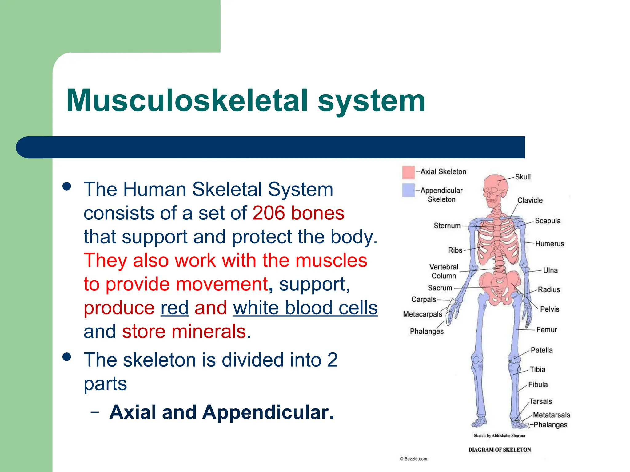
TheHuman Skeletal System
consists of a set of 206 bones
that support and protect the body.
They also work with the muscles
to provide movement, support,
produce red and white blood cells
and store minerals.
The skeleton is divided into 2
parts
– Axial and Appendicular.
4.
Axial Skeleton
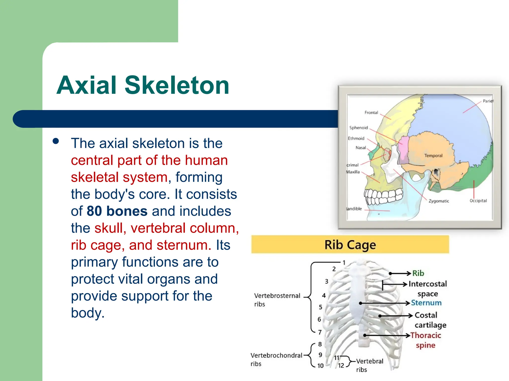
Theaxial skeleton is the
central part of the human
skeletal system, forming
the body's core. It consists
of 80 bones and includes
the skull, vertebral column,
rib cage, and sternum. Its
primary functions are to
protect vital organs and
provide support for the
body.
5.
Vertebral Column /Spine
The vertebral column,
also known as the
spinal column or spine,
is a flexible column of
33 vertebrae that
extends from the skull
to the pelvis.
6.
Appendicular Skeleton
Theappendicular skeleton is
composed of bones that anchor the
appendages to the axial skeleton.
– The Upper Extremities
– The Lower Extremities
– The Shoulder Girdle (pectoral
girdle)
– The Pelvic Girdle (the sacrum
and coccyx are considered part
of the vertebral column)
Tissues involved
Thereare 5 basic tissues comprising the
musculoskeletal system:
– bones,
– ligaments (attaching bone to bone)
– cartilage (protective gel-like substance lining the
joints and intervertebral discs),
– skeletal muscles
– tendons (attaching muscle to bone).
9.
Composition of bones
Bone is not a uniformly
solid material, but
rather has some
spaces between its
hard elements.
Two types of bone
tissue
– Compact
– Spongy
10.
Compact Bone
Thehard outer layer of bones is composed
of compact bone tissue, so-called due to its
minimal gaps and spaces.
This tissue gives bones their smooth, white,
and solid appearance, and accounts for 80%
of the total bone mass of an adult skeleton.
11.
Spongy Bone
Fillingthe interior of the organ is the trabecular bone
tissue (an open cell porous network also called
cancellous or spongy bone) which is composed of a
network of rod- and plate-like elements that make
the overall organ lighter and allowing room for blood
vessels and marrow.
Trabecular bone accounts for the remaining 20% of
total bone mass, but has nearly ten times the surface
area of compact bone.
12.
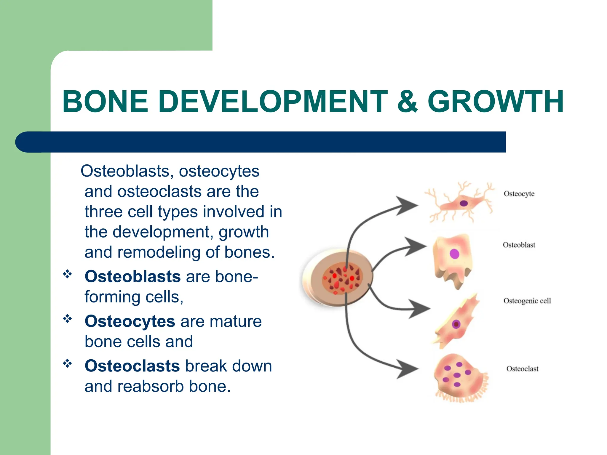
BONE DEVELOPMENT &GROWTH
Osteoblasts, osteocytes
and osteoclasts are the
three cell types involved in
the development, growth
and remodeling of bones.
Osteoblasts are bone-
forming cells,
Osteocytes are mature
bone cells and
Osteoclasts break down
and reabsorb bone.
13.
Five different typesof bones
Long (femur)
Short (carpus)
Flat (sternum)
Irregular (vertebrae)
Sesamoid (embedded
in tendon)
14.
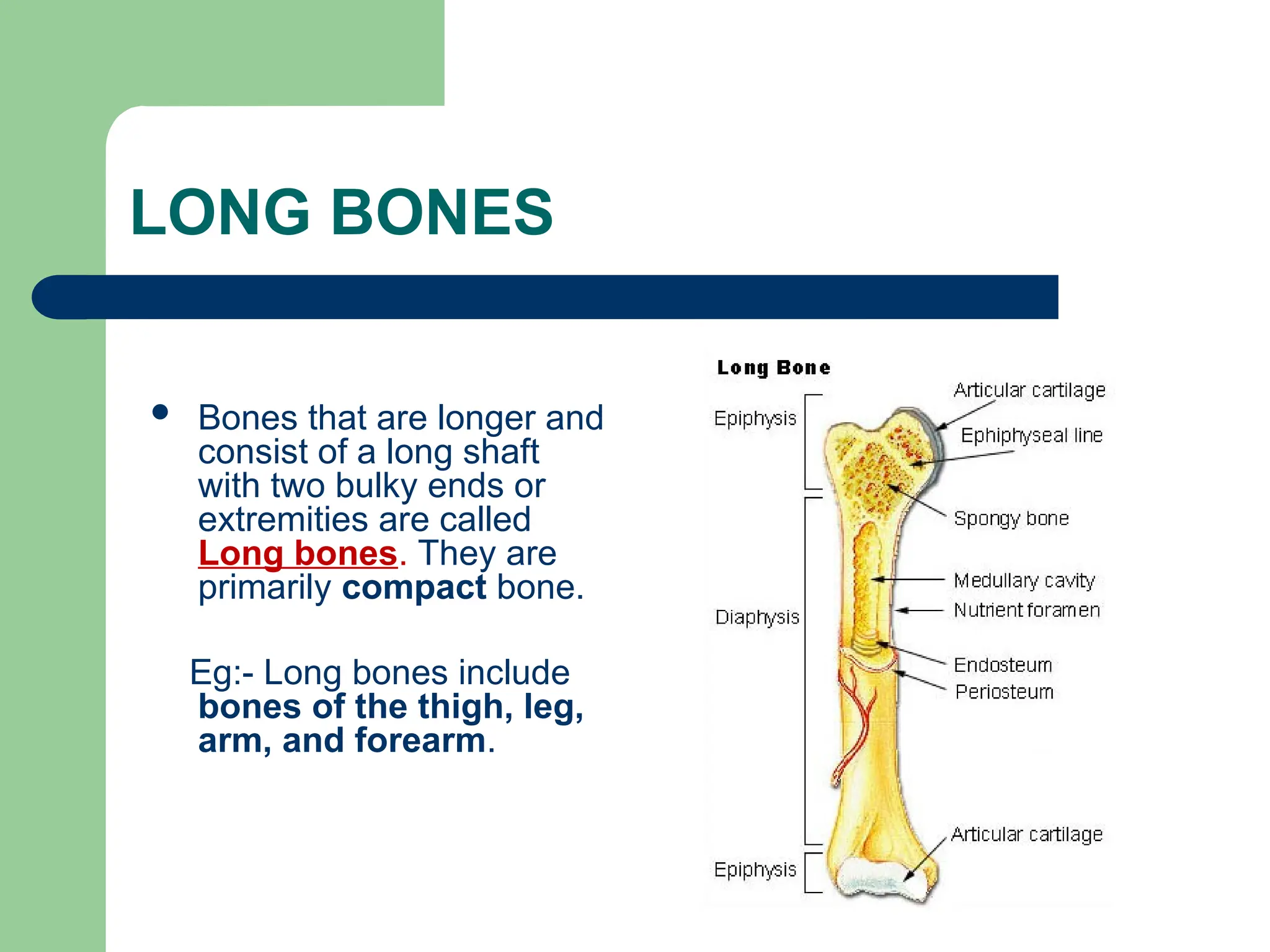
LONG BONES
Bonesthat are longer and
consist of a long shaft
with two bulky ends or
extremities are called
Long bones. They are
primarily compact bone.
Eg:- Long bones include
bones of the thigh, leg,
arm, and forearm.
15.
SHORT BONES
Shortbones consist
primarily of spongy
bone, which is
covered by a thin layer
of compact bone.
Eg:- Short bones include
the bones of the
wrist and ankle.
16.
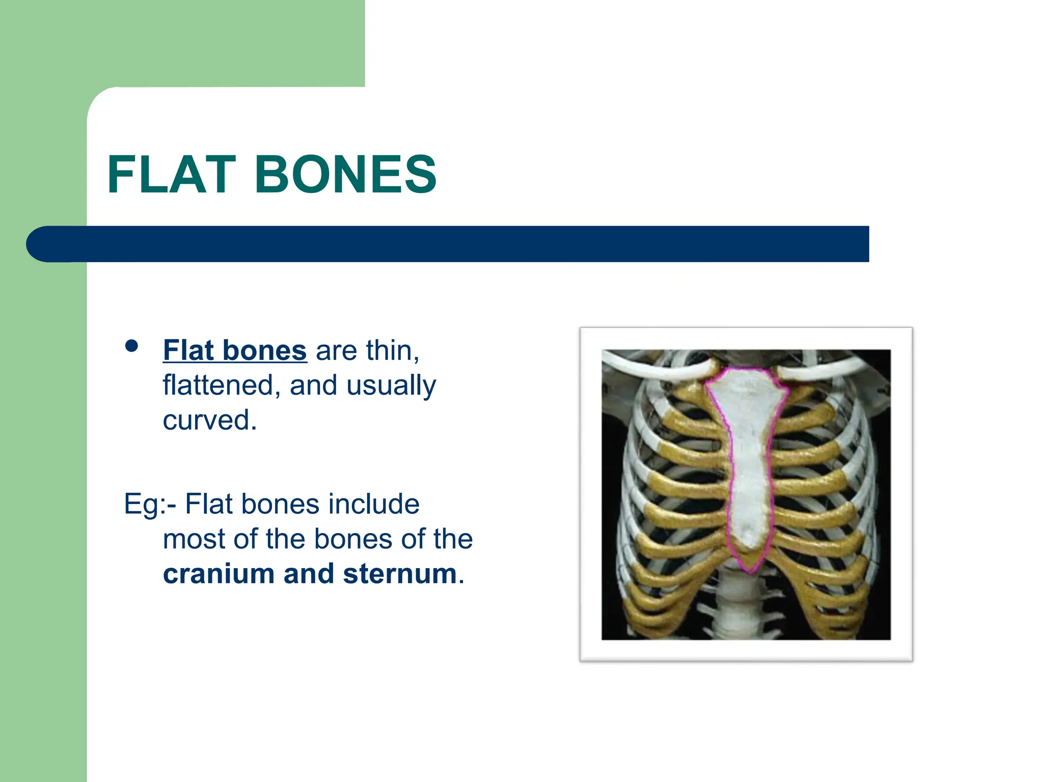
FLAT BONES
Flatbones are thin,
flattened, and usually
curved.
Eg:- Flat bones include
most of the bones of the
cranium and sternum.
17.
IRREGULAR BONES
Bonesthat are not in any
of the above three
categories are classified
as Irregular bones.
They are primarily
spongy bone that is
covered with a thin layer
of compact bone.
Eg:- The vertebrae and
some of the bones in the
skull are irregular bones.
18.
THE JOINTS
A Jointis the point where two or more bones meet.
There are three main types:
Fibrous (immoveable)- Eg:- Skull Joint.
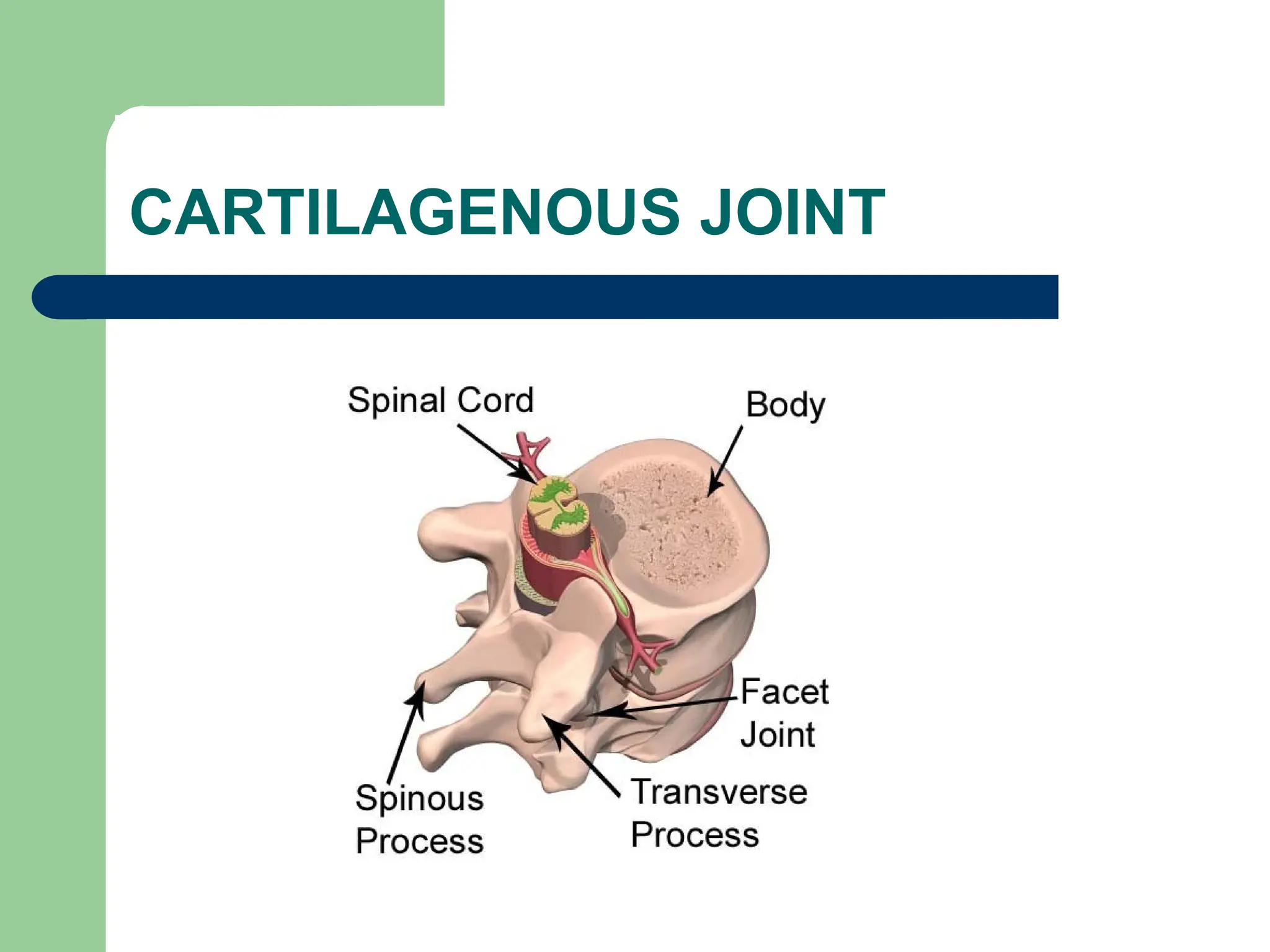
Cartilagenous (partially moveable)-
Eg:- Intervertebral disc of spinal column.
Synovial (freely moveable) joint.
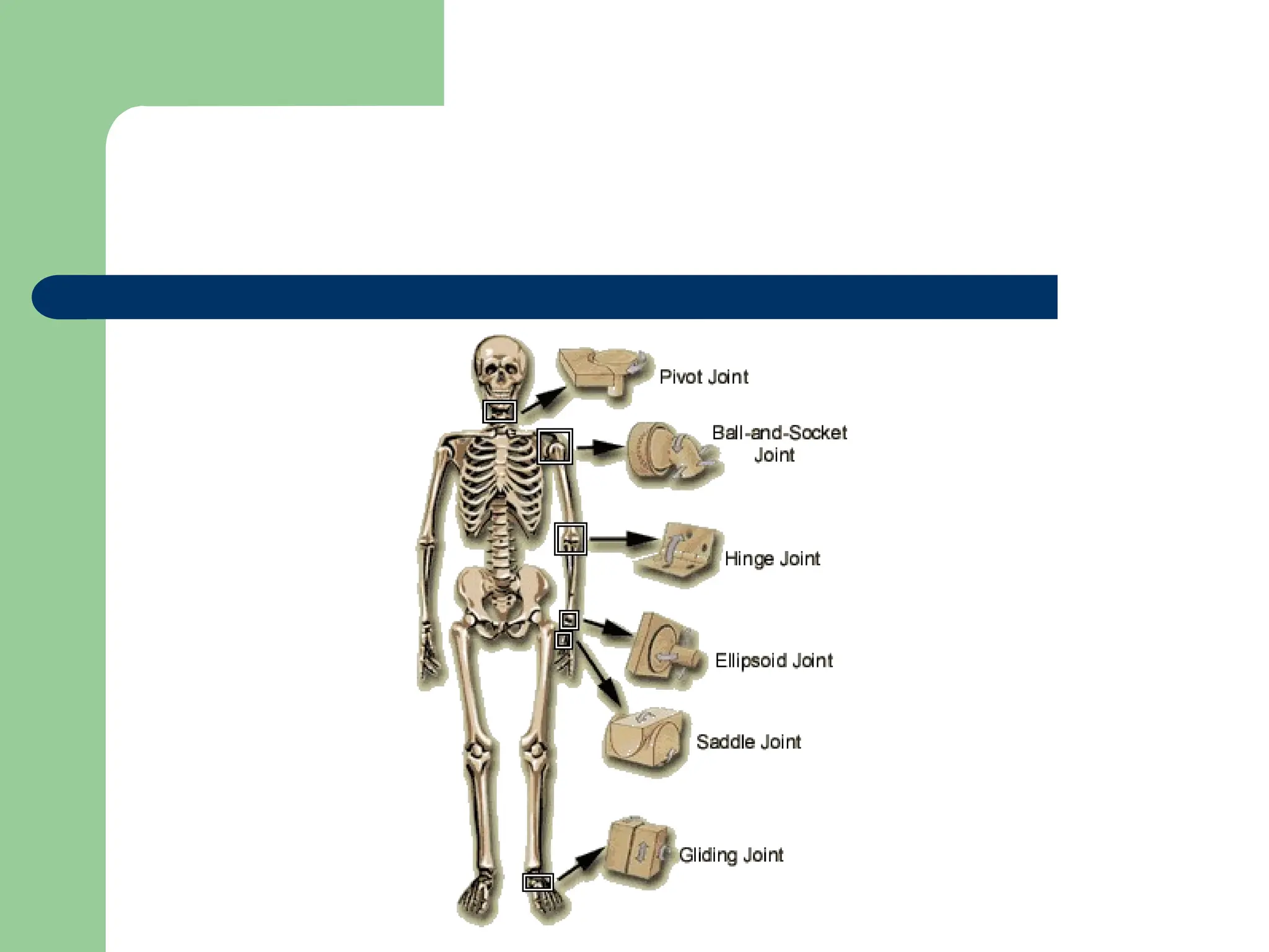
SYNOVIAL JOINT
There aresix types of synovial joints:
1) Pivot joint
2) Ball-and-socket joint
3) Hinge joint
4) Condyloid joint
5) Saddle joint
6) Gliding joint
22.
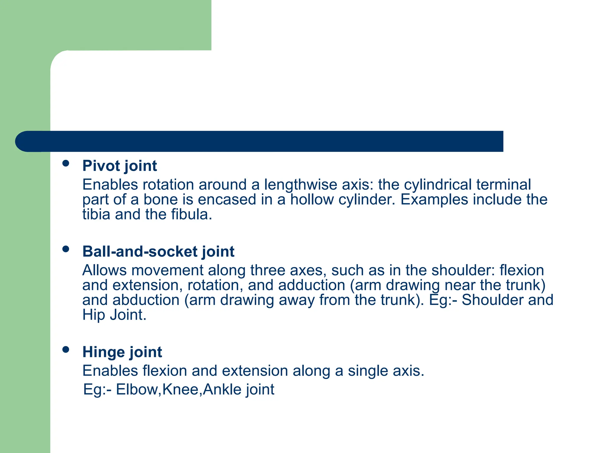
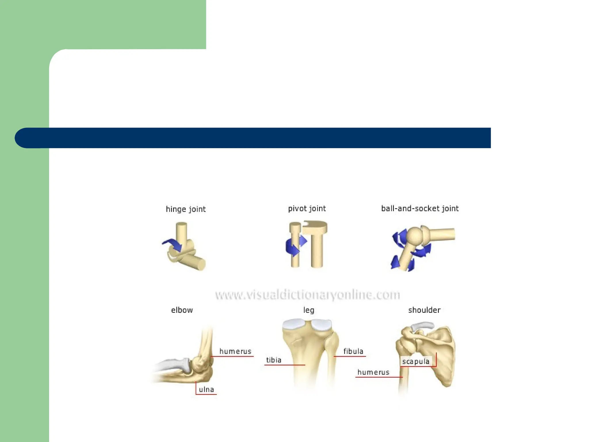
Pivot joint
Enablesrotation around a lengthwise axis: the cylindrical terminal
part of a bone is encased in a hollow cylinder. Examples include the
tibia and the fibula.
Ball-and-socket joint
Allows movement along three axes, such as in the shoulder: flexion
and extension, rotation, and adduction (arm drawing near the trunk)
and abduction (arm drawing away from the trunk). Eg:- Shoulder and
Hip Joint.
Hinge joint
Enables flexion and extension along a single axis.
Eg:- Elbow,Knee,Ankle joint
24.
Condyloid joint
Anexample is the wrist, which the hand can move on two axes:
flexion and extension; it can also be tilted sideways. Eg:- Wrist
joint.
Saddle joint
Resembles the condyloid joint but allows a wider range of
motion; this type of joint is rare. Eg:- Thumb.
Gliding joint
Surfaces of these joints are relatively flat and not very mobile;
they allow only a narrow gliding range.
Eg:- Vertebrae, certain bones of the wrist and ankle).
27.
THE TENDONS
Atendon is a tough but flexible
structure made of fibrous tissue
that joins a bone to a muscle.
When a muscle contracts it
pulls on a bone to cause
movement. The tendon
transmits the force from the
muscle to the bone.
The tendonitis is the
inflammation of a tendon.
28.
THE LIGAMENTS
Ligaments arebands of connective
tissues that link two or more bones to
make joints stable and prevent from
excessive movements.
29.
THE MUSCULAR SYSTEM
Our Skeletal has more than 650
muscles, most of them disposed in
pairs to provide movement.
30.
Three Types ofMuscles
The three types of muscles tissues are:
– Smooth
– Skeletal
– Cardiac
31.
Smooth Muscle
smooth(or visceral) muscle-
– forms the muscle layers in the walls of
the digestive tract, bladder, various
ducts, arteries and veins, and other
internal organs.
– Smooth- muscle cells are elongated and
thin, have only one nucleus, and form
sheets rather than bundles of muscles.
– Smooth muscle is controlled by the
autonomic nervous system (ANS).
32.
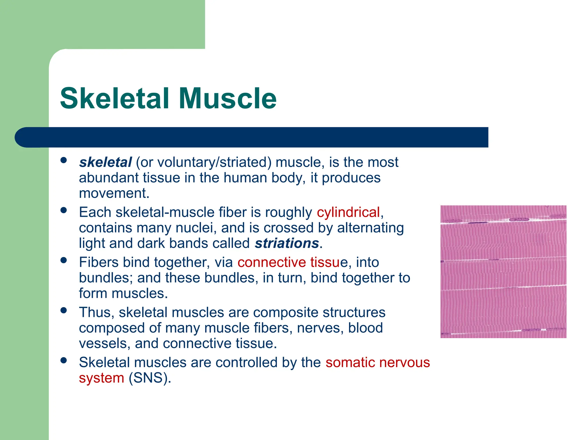
Skeletal Muscle
skeletal(or voluntary/striated) muscle, is the most
abundant tissue in the human body, it produces
movement.
Each skeletal-muscle fiber is roughly cylindrical,
contains many nuclei, and is crossed by alternating
light and dark bands called striations.
Fibers bind together, via connective tissue, into
bundles; and these bundles, in turn, bind together to
form muscles.
Thus, skeletal muscles are composite structures
composed of many muscle fibers, nerves, blood
vessels, and connective tissue.
Skeletal muscles are controlled by the somatic nervous
system (SNS).
33.
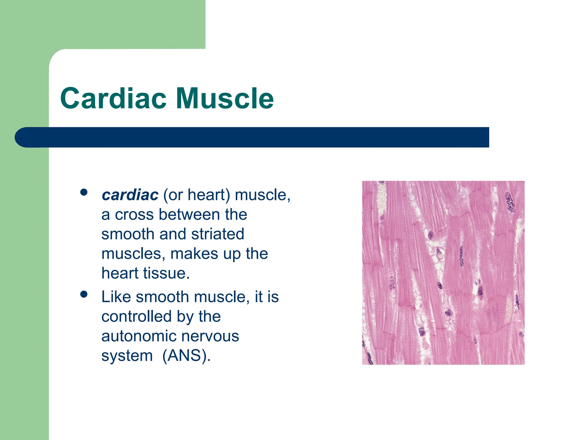
Cardiac Muscle
cardiac(or heart) muscle,
a cross between the
smooth and striated
muscles, makes up the
heart tissue.
Like smooth muscle, it is
controlled by the
autonomic nervous
system (ANS).
34.
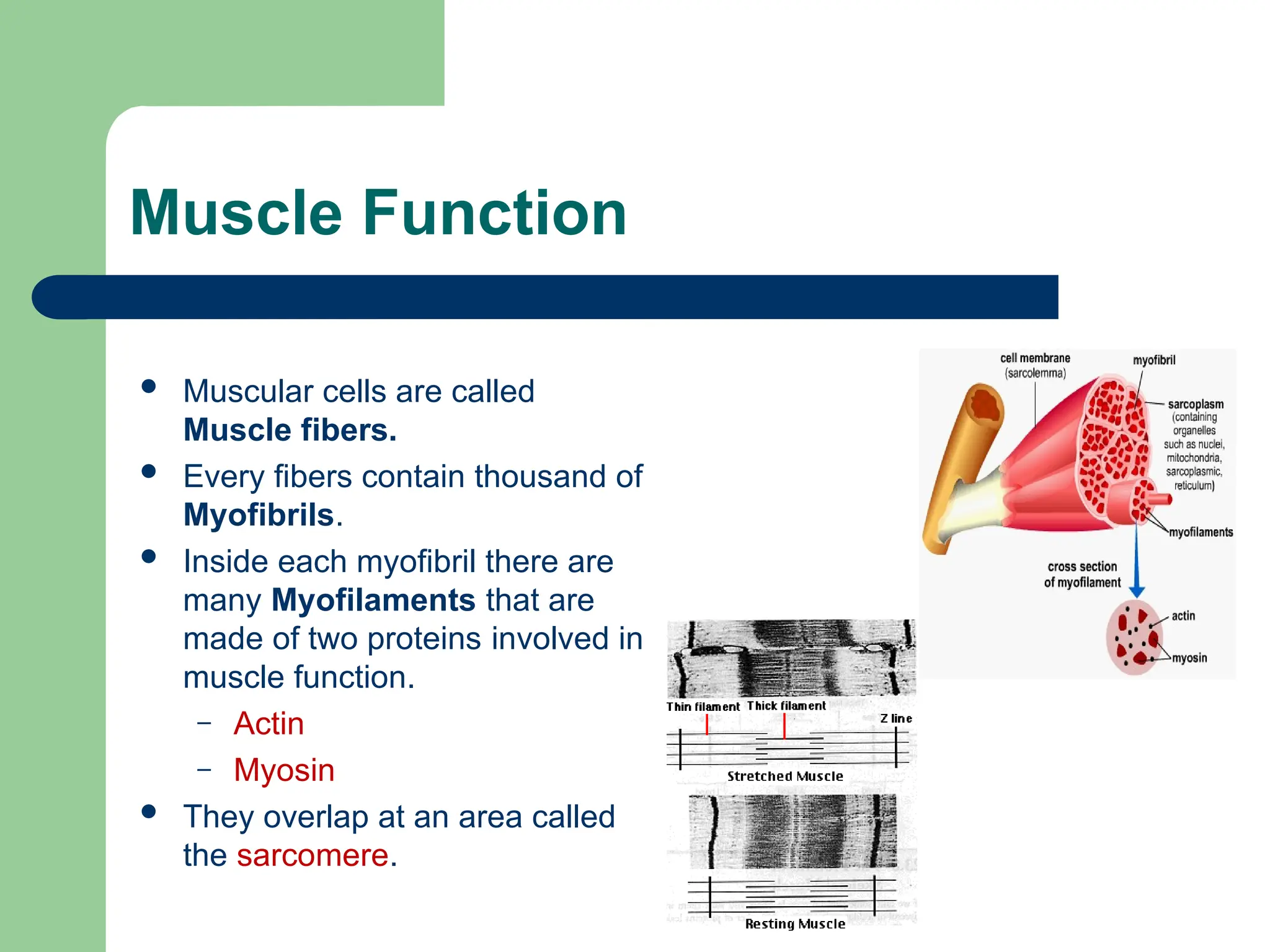
Muscle Function
Muscularcells are called
Muscle fibers.
Every fibers contain thousand of
Myofibrils.
Inside each myofibril there are
many Myofilaments that are
made of two proteins involved in
muscle function.
– Actin
– Myosin
They overlap at an area called
the sarcomere.