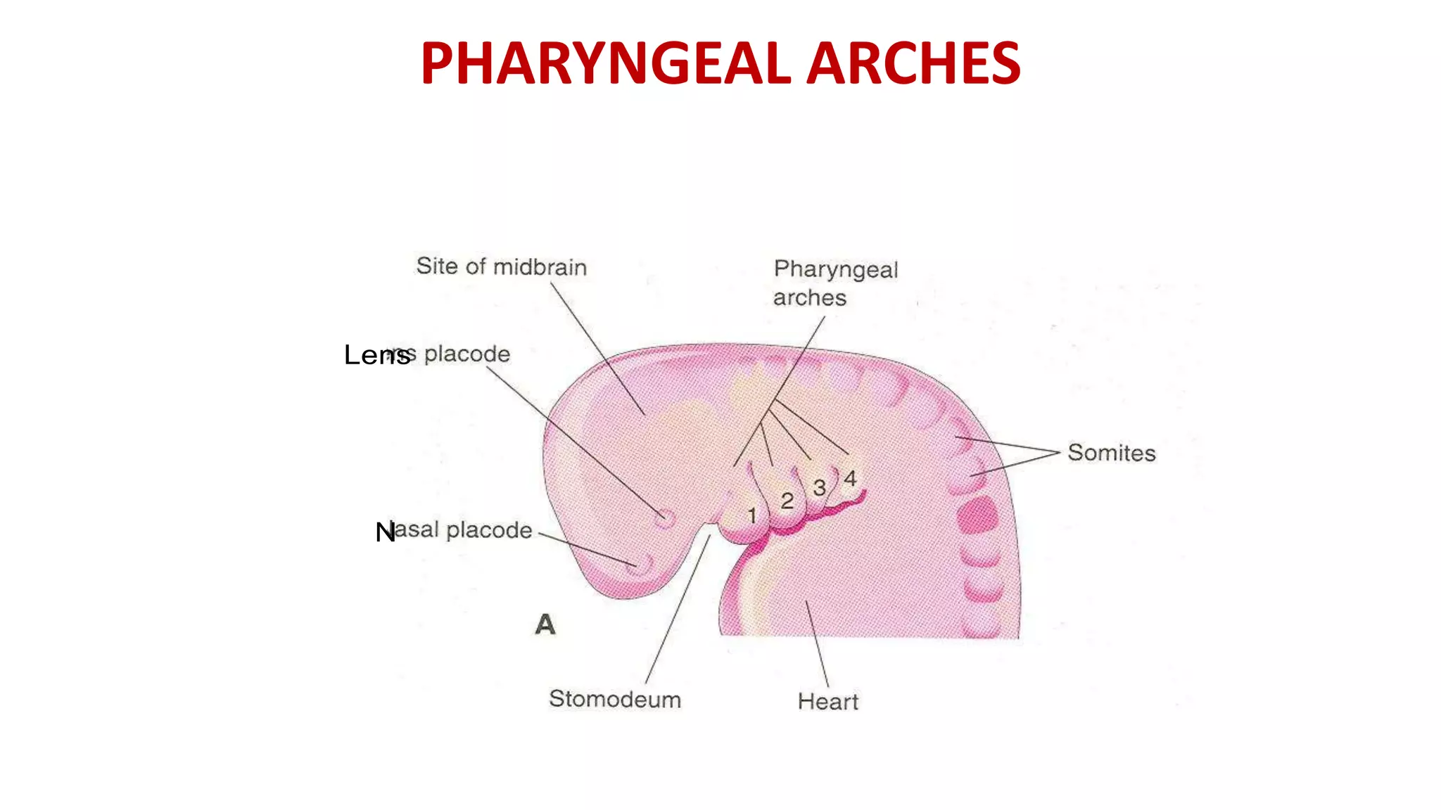
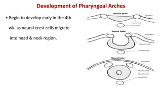
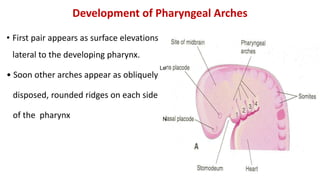
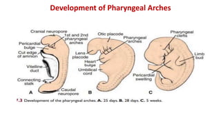
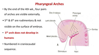
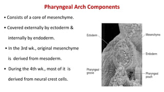
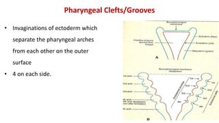
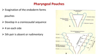
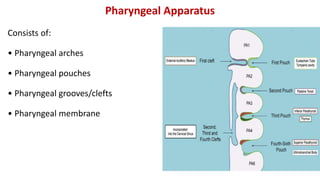
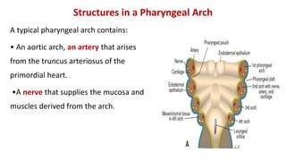
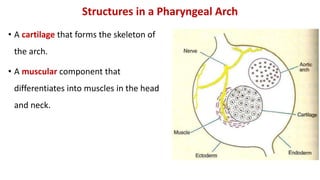
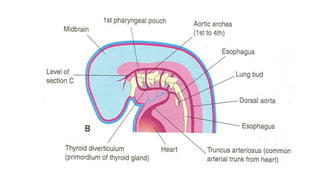
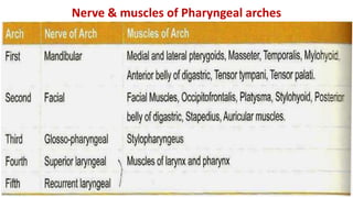
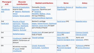
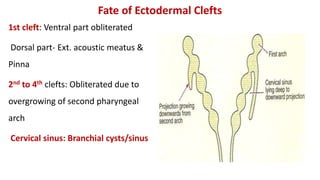
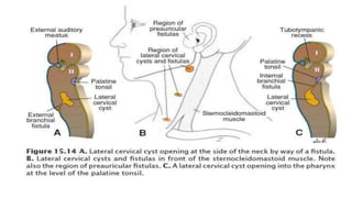
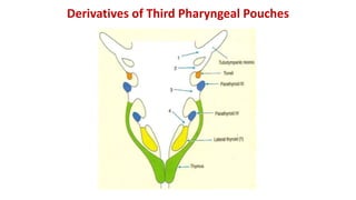
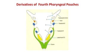
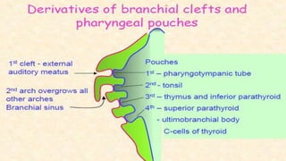
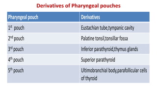
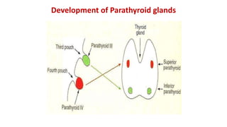
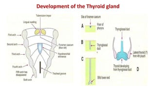
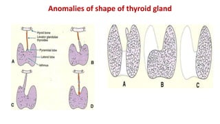
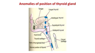
PHARYNGEAL ARCHES
What is Pharyngeal Arch?
• Rod-like thickenings of mesoderm present in the wall of the foregut.
• They appear in 4th-5th weeks of development.
• Contribute to the characteristic external appearance of the embryo.
• As its development resembles with gills ( Branchia: Greek word) in fishes
& amphibians, therefore also called as Branchial arch
Development of Pharyngeal Arches
• Begin to develop early in the 4th
wk. as neural crest cells migrate
into head & neck region.
• First pair appears as surface elevations
lateral to the developing pharynx.
• Soon other arches appear as obliquely
disposed, rounded ridges on each side
of the pharynx
Development of Pharyngeal Arches
Le
N
Development of Pharyngeal Arches
Pharyngeal Arches
• By the end of the 4th wk., four pairs
of arches are visible externally.
• 5th & 6th are rudimentary & not
visible on the surface of embryo.
• 5th arch does not develop in
humans
• Numbered in craniocaudal
sequence.
Le
N
Pharyngeal Arch Components
• Consists of a core of mesenchyme.
• Covered externally by ectoderm &
internally by endoderm.
• In the 3rd wk., original mesenchyme
is derived from mesoderm.
• During the 4th wk., most of it is
derived from neural crest cells.
• Invaginations of ectoderm which
separate the pharyngeal arches
from each other on the outer
surface
• 4 on each side.
Pharyngeal Clefts/Grooves
Pharyngeal Pouches
 Evagination of the endoderm forms
pouches
 Develop in a craniocaudal sequence
 4 on each side
 5th pair is absent or rudimentary
Pharyngeal Apparatus
Consists of:
• Pharyngeal arches
• Pharyngeal pouches
• Pharyngeal grooves/clefts
• Pharyngeal membrane
Grevellec and Tucker, 2010
Structures in a Pharyngeal Arch
A typical pharyngeal arch contains:
• An aortic arch, an artery that arises
from the truncus arteriosus of the
primordial heart.
•A nerve that supplies the mucosa and
muscles derived from the arch.
Contents
Structures in a Pharyngeal Arch
• A cartilage that forms the skeleton of
the arch.
• A muscular component that
differentiates into muscles in the head
and neck.
Arrangement of nerves supplying the pharyngeal arch
(in lower animals)
1st Arch: Mandibular branch of V nerve
2nd Arch : Facial
3rd Arch: Glossopharyngeal
4th Arch : Superior laryngeal branch
of Vagus
6th Arch: Recurrent laryngeal branch
of Vagus
Nerves of Pharyngeal Arches
First Pharyngeal Arch (Mandibular Arch)
Subdivides into dorsal and ventral parts
Dorsal : Maxillary Process
Ventral : Mandibular Process
Maxillary process forms
Maxilla
Zygomatic
Vomer
Squamous part of Temporal bone
First Pharyngeal Arch (Mandibular Arch)
• Cartilage : Meckel's cartilage
• Ventral part gets absorbed into the
developing mandible.
• Mesenchyme of mandibular process
gives rise to mandible by IM
ossification.
• Dorsal part gives rise to 2 bones
and 2 ligaments
First Pharyngeal Arch (Mandibular Arch)
Muscles of First Pharyngeal Arch
• Masseter
• Temporalis
• Medial pterygoid
• Lateral pterygoid
• Ant belly of digastric
• Mylohyoid
• Tensor tympani
• Tensor veli palatini
First Pharyngeal Arch
• Nerve : Mandibular branch of trigeminal neve (V) ( post trematic
nerve)
• Chorda tympani of facial nerve is pretrematic nerve
• Artery : Maxillary artery
Second Pharyngeal Arch (Hyoid Arch)
Cartilage: Reichert's cartilage.
It forms:
• Stapes
• Styloid Process
• Stylohyoid Ligament
• Smaller Cornu of Hyoid
• Superior half of body of Hyoid
Muscles of Second Pharyngeal Arch
• Muscles of facial expression
• Occipito frontalis
• Platysma
• Stylohyoid
• Post belly of digastric
• Stapedius
• Auricular muscles
Second Pharyngeal Arch
Nerve :Facial nerve
Artery: Stapedial artery (only during fetal life)
Third Pharyngeal Arch
Cartilage forms:
• Greater cornu
• Lower half of the body of hyoid bone.
Muscle : Stylopharyngeus
Nerve: Glossopharyngeal nerve
Artery : CCA,ICA
Fourth Pharyngeal Arch
Cartilage: Thyroid cartilage
Muscles :
Constrictors of pharynx
Cricothyroid
Levator veli palatini
Nerve: Sup laryngeal nerve
Artery : Arch of aorta
Rt SCA
Sixth Pharyngeal Arch
Cartilage: All cartilages of larynx except
Epiglottis
Muscles:
All the intrinsic muscles of larynx except
Cricothyroid
Nerve : Recurrent laryngeal branch of Vagus
nerve
Artery: Pulmonary Arteries & Ductus Arteriosus
Nerve & muscles of Pharyngeal arches
Pharyngeal
arch
Muscular
contributions
Skeletal contributions Nerve Artery
1st
Mandibular
arch
Muscles of mastication,
Anterior belly
of digastric, Mylohyoid,
Tensor
tympani, Tensor palatini
Premaxilla, Maxilla,
Zygomatic, Squamous part
of Temporal
Mandible, Malleus , Incus,
Ant Lig of Malleus,
Sphenomandibular ligament.
Mandibular division of
Trigeminal(V)
Maxillary artery
2nd
Hyoid arch
Muscles of facial
expression, Platysma,
Stapedius, Stylohyoid,
Posterior belly of
Digastric, Auricular
Reichert's cartilage
Stapes, Styloid Process, Stylohyoid
ligament, Lesser horns & Upper
part of body of Hyoid
Facial nerve (VII) Stapedial artery
3rd
Stylopharyngeus
Greater horns & Lower part of
body of hyoid
Glossopharyngeal
nerve (IX)
Common Carotid,
Internal carotid
4th Cricothyroid ,Levator veli
palatini, Constrictors of
pharynx
Thyroid cartilage Vagus nerve (X),
Superior laryngeal nerve
Aortic arch
Subclavian artery
6th
All intrinsic muscles
of Larynx except
Cricothyroid
Cricoid, Arytenoid,
Corniculate , Cuneiform
Vagus nerve (X),
Recurrent laryngeal
nerve
Pulmonary arteries
& Ductus arteriosus
Fate of Ectodermal Clefts
1st cleft: Ventral part obliterated
Dorsal part- Ext. acoustic meatus &
Pinna
2nd to 4th clefts: Obliterated due to
overgrowing of second pharyngeal
arch
Cervical sinus: Branchial cysts/sinus
Pharyngeal Membranes
Endoderm of pouches & Ectoderm
of grooves are separated by
mesenchyme except bet first &
second pouch where they approach
each other: Pharyngeal Membrane
This becomes the tympanic
membrane lined on outside by
ectoderm & inside by endoderm
Derivatives of First Pharyngeal Pouch
First pharyngeal pouch expands:
Tubo tympanic Recess
Expanded distal part forms tympanic
cavity and mastoid antrum
Proximal part forms the auditory
tube
Pharyngeal pouchesDerivatives of First Pharyngeal Pouch
Derivatives of Second Pharyngeal Pouches
Derivatives of Third Pharyngeal Pouches
Derivatives of Fourth Pharyngeal Pouches
Pharyngealpouch Derivatives
1st pouch Eustachiantube,tympaniccavity
2nd pouch Palatinetonsil,tonsillarfossa
3rd pouch Inferiorparathyroid,thymusglands
4th pouch Superiorparathyroid
5th pouch Ultimobranchialbody,parafollicular cells
ofthyroid
Derivatives of Pharyngeal pouches
Development of Parathyroid glands
Development of the Thyroid gland
Anomalies of shape of thyroid gland
Anomalies of position of thyroid gland
Other anomalies of thyroid gland
• Ectopic thyroid tissue
• Remnants of thyroglossal duct:
(a) Thyroglossal cysts
(b) Thyroglossal fistula
(c) Carcinoma

Pharyngeal arches

  • 1.
  • 2.
    What is PharyngealArch? • Rod-like thickenings of mesoderm present in the wall of the foregut. • They appear in 4th-5th weeks of development. • Contribute to the characteristic external appearance of the embryo. • As its development resembles with gills ( Branchia: Greek word) in fishes & amphibians, therefore also called as Branchial arch
  • 3.
    Development of PharyngealArches • Begin to develop early in the 4th wk. as neural crest cells migrate into head & neck region.
  • 4.
    • First pairappears as surface elevations lateral to the developing pharynx. • Soon other arches appear as obliquely disposed, rounded ridges on each side of the pharynx Development of Pharyngeal Arches Le N
  • 5.
  • 6.
    Pharyngeal Arches • Bythe end of the 4th wk., four pairs of arches are visible externally. • 5th & 6th are rudimentary & not visible on the surface of embryo. • 5th arch does not develop in humans • Numbered in craniocaudal sequence. Le N
  • 7.
    Pharyngeal Arch Components •Consists of a core of mesenchyme. • Covered externally by ectoderm & internally by endoderm. • In the 3rd wk., original mesenchyme is derived from mesoderm. • During the 4th wk., most of it is derived from neural crest cells.
  • 8.
    • Invaginations ofectoderm which separate the pharyngeal arches from each other on the outer surface • 4 on each side. Pharyngeal Clefts/Grooves
  • 9.
    Pharyngeal Pouches  Evaginationof the endoderm forms pouches  Develop in a craniocaudal sequence  4 on each side  5th pair is absent or rudimentary
  • 10.
    Pharyngeal Apparatus Consists of: •Pharyngeal arches • Pharyngeal pouches • Pharyngeal grooves/clefts • Pharyngeal membrane Grevellec and Tucker, 2010
  • 11.
    Structures in aPharyngeal Arch A typical pharyngeal arch contains: • An aortic arch, an artery that arises from the truncus arteriosus of the primordial heart. •A nerve that supplies the mucosa and muscles derived from the arch. Contents
  • 12.
    Structures in aPharyngeal Arch • A cartilage that forms the skeleton of the arch. • A muscular component that differentiates into muscles in the head and neck.
  • 14.
    Arrangement of nervessupplying the pharyngeal arch (in lower animals)
  • 15.
    1st Arch: Mandibularbranch of V nerve 2nd Arch : Facial 3rd Arch: Glossopharyngeal 4th Arch : Superior laryngeal branch of Vagus 6th Arch: Recurrent laryngeal branch of Vagus Nerves of Pharyngeal Arches
  • 16.
    First Pharyngeal Arch(Mandibular Arch) Subdivides into dorsal and ventral parts Dorsal : Maxillary Process Ventral : Mandibular Process Maxillary process forms Maxilla Zygomatic Vomer Squamous part of Temporal bone
  • 17.
    First Pharyngeal Arch(Mandibular Arch)
  • 18.
    • Cartilage :Meckel's cartilage • Ventral part gets absorbed into the developing mandible. • Mesenchyme of mandibular process gives rise to mandible by IM ossification. • Dorsal part gives rise to 2 bones and 2 ligaments First Pharyngeal Arch (Mandibular Arch)
  • 19.
    Muscles of FirstPharyngeal Arch • Masseter • Temporalis • Medial pterygoid • Lateral pterygoid • Ant belly of digastric • Mylohyoid • Tensor tympani • Tensor veli palatini
  • 20.
    First Pharyngeal Arch •Nerve : Mandibular branch of trigeminal neve (V) ( post trematic nerve) • Chorda tympani of facial nerve is pretrematic nerve • Artery : Maxillary artery
  • 21.
    Second Pharyngeal Arch(Hyoid Arch) Cartilage: Reichert's cartilage. It forms: • Stapes • Styloid Process • Stylohyoid Ligament • Smaller Cornu of Hyoid • Superior half of body of Hyoid
  • 22.
    Muscles of SecondPharyngeal Arch • Muscles of facial expression • Occipito frontalis • Platysma • Stylohyoid • Post belly of digastric • Stapedius • Auricular muscles
  • 23.
    Second Pharyngeal Arch Nerve:Facial nerve Artery: Stapedial artery (only during fetal life)
  • 24.
    Third Pharyngeal Arch Cartilageforms: • Greater cornu • Lower half of the body of hyoid bone. Muscle : Stylopharyngeus Nerve: Glossopharyngeal nerve Artery : CCA,ICA
  • 25.
    Fourth Pharyngeal Arch Cartilage:Thyroid cartilage Muscles : Constrictors of pharynx Cricothyroid Levator veli palatini Nerve: Sup laryngeal nerve Artery : Arch of aorta Rt SCA
  • 26.
    Sixth Pharyngeal Arch Cartilage:All cartilages of larynx except Epiglottis Muscles: All the intrinsic muscles of larynx except Cricothyroid Nerve : Recurrent laryngeal branch of Vagus nerve Artery: Pulmonary Arteries & Ductus Arteriosus
  • 27.
    Nerve & musclesof Pharyngeal arches
  • 28.
    Pharyngeal arch Muscular contributions Skeletal contributions NerveArtery 1st Mandibular arch Muscles of mastication, Anterior belly of digastric, Mylohyoid, Tensor tympani, Tensor palatini Premaxilla, Maxilla, Zygomatic, Squamous part of Temporal Mandible, Malleus , Incus, Ant Lig of Malleus, Sphenomandibular ligament. Mandibular division of Trigeminal(V) Maxillary artery 2nd Hyoid arch Muscles of facial expression, Platysma, Stapedius, Stylohyoid, Posterior belly of Digastric, Auricular Reichert's cartilage Stapes, Styloid Process, Stylohyoid ligament, Lesser horns & Upper part of body of Hyoid Facial nerve (VII) Stapedial artery 3rd Stylopharyngeus Greater horns & Lower part of body of hyoid Glossopharyngeal nerve (IX) Common Carotid, Internal carotid 4th Cricothyroid ,Levator veli palatini, Constrictors of pharynx Thyroid cartilage Vagus nerve (X), Superior laryngeal nerve Aortic arch Subclavian artery 6th All intrinsic muscles of Larynx except Cricothyroid Cricoid, Arytenoid, Corniculate , Cuneiform Vagus nerve (X), Recurrent laryngeal nerve Pulmonary arteries & Ductus arteriosus
  • 29.
    Fate of EctodermalClefts 1st cleft: Ventral part obliterated Dorsal part- Ext. acoustic meatus & Pinna 2nd to 4th clefts: Obliterated due to overgrowing of second pharyngeal arch Cervical sinus: Branchial cysts/sinus
  • 31.
    Pharyngeal Membranes Endoderm ofpouches & Ectoderm of grooves are separated by mesenchyme except bet first & second pouch where they approach each other: Pharyngeal Membrane This becomes the tympanic membrane lined on outside by ectoderm & inside by endoderm
  • 32.
    Derivatives of FirstPharyngeal Pouch First pharyngeal pouch expands: Tubo tympanic Recess Expanded distal part forms tympanic cavity and mastoid antrum Proximal part forms the auditory tube
  • 33.
    Pharyngeal pouchesDerivatives ofFirst Pharyngeal Pouch
  • 34.
    Derivatives of SecondPharyngeal Pouches
  • 35.
    Derivatives of ThirdPharyngeal Pouches
  • 36.
    Derivatives of FourthPharyngeal Pouches
  • 38.
    Pharyngealpouch Derivatives 1st pouchEustachiantube,tympaniccavity 2nd pouch Palatinetonsil,tonsillarfossa 3rd pouch Inferiorparathyroid,thymusglands 4th pouch Superiorparathyroid 5th pouch Ultimobranchialbody,parafollicular cells ofthyroid Derivatives of Pharyngeal pouches
  • 39.
  • 40.
    Development of theThyroid gland
  • 41.
    Anomalies of shapeof thyroid gland
  • 42.
    Anomalies of positionof thyroid gland
  • 43.
    Other anomalies ofthyroid gland • Ectopic thyroid tissue • Remnants of thyroglossal duct: (a) Thyroglossal cysts (b) Thyroglossal fistula (c) Carcinoma