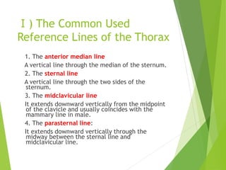
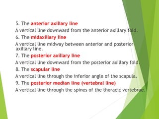
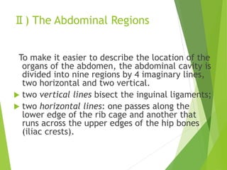
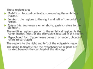
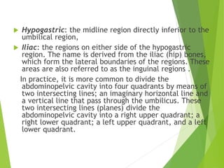
SlideShare a Scribd company logo
No. 3
1. Introduction of Myology
2. Introduction of Splanchnology
THE ROLE OF PATHOLOGY IN DIAGNOSTIC
PROCESS: FROM CLINICAL REASONING TO
MOLECULAR BIOLOGY.
 patients present with symptoms and
clinical examination reveals signs
which suggest a diagnosis
 -examination of various specimens in
pathology laboratories helps to
establish and confirm diagnosis, and
monitor the treatment
 Diagnosis is the act of identifying a
disease in an individual patient and is
based on clinical history, physical
examination and pathology
investigation
Diagnosis
 Diagnosis involves skills and laboratory tests
 Special pathology techniques are used
 Special stains, immunohistochemistry and molecular
biology techniques are routinely used
The role of pathologist:
 Looking at samples of tissues
(biopsies)
 Using the range of special laboratory
techniques
 Histology
 Autopsy
 Special stains
 Immunohistochemistry
 Electron microscopy
 Molecular biology techniques
Chapter 3
Myology
Section 1 Introduction
 Muscle tissue constitutes almost one-half of
the total body weight. Much of the body’s form
is due to the numerous muscles that attach to
the skeleton and underlie the skin. Other
muscles are located within the walls of hollow
organs and blood vessels.
 Muscle types:
The body contains three types of muscles--
skeletal muscle,
smooth muscle,
cardiac muscle.
 Muscles can be further classified as voluntary
and involuntary, on the basis of the type of
control exercised over their activity.
 Voluntary muscles: The contractions of
voluntary muscles are normally under the
conscious control of the individual. Voluntary
muscles are controlled by the portion of the
nervous system known as the somatic nervous
system.
 Involuntary muscles: The contractions of
involuntary muscles are generally not under
the conscious control of the individual. Rather,
they are governed by the portion of the
nervous system known as the autonomic
nervous system, as well as by hormones and
by factors intrinsic to the muscles themselves.
Skeletal muscles:
The muscles of locomotor apparatus are the
skeletal (or striated) muscles. All of them are
attached by at least one end to some part of
the skeleton.
The skeletal muscle is voluntary muscle
because its contraction is under the voluntary
control. About 40% of the body weight is
shared by the skeletal muscle.
Each skeletal muscle possesses a definite
shape, structure, location and accessory
apparatus, and is supplied by abundant blood
vessels, lymphatics and nerves, so that, it can
be regarded as an organ.
 Smooth muscles: Smooth muscle is so named
because its cells lack the striations evident in
skeletal muscle cells. It is also called visceral
muscle because it is found in the walls of
hollow organs and tubes such as the stomach,
intestines, and blood vessels.
Smooth muscle is involuntary muscle, and its
contractions govern the movement of
materials through the organ systems of the
body.
 Cardiac muscles: Cardiac muscle is a
specialized type of muscle that forms the wall
of the heart. It is involuntary, like smooth
muscle, and stiated, like skeletal muscle.
Ⅰ. The Morphology of Skeletal Muscle
 Muscle fiber: Each muscle is composed of a collection of muscle fibers
(cells).
 The muscle fiber, bundle of muscle fibers and the muscle are wrapped by
connective tissue.
1. Connective-tissue
coverings
 Muscle fibers are held together by thin
sheets of fibrous connective-tissue
membranes called fascia. The fascia that
envelops an entire muscle is called the
epimysium. Fascia also penetrates
muscle, separating the muscle fibers into
bundles called the perimysium. We can
say that the perimysium is the connective
tissue that surrounds each bundle of
muscle fibers. The endomysium envelops
the cell membrane of each muscle fiber.
2. Belly and tendon
 The fleshy part of a muscle is often called the
belly. At each end of a skeletal muscle the
connective tissue blends with the strong
collagen bundles to form the tendon that
anchors it to the bone, cartilage or articular
capsule.
 The muscles vary extremely in their shape. It
may be long, short, broad and circular band-
like (sphincter).
 Some muscles have more than one belly or
one origin. The fibers of some muscles are
oblique to the long axis of the muscle.
Because of their resemblance to feathers,
they are called pinnate muscles.
Ⅱ. The Origin, Insertion and Action of
Skeletal Muscle
1. Origin and Insertion:
Most of muscles are attached either directly or by
means of their tendons of aponeuroses to bones,
cartilages, ligaments, fasciae or to some combination
of these. Some muscles are attached to organs, such as
the eyeball, and some are attached to skin or mucous
membrane.
When a muscle contracts and becomes short, one of its
attachments usually remains fixed and the other one
moves.
The fixed attachment is called the origin, the movable
one the insertion.
In the limbs, the more distal parts are
usually mobile. Therefore the distal
attachment is usually called the insertion.
However, the terms origin and insertion are
convenient merely for purposes of
description.
Sometimes the anatomical insertion
remains fixed and the origin moves.
Sometimes both ends remain fixed, the
muscle then stabilizes a joint.
2. Prime movers (agonists) and antagonist, synergist and
fixators:
 During movements of the body, certain principal muscles are
called into action. These muscles, called prime movers or
agonists, contract actively (shorten and thicken) and
produce the desired movement.
 A muscle that opposes the action of a prime mover is called
an antagonist.
 The muscles which cooperate in performing an action are
called synergist.
 Other muscles, called fixators, steady the proximal parts of
a limb while movements are occurring in distal parts.
A muscle may act as a prime mover, antagonist, synergist or
fixator under different conditions.
Ⅲ. The Nomenclature of
Muscle
The names of muscles usually indicated some
structural or functional feature.
 A name may indicate shape, e.g., rhomboid,
trapezius, or gracilis.
 A name may refer to location, e.g., tibialis
posterior.
 The number of heads of origin is indicated by the
terms biceps, triceps, and quadriceps.
 The names of some muscles, such as levator
scapulae and extensor digitorum, depend on their
actions.
 Action and shape are combined in the term
pronator quadratus,.
 The action and location are combined in the term
flexor digitorum profundus.
Ⅳ. The Supplementary Structures
of Muscles
Ⅰ) The Fascia
 It is formed by connective tissue and can be divided, from where it is
found, into two groups, superficial and deep.
1. The superficial fascia
It is found immediately beneath the cutis,
covering almost the entire body.
It is a layer of loose connective tissue
containing fat in varying quantity. The
superficial fascia varies in thickness in
different individuals and different parts of
the body. It is very dense in the scalp, in the
palm of hand and the sole of foot. In the
groin it is so thick that is capable of being
subdivided into several laminae. Beneath the
fatty layer of the superficial fascia, there is
generally another layer of the same structure,
comparatively devoid of adipose (fatty) tissue,
in which the trunks of the subcutaneous
vessels and nerves, the superficial lymph
nodes, the mammary gland and certain
cutaneous muscles are found.
2. The deep fascia (proper
fascia)
 It is a dense, inelastic fibrous membrane,
forming a strong investment, which not only
binds down collectively the muscles in each
region, but also gives a separate sheath to
each, as well as to the vessels and nerves.
 In the limbs, the fasciae give off septa which
separate the groups of muscle and attached
to the periosteum. These prolongations of
fasciae are usually called the intermuscular
septa. In certain situations the deep fascia
affords attachment for muscles. It is
thickened at the wrist and ankle to form the
retinaculum that maintains its underlying
tendons.
Ⅱ) The Synovial Bursa
 It is a connetive tissue sac with a slippery inner surface, and filled with
synovial fluid. Bursa presents in the place where tendon rubs against bone,
ligament, or other tendon, or where skin moves over a body prominence.
Ⅲ) The Tendinous Sheath
 The tendinous sheath is usually located
in the hand and foot in which the tendon
runs. It constitutes fibrous layer (fibrous
sheath of tendon) and synovial layer
(synovial sheath of tendon).
 Synovial sheath of tendon is a double
layered synovial sheath. The outer layer
is parietal layer and the inner layer is
visceral layer. The mesotendom forms
the continuity between the inner and
outer synovial layers and carries blood
vessels to the tendon. The inner layer of
the synovial sheath is fused with the
tendon. The fluid in the cavity of the
sheath is similar to synovial fluid of the
joint cavity and facilitates movement by
minimizing friction.
Ⅳ) The Sesamoid Bones
 They are developed in those tendons exert a great amount of pressure
upon the part over which they glide. They minimize the friction, change
the direction of pulling force and stengthen the force of the muscle.
PART Ⅱ
SPLANCHNOLOGY
Chapter 1 Introduction
Viscera and Splanchnology:
 Viscera are the organs that are mainly
situated in the thoracic, abdominal and pelvic
cavities and fulfill the metabolism and
maintain the life of the species.
They are associated with the pleura and
peritoneum.
According to the functions which they perform,
the viscera can be arranged in the
alimentary system, respiratory system,
urinary system and genital system.
 Splanchnology means the study of viscera.
Functions:
The main functions of the viscera are to fulfill the
metabolism and maintain the life of the species.
 The alimentary system is adapted to ingest foods, to
secrete enzymes that modify the sizes of food molecules, to
absorb the products of this digestive action, and to eliminate
the unused residues.
 The respiratory system is to carry out the gas exchanges-
supply of oxygen for the living cells and remove of carbon
dioxide resulting from cell metabolism.
 The primary function of the urinary system is to keep the
body in homeostasis by removing and restoring selected
amount of water and solutes. It also excretes selected
amount of various wastes.
 The functions of the genital system are to produce germ
cells and to secrete some hormones.
Ⅰ. General Structure of
Viscera
 According to their general structures, in each of the
three chief subdivisions, the alimentary, respiratory and
the urogenital system, two principal constituents may
be recognized, i.e., a tubular canal and a series of non-
tubular so called parenchymatous organs.
Ⅰ) The Tubular Viscus
 The basic arrangement of tissue layers in the wall of tube of viscera is
best seen in the alimentary system.
The wall of the alimentary tract, particularly those
from the esophagus to the anal canal, consists of:
 Mucous membrane (mucosa): possessing the functions
of secreting and absorbing.
 Submucosa: external to the mucosa, consisting of
areolar tissue.
 Muscular coat: outside the submucosa, with two
muscular coats—an outer longitudinal one and an inner
circular one.
 Serosa (adventitia): It is the outermost coat of the
tract and is composed of a thin layer of connective
tissue and a mesothelium on those organs that lie in the
abdominal and pelvic cavities.
 The respiratory tract consists mainly of the mucosa,
submucosa and outermost coat, the latter is composed
of cartilages and connective tissue.
 The wall of the urogenital tract consists of three layers
of tissues—the mucous coat, the muscular coat, and the
fibrous coat.
Ⅱ) The Parenchymatous
Organ
 The parenchymatous organ is encased in a
thin, fibrous capsule from which fibrous
partitions pass into the interior to divide
the organ into units known as lobules.
 There is a somewhat depression or slit on
the surface of the viscus named the hilum
or porta, such as the hilum of lung, the
porta hepatis and the hilum of kidney,
where the structures which form the root
of the viscus enter and leave the viscus.
Ⅱ. Reference Lines and Abdominal
Regions
 In order to observe the relative locations and the arrangements of internal
organs, it is necessary to mark reference lines on the surface of thorax
and to subdivide abdomen into regions.
Ⅰ) The Common Used
Reference Lines of the Thorax
1. The anterior median line
A vertical line through the median of the sternum.
2. The sternal line
A vertical line through the two sides of the
sternum.
3. The midclavicular line
It extends downward vertically from the midpoint
of the clavicle and usually coincides with the
mammary line in male.
4. The parasternal line:
It extends downward vertically through the
midway between the sternal line and
midclavicular line.
5. The anterior axillary line
A vertical line downward from the anterior axillary fold.
6. The midaxillary line
A vertical line midway between anterior and posterior
axillary line.
7. The posterior axillary line
A vertical line downward from the posterior axillary fold.
8. The scapular line
A vertical line through the inferior angle of the scapula.
9. The posterior median line (vertebral line)
A vertical line through the spines of the thoracic vertebrae.
Ⅱ) The Abdominal Regions
To make it easier to describe the location of the
organs of the abdomen, the abdominal cavity is
divided into nine regions by 4 imaginary lines,
two horizontal and two vertical.
 two vertical lines bisect the inguinal ligaments;
 two horizontal lines: one passes along the
lower edge of the rib cage and another that
runs across the upper edges of the hip bones
(iliac crests).
These regions are:
 Umbilical: located centrally, surrounding the umbilicus
(navel).
 Lumbar: the regions to the right and left of the umbilical
region.
 Epigastric: (epi-means on or above; gastric-refers to the
stomach).
The midline region superior to the umbilical region. As the
name implies, most of the stomach is located in this region.
 Hypochondriac: (hypo-means beneath or under; chondral
refers to cartilage)
The regions to the right and left of the epigastric region.
The name indicates that the hypochondriac regions are
located beneath the cartilage of the rib cage.
 Hypogastric: the midline region directly inferior to the
umbilical region,
 Iliac: the regions on either side of the hypogastric
region. The name is derived from the iliac (hip) bones,
which form the lateral boundaries of the regions. These
areas are also referred to as the inguinal regions .
In practice, it is more common to divide the
abdominopelvic cavity into four quadrants by means of
two intersecting lines; an imaginary horizontal line and
a vertical line that pass through the umbilicus. These
two intersecting lines (planes) divide the
abdominopelvic cavity into a right upper quadrant; a
right lower quadrant; a left upper quadrant, and a left
lower quadrant.

More Related Content

Similar to myology.ppt

Basic Structures in human body
Basic Structures in human bodyBasic Structures in human body
Basic Structures in human body
ssuseraf61fb
 
Muscular system.pptx
Muscular system.pptxMuscular system.pptx
Muscular system.pptx
Maxinefoye
 
Muscle in human body
Muscle in human bodyMuscle in human body
Muscle in human body
Dr Usha (Physio)
 
Human Anatomy and Physiology 1 - Chapter 6.pptx
Human Anatomy and Physiology 1 - Chapter 6.pptxHuman Anatomy and Physiology 1 - Chapter 6.pptx
Human Anatomy and Physiology 1 - Chapter 6.pptx
RuchithChandeepa
 
Prelim PDHPE Core 2: Body in Motion
Prelim PDHPE Core 2: Body in MotionPrelim PDHPE Core 2: Body in Motion
Prelim PDHPE Core 2: Body in Motion
Vas Ratusau
 
Muscular system
Muscular system Muscular system
Muscular system
Soneeshah
 
Muscles anatomy and physiology
Muscles anatomy and physiology Muscles anatomy and physiology
Muscles anatomy and physiology
Manthan Kapadiya
 
Introduction to Muscle & Its Types
Introduction to Muscle & Its Types Introduction to Muscle & Its Types
Introduction to Muscle & Its Types
Dr Syed Altaf
 
10 Muscle Tissue And Organization
10 Muscle Tissue And Organization10 Muscle Tissue And Organization
10 Muscle Tissue And Organization
Kevin Young
 
Muscular system in insects
Muscular system in insectsMuscular system in insects
Muscular system in insects
Jayantyadav94
 
muscular system
muscular systemmuscular system
Muscles of mastication by DR. C.P. ARYA ( B.Sc. ;B.D.S. ;M.D.S. ;P.M.S. ;R.N....
Muscles of mastication by DR. C.P. ARYA ( B.Sc. ;B.D.S. ;M.D.S. ;P.M.S. ;R.N....Muscles of mastication by DR. C.P. ARYA ( B.Sc. ;B.D.S. ;M.D.S. ;P.M.S. ;R.N....
Muscles of mastication by DR. C.P. ARYA ( B.Sc. ;B.D.S. ;M.D.S. ;P.M.S. ;R.N....
DR. C. P. ARYA
 
Myology
MyologyMyology
Myology
xayouluma
 
Skeletal system
Skeletal systemSkeletal system
Skeletal system
Yogeshwary Bhongade
 
Bio13 .ppt;10 muscle-tissue-and-organization ppt zeny
Bio13 .ppt;10 muscle-tissue-and-organization ppt zenyBio13 .ppt;10 muscle-tissue-and-organization ppt zeny
Bio13 .ppt;10 muscle-tissue-and-organization ppt zenyDevron Jeko
 
10 Muscle Tissue And Organization
10 Muscle Tissue And Organization10 Muscle Tissue And Organization
10 Muscle Tissue And Organizationguest334add
 
Locomotor system
Locomotor systemLocomotor system
Locomotor system
Kamal Deen
 
Muscles
MusclesMuscles
Muscles
Aarif Kanadia
 
Histology of muscle
Histology of muscle Histology of muscle
Histology of muscle
syed shahzaib
 

Similar to myology.ppt (20)

Basic Structures in human body
Basic Structures in human bodyBasic Structures in human body
Basic Structures in human body
 
L4 Muscles1
L4 Muscles1L4 Muscles1
L4 Muscles1
 
Muscular system.pptx
Muscular system.pptxMuscular system.pptx
Muscular system.pptx
 
Muscle in human body
Muscle in human bodyMuscle in human body
Muscle in human body
 
Human Anatomy and Physiology 1 - Chapter 6.pptx
Human Anatomy and Physiology 1 - Chapter 6.pptxHuman Anatomy and Physiology 1 - Chapter 6.pptx
Human Anatomy and Physiology 1 - Chapter 6.pptx
 
Prelim PDHPE Core 2: Body in Motion
Prelim PDHPE Core 2: Body in MotionPrelim PDHPE Core 2: Body in Motion
Prelim PDHPE Core 2: Body in Motion
 
Muscular system
Muscular system Muscular system
Muscular system
 
Muscles anatomy and physiology
Muscles anatomy and physiology Muscles anatomy and physiology
Muscles anatomy and physiology
 
Introduction to Muscle & Its Types
Introduction to Muscle & Its Types Introduction to Muscle & Its Types
Introduction to Muscle & Its Types
 
10 Muscle Tissue And Organization
10 Muscle Tissue And Organization10 Muscle Tissue And Organization
10 Muscle Tissue And Organization
 
Muscular system in insects
Muscular system in insectsMuscular system in insects
Muscular system in insects
 
muscular system
muscular systemmuscular system
muscular system
 
Muscles of mastication by DR. C.P. ARYA ( B.Sc. ;B.D.S. ;M.D.S. ;P.M.S. ;R.N....
Muscles of mastication by DR. C.P. ARYA ( B.Sc. ;B.D.S. ;M.D.S. ;P.M.S. ;R.N....Muscles of mastication by DR. C.P. ARYA ( B.Sc. ;B.D.S. ;M.D.S. ;P.M.S. ;R.N....
Muscles of mastication by DR. C.P. ARYA ( B.Sc. ;B.D.S. ;M.D.S. ;P.M.S. ;R.N....
 
Myology
MyologyMyology
Myology
 
Skeletal system
Skeletal systemSkeletal system
Skeletal system
 
Bio13 .ppt;10 muscle-tissue-and-organization ppt zeny
Bio13 .ppt;10 muscle-tissue-and-organization ppt zenyBio13 .ppt;10 muscle-tissue-and-organization ppt zeny
Bio13 .ppt;10 muscle-tissue-and-organization ppt zeny
 
10 Muscle Tissue And Organization
10 Muscle Tissue And Organization10 Muscle Tissue And Organization
10 Muscle Tissue And Organization
 
Locomotor system
Locomotor systemLocomotor system
Locomotor system
 
Muscles
MusclesMuscles
Muscles
 
Histology of muscle
Histology of muscle Histology of muscle
Histology of muscle
 

More from fgcpkinaeehilrjwvw

Introduction. Hypoxia is a state in whic
Introduction. Hypoxia is a state in whicIntroduction. Hypoxia is a state in whic
Introduction. Hypoxia is a state in whic
fgcpkinaeehilrjwvw
 
altitude.pptx
altitude.pptxaltitude.pptx
altitude.pptx
fgcpkinaeehilrjwvw
 
Renal therapy.pptx
Renal therapy.pptxRenal therapy.pptx
Renal therapy.pptx
fgcpkinaeehilrjwvw
 
Pathology.ppt
Pathology.pptPathology.ppt
Pathology.ppt
fgcpkinaeehilrjwvw
 
Action Potential Generation.ppt
Action Potential Generation.pptAction Potential Generation.ppt
Action Potential Generation.ppt
fgcpkinaeehilrjwvw
 
UNDERSTANDING Of ANAPHYLAXIS.pptx
UNDERSTANDING  Of  ANAPHYLAXIS.pptxUNDERSTANDING  Of  ANAPHYLAXIS.pptx
UNDERSTANDING Of ANAPHYLAXIS.pptx
fgcpkinaeehilrjwvw
 
action potential nd hypoxia.ppt
action potential nd hypoxia.pptaction potential nd hypoxia.ppt
action potential nd hypoxia.ppt
fgcpkinaeehilrjwvw
 
Heart failure.ppt
Heart failure.pptHeart failure.ppt
Heart failure.ppt
fgcpkinaeehilrjwvw
 
Introduction to pathology & causes of diseases.ppt
Introduction to pathology & causes of diseases.pptIntroduction to pathology & causes of diseases.ppt
Introduction to pathology & causes of diseases.ppt
fgcpkinaeehilrjwvw
 
THORAX BONES.ppt
THORAX BONES.pptTHORAX BONES.ppt
THORAX BONES.ppt
fgcpkinaeehilrjwvw
 
Regions of the lower limb.pptx
Regions of the lower limb.pptxRegions of the lower limb.pptx
Regions of the lower limb.pptx
fgcpkinaeehilrjwvw
 
diseases.ppt
diseases.pptdiseases.ppt
diseases.ppt
fgcpkinaeehilrjwvw
 
Allergies & Anaphylaxis.ppt
Allergies & Anaphylaxis.pptAllergies & Anaphylaxis.ppt
Allergies & Anaphylaxis.ppt
fgcpkinaeehilrjwvw
 
Pathogenic effect of environmental factors.ppt
Pathogenic effect of environmental factors.pptPathogenic effect of environmental factors.ppt
Pathogenic effect of environmental factors.ppt
fgcpkinaeehilrjwvw
 
16959631.ppt
16959631.ppt16959631.ppt
16959631.ppt
fgcpkinaeehilrjwvw
 
Vertebral Column.ppt
Vertebral Column.pptVertebral Column.ppt
Vertebral Column.ppt
fgcpkinaeehilrjwvw
 

More from fgcpkinaeehilrjwvw (16)

Introduction. Hypoxia is a state in whic
Introduction. Hypoxia is a state in whicIntroduction. Hypoxia is a state in whic
Introduction. Hypoxia is a state in whic
 
altitude.pptx
altitude.pptxaltitude.pptx
altitude.pptx
 
Renal therapy.pptx
Renal therapy.pptxRenal therapy.pptx
Renal therapy.pptx
 
Pathology.ppt
Pathology.pptPathology.ppt
Pathology.ppt
 
Action Potential Generation.ppt
Action Potential Generation.pptAction Potential Generation.ppt
Action Potential Generation.ppt
 
UNDERSTANDING Of ANAPHYLAXIS.pptx
UNDERSTANDING  Of  ANAPHYLAXIS.pptxUNDERSTANDING  Of  ANAPHYLAXIS.pptx
UNDERSTANDING Of ANAPHYLAXIS.pptx
 
action potential nd hypoxia.ppt
action potential nd hypoxia.pptaction potential nd hypoxia.ppt
action potential nd hypoxia.ppt
 
Heart failure.ppt
Heart failure.pptHeart failure.ppt
Heart failure.ppt
 
Introduction to pathology & causes of diseases.ppt
Introduction to pathology & causes of diseases.pptIntroduction to pathology & causes of diseases.ppt
Introduction to pathology & causes of diseases.ppt
 
THORAX BONES.ppt
THORAX BONES.pptTHORAX BONES.ppt
THORAX BONES.ppt
 
Regions of the lower limb.pptx
Regions of the lower limb.pptxRegions of the lower limb.pptx
Regions of the lower limb.pptx
 
diseases.ppt
diseases.pptdiseases.ppt
diseases.ppt
 
Allergies & Anaphylaxis.ppt
Allergies & Anaphylaxis.pptAllergies & Anaphylaxis.ppt
Allergies & Anaphylaxis.ppt
 
Pathogenic effect of environmental factors.ppt
Pathogenic effect of environmental factors.pptPathogenic effect of environmental factors.ppt
Pathogenic effect of environmental factors.ppt
 
16959631.ppt
16959631.ppt16959631.ppt
16959631.ppt
 
Vertebral Column.ppt
Vertebral Column.pptVertebral Column.ppt
Vertebral Column.ppt
 

Recently uploaded

Secret Tantric VIP Erotic Massage London
Secret Tantric VIP Erotic Massage LondonSecret Tantric VIP Erotic Massage London
Secret Tantric VIP Erotic Massage London
Secret Tantric - VIP Erotic Massage London
 
BOWEL ELIMINATION BY ANUSHRI SRIVASTAVA.pptx
BOWEL ELIMINATION BY ANUSHRI SRIVASTAVA.pptxBOWEL ELIMINATION BY ANUSHRI SRIVASTAVA.pptx
BOWEL ELIMINATION BY ANUSHRI SRIVASTAVA.pptx
AnushriSrivastav
 
What Are Homeopathic Treatments for Migraines.pdf
What Are Homeopathic Treatments for Migraines.pdfWhat Are Homeopathic Treatments for Migraines.pdf
What Are Homeopathic Treatments for Migraines.pdf
Dharma Homoeopathy
 
POLYCYSTIC OVARIAN SYNDROME (PCOS)......
POLYCYSTIC OVARIAN SYNDROME (PCOS)......POLYCYSTIC OVARIAN SYNDROME (PCOS)......
POLYCYSTIC OVARIAN SYNDROME (PCOS)......
Ameena Kadar
 
Preventing Pickleball Injuries & Treatment
Preventing Pickleball Injuries & TreatmentPreventing Pickleball Injuries & Treatment
Preventing Pickleball Injuries & Treatment
LAB Sports Therapy
 
Overcome Your Phobias with Hypnotherapy.pptx
Overcome Your Phobias with Hypnotherapy.pptxOvercome Your Phobias with Hypnotherapy.pptx
Overcome Your Phobias with Hypnotherapy.pptx
renewlifehypnosis
 
Artificial Intelligence to Optimize Cardiovascular Therapy
Artificial Intelligence to Optimize Cardiovascular TherapyArtificial Intelligence to Optimize Cardiovascular Therapy
Artificial Intelligence to Optimize Cardiovascular Therapy
Iris Thiele Isip-Tan
 
Navigating Challenges: Mental Health, Legislation, and the Prison System in B...
Navigating Challenges: Mental Health, Legislation, and the Prison System in B...Navigating Challenges: Mental Health, Legislation, and the Prison System in B...
Navigating Challenges: Mental Health, Legislation, and the Prison System in B...
Guillermo Rivera
 
CHAPTER 1 SEMESTER V PREVENTIVE-PEDIATRICS.pdf
CHAPTER 1 SEMESTER V PREVENTIVE-PEDIATRICS.pdfCHAPTER 1 SEMESTER V PREVENTIVE-PEDIATRICS.pdf
CHAPTER 1 SEMESTER V PREVENTIVE-PEDIATRICS.pdf
Sachin Sharma
 
ABDOMINAL COMPARTMENT SYSNDROME
ABDOMINAL COMPARTMENT SYSNDROMEABDOMINAL COMPARTMENT SYSNDROME
ABDOMINAL COMPARTMENT SYSNDROME
Rommel Luis III Israel
 
CANCER CANCER CANCER CANCER CANCER CANCER
CANCER  CANCER  CANCER  CANCER  CANCER CANCERCANCER  CANCER  CANCER  CANCER  CANCER CANCER
CANCER CANCER CANCER CANCER CANCER CANCER
KRISTELLEGAMBOA2
 
Introduction to Forensic Pathology course
Introduction to Forensic Pathology courseIntroduction to Forensic Pathology course
Introduction to Forensic Pathology course
fprxsqvnz5
 
GLOBAL WARMING BY PRIYA BHOJWANI @..pptx
GLOBAL WARMING BY PRIYA BHOJWANI @..pptxGLOBAL WARMING BY PRIYA BHOJWANI @..pptx
GLOBAL WARMING BY PRIYA BHOJWANI @..pptx
priyabhojwani1200
 
Deepfake Detection_Using Machine Learning .pptx
Deepfake Detection_Using Machine Learning .pptxDeepfake Detection_Using Machine Learning .pptx
Deepfake Detection_Using Machine Learning .pptx
mahalsuraj389
 
Demystifying-Gene-Editing-The-Promise-and-Peril-of-CRISPR.pdf
Demystifying-Gene-Editing-The-Promise-and-Peril-of-CRISPR.pdfDemystifying-Gene-Editing-The-Promise-and-Peril-of-CRISPR.pdf
Demystifying-Gene-Editing-The-Promise-and-Peril-of-CRISPR.pdf
SasikiranMarri
 
Navigating Healthcare with Telemedicine
Navigating Healthcare with  TelemedicineNavigating Healthcare with  Telemedicine
Navigating Healthcare with Telemedicine
Iris Thiele Isip-Tan
 
Medical Technology Tackles New Health Care Demand - Research Report - March 2...
Medical Technology Tackles New Health Care Demand - Research Report - March 2...Medical Technology Tackles New Health Care Demand - Research Report - March 2...
Medical Technology Tackles New Health Care Demand - Research Report - March 2...
pchutichetpong
 
Contact Now 89011**83002 Dehradun ℂall Girls By Full Service ℂall Girl In De...
Contact Now  89011**83002 Dehradun ℂall Girls By Full Service ℂall Girl In De...Contact Now  89011**83002 Dehradun ℂall Girls By Full Service ℂall Girl In De...
Contact Now 89011**83002 Dehradun ℂall Girls By Full Service ℂall Girl In De...
aunty1x2
 
Jaipur ❤cALL gIRLS 89O1183002 ❤ℂall Girls IN JaiPuR ESCORT SERVICE
Jaipur ❤cALL gIRLS 89O1183002 ❤ℂall Girls IN JaiPuR ESCORT SERVICEJaipur ❤cALL gIRLS 89O1183002 ❤ℂall Girls IN JaiPuR ESCORT SERVICE
Jaipur ❤cALL gIRLS 89O1183002 ❤ℂall Girls IN JaiPuR ESCORT SERVICE
ranishasharma67
 
Navigating the Health Insurance Market_ Understanding Trends and Options.pdf
Navigating the Health Insurance Market_ Understanding Trends and Options.pdfNavigating the Health Insurance Market_ Understanding Trends and Options.pdf
Navigating the Health Insurance Market_ Understanding Trends and Options.pdf
Enterprise Wired
 

Recently uploaded (20)

Secret Tantric VIP Erotic Massage London
Secret Tantric VIP Erotic Massage LondonSecret Tantric VIP Erotic Massage London
Secret Tantric VIP Erotic Massage London
 
BOWEL ELIMINATION BY ANUSHRI SRIVASTAVA.pptx
BOWEL ELIMINATION BY ANUSHRI SRIVASTAVA.pptxBOWEL ELIMINATION BY ANUSHRI SRIVASTAVA.pptx
BOWEL ELIMINATION BY ANUSHRI SRIVASTAVA.pptx
 
What Are Homeopathic Treatments for Migraines.pdf
What Are Homeopathic Treatments for Migraines.pdfWhat Are Homeopathic Treatments for Migraines.pdf
What Are Homeopathic Treatments for Migraines.pdf
 
POLYCYSTIC OVARIAN SYNDROME (PCOS)......
POLYCYSTIC OVARIAN SYNDROME (PCOS)......POLYCYSTIC OVARIAN SYNDROME (PCOS)......
POLYCYSTIC OVARIAN SYNDROME (PCOS)......
 
Preventing Pickleball Injuries & Treatment
Preventing Pickleball Injuries & TreatmentPreventing Pickleball Injuries & Treatment
Preventing Pickleball Injuries & Treatment
 
Overcome Your Phobias with Hypnotherapy.pptx
Overcome Your Phobias with Hypnotherapy.pptxOvercome Your Phobias with Hypnotherapy.pptx
Overcome Your Phobias with Hypnotherapy.pptx
 
Artificial Intelligence to Optimize Cardiovascular Therapy
Artificial Intelligence to Optimize Cardiovascular TherapyArtificial Intelligence to Optimize Cardiovascular Therapy
Artificial Intelligence to Optimize Cardiovascular Therapy
 
Navigating Challenges: Mental Health, Legislation, and the Prison System in B...
Navigating Challenges: Mental Health, Legislation, and the Prison System in B...Navigating Challenges: Mental Health, Legislation, and the Prison System in B...
Navigating Challenges: Mental Health, Legislation, and the Prison System in B...
 
CHAPTER 1 SEMESTER V PREVENTIVE-PEDIATRICS.pdf
CHAPTER 1 SEMESTER V PREVENTIVE-PEDIATRICS.pdfCHAPTER 1 SEMESTER V PREVENTIVE-PEDIATRICS.pdf
CHAPTER 1 SEMESTER V PREVENTIVE-PEDIATRICS.pdf
 
ABDOMINAL COMPARTMENT SYSNDROME
ABDOMINAL COMPARTMENT SYSNDROMEABDOMINAL COMPARTMENT SYSNDROME
ABDOMINAL COMPARTMENT SYSNDROME
 
CANCER CANCER CANCER CANCER CANCER CANCER
CANCER  CANCER  CANCER  CANCER  CANCER CANCERCANCER  CANCER  CANCER  CANCER  CANCER CANCER
CANCER CANCER CANCER CANCER CANCER CANCER
 
Introduction to Forensic Pathology course
Introduction to Forensic Pathology courseIntroduction to Forensic Pathology course
Introduction to Forensic Pathology course
 
GLOBAL WARMING BY PRIYA BHOJWANI @..pptx
GLOBAL WARMING BY PRIYA BHOJWANI @..pptxGLOBAL WARMING BY PRIYA BHOJWANI @..pptx
GLOBAL WARMING BY PRIYA BHOJWANI @..pptx
 
Deepfake Detection_Using Machine Learning .pptx
Deepfake Detection_Using Machine Learning .pptxDeepfake Detection_Using Machine Learning .pptx
Deepfake Detection_Using Machine Learning .pptx
 
Demystifying-Gene-Editing-The-Promise-and-Peril-of-CRISPR.pdf
Demystifying-Gene-Editing-The-Promise-and-Peril-of-CRISPR.pdfDemystifying-Gene-Editing-The-Promise-and-Peril-of-CRISPR.pdf
Demystifying-Gene-Editing-The-Promise-and-Peril-of-CRISPR.pdf
 
Navigating Healthcare with Telemedicine
Navigating Healthcare with  TelemedicineNavigating Healthcare with  Telemedicine
Navigating Healthcare with Telemedicine
 
Medical Technology Tackles New Health Care Demand - Research Report - March 2...
Medical Technology Tackles New Health Care Demand - Research Report - March 2...Medical Technology Tackles New Health Care Demand - Research Report - March 2...
Medical Technology Tackles New Health Care Demand - Research Report - March 2...
 
Contact Now 89011**83002 Dehradun ℂall Girls By Full Service ℂall Girl In De...
Contact Now  89011**83002 Dehradun ℂall Girls By Full Service ℂall Girl In De...Contact Now  89011**83002 Dehradun ℂall Girls By Full Service ℂall Girl In De...
Contact Now 89011**83002 Dehradun ℂall Girls By Full Service ℂall Girl In De...
 
Jaipur ❤cALL gIRLS 89O1183002 ❤ℂall Girls IN JaiPuR ESCORT SERVICE
Jaipur ❤cALL gIRLS 89O1183002 ❤ℂall Girls IN JaiPuR ESCORT SERVICEJaipur ❤cALL gIRLS 89O1183002 ❤ℂall Girls IN JaiPuR ESCORT SERVICE
Jaipur ❤cALL gIRLS 89O1183002 ❤ℂall Girls IN JaiPuR ESCORT SERVICE
 
Navigating the Health Insurance Market_ Understanding Trends and Options.pdf
Navigating the Health Insurance Market_ Understanding Trends and Options.pdfNavigating the Health Insurance Market_ Understanding Trends and Options.pdf
Navigating the Health Insurance Market_ Understanding Trends and Options.pdf
 

myology.ppt

  • 1. No. 3 1. Introduction of Myology 2. Introduction of Splanchnology
  • 2. THE ROLE OF PATHOLOGY IN DIAGNOSTIC PROCESS: FROM CLINICAL REASONING TO MOLECULAR BIOLOGY.  patients present with symptoms and clinical examination reveals signs which suggest a diagnosis  -examination of various specimens in pathology laboratories helps to establish and confirm diagnosis, and monitor the treatment  Diagnosis is the act of identifying a disease in an individual patient and is based on clinical history, physical examination and pathology investigation
  • 3. Diagnosis  Diagnosis involves skills and laboratory tests  Special pathology techniques are used  Special stains, immunohistochemistry and molecular biology techniques are routinely used
  • 4. The role of pathologist:  Looking at samples of tissues (biopsies)  Using the range of special laboratory techniques  Histology  Autopsy  Special stains  Immunohistochemistry  Electron microscopy  Molecular biology techniques
  • 6. Section 1 Introduction  Muscle tissue constitutes almost one-half of the total body weight. Much of the body’s form is due to the numerous muscles that attach to the skeleton and underlie the skin. Other muscles are located within the walls of hollow organs and blood vessels.  Muscle types: The body contains three types of muscles-- skeletal muscle, smooth muscle, cardiac muscle.  Muscles can be further classified as voluntary and involuntary, on the basis of the type of control exercised over their activity.
  • 7.  Voluntary muscles: The contractions of voluntary muscles are normally under the conscious control of the individual. Voluntary muscles are controlled by the portion of the nervous system known as the somatic nervous system.  Involuntary muscles: The contractions of involuntary muscles are generally not under the conscious control of the individual. Rather, they are governed by the portion of the nervous system known as the autonomic nervous system, as well as by hormones and by factors intrinsic to the muscles themselves.
  • 8. Skeletal muscles: The muscles of locomotor apparatus are the skeletal (or striated) muscles. All of them are attached by at least one end to some part of the skeleton. The skeletal muscle is voluntary muscle because its contraction is under the voluntary control. About 40% of the body weight is shared by the skeletal muscle. Each skeletal muscle possesses a definite shape, structure, location and accessory apparatus, and is supplied by abundant blood vessels, lymphatics and nerves, so that, it can be regarded as an organ.
  • 9.
  • 10.  Smooth muscles: Smooth muscle is so named because its cells lack the striations evident in skeletal muscle cells. It is also called visceral muscle because it is found in the walls of hollow organs and tubes such as the stomach, intestines, and blood vessels. Smooth muscle is involuntary muscle, and its contractions govern the movement of materials through the organ systems of the body.  Cardiac muscles: Cardiac muscle is a specialized type of muscle that forms the wall of the heart. It is involuntary, like smooth muscle, and stiated, like skeletal muscle.
  • 11. Ⅰ. The Morphology of Skeletal Muscle  Muscle fiber: Each muscle is composed of a collection of muscle fibers (cells).  The muscle fiber, bundle of muscle fibers and the muscle are wrapped by connective tissue.
  • 12. 1. Connective-tissue coverings  Muscle fibers are held together by thin sheets of fibrous connective-tissue membranes called fascia. The fascia that envelops an entire muscle is called the epimysium. Fascia also penetrates muscle, separating the muscle fibers into bundles called the perimysium. We can say that the perimysium is the connective tissue that surrounds each bundle of muscle fibers. The endomysium envelops the cell membrane of each muscle fiber.
  • 13.
  • 14. 2. Belly and tendon  The fleshy part of a muscle is often called the belly. At each end of a skeletal muscle the connective tissue blends with the strong collagen bundles to form the tendon that anchors it to the bone, cartilage or articular capsule.  The muscles vary extremely in their shape. It may be long, short, broad and circular band- like (sphincter).  Some muscles have more than one belly or one origin. The fibers of some muscles are oblique to the long axis of the muscle. Because of their resemblance to feathers, they are called pinnate muscles.
  • 15.
  • 16.
  • 17. Ⅱ. The Origin, Insertion and Action of Skeletal Muscle 1. Origin and Insertion: Most of muscles are attached either directly or by means of their tendons of aponeuroses to bones, cartilages, ligaments, fasciae or to some combination of these. Some muscles are attached to organs, such as the eyeball, and some are attached to skin or mucous membrane. When a muscle contracts and becomes short, one of its attachments usually remains fixed and the other one moves. The fixed attachment is called the origin, the movable one the insertion.
  • 18. In the limbs, the more distal parts are usually mobile. Therefore the distal attachment is usually called the insertion. However, the terms origin and insertion are convenient merely for purposes of description. Sometimes the anatomical insertion remains fixed and the origin moves. Sometimes both ends remain fixed, the muscle then stabilizes a joint.
  • 19. 2. Prime movers (agonists) and antagonist, synergist and fixators:  During movements of the body, certain principal muscles are called into action. These muscles, called prime movers or agonists, contract actively (shorten and thicken) and produce the desired movement.  A muscle that opposes the action of a prime mover is called an antagonist.  The muscles which cooperate in performing an action are called synergist.  Other muscles, called fixators, steady the proximal parts of a limb while movements are occurring in distal parts. A muscle may act as a prime mover, antagonist, synergist or fixator under different conditions.
  • 20. Ⅲ. The Nomenclature of Muscle The names of muscles usually indicated some structural or functional feature.  A name may indicate shape, e.g., rhomboid, trapezius, or gracilis.  A name may refer to location, e.g., tibialis posterior.  The number of heads of origin is indicated by the terms biceps, triceps, and quadriceps.  The names of some muscles, such as levator scapulae and extensor digitorum, depend on their actions.  Action and shape are combined in the term pronator quadratus,.  The action and location are combined in the term flexor digitorum profundus.
  • 21. Ⅳ. The Supplementary Structures of Muscles Ⅰ) The Fascia  It is formed by connective tissue and can be divided, from where it is found, into two groups, superficial and deep.
  • 22. 1. The superficial fascia It is found immediately beneath the cutis, covering almost the entire body. It is a layer of loose connective tissue containing fat in varying quantity. The superficial fascia varies in thickness in different individuals and different parts of the body. It is very dense in the scalp, in the palm of hand and the sole of foot. In the groin it is so thick that is capable of being subdivided into several laminae. Beneath the fatty layer of the superficial fascia, there is generally another layer of the same structure, comparatively devoid of adipose (fatty) tissue, in which the trunks of the subcutaneous vessels and nerves, the superficial lymph nodes, the mammary gland and certain cutaneous muscles are found.
  • 23.
  • 24. 2. The deep fascia (proper fascia)  It is a dense, inelastic fibrous membrane, forming a strong investment, which not only binds down collectively the muscles in each region, but also gives a separate sheath to each, as well as to the vessels and nerves.  In the limbs, the fasciae give off septa which separate the groups of muscle and attached to the periosteum. These prolongations of fasciae are usually called the intermuscular septa. In certain situations the deep fascia affords attachment for muscles. It is thickened at the wrist and ankle to form the retinaculum that maintains its underlying tendons.
  • 25. Ⅱ) The Synovial Bursa  It is a connetive tissue sac with a slippery inner surface, and filled with synovial fluid. Bursa presents in the place where tendon rubs against bone, ligament, or other tendon, or where skin moves over a body prominence.
  • 26. Ⅲ) The Tendinous Sheath  The tendinous sheath is usually located in the hand and foot in which the tendon runs. It constitutes fibrous layer (fibrous sheath of tendon) and synovial layer (synovial sheath of tendon).  Synovial sheath of tendon is a double layered synovial sheath. The outer layer is parietal layer and the inner layer is visceral layer. The mesotendom forms the continuity between the inner and outer synovial layers and carries blood vessels to the tendon. The inner layer of the synovial sheath is fused with the tendon. The fluid in the cavity of the sheath is similar to synovial fluid of the joint cavity and facilitates movement by minimizing friction.
  • 27. Ⅳ) The Sesamoid Bones  They are developed in those tendons exert a great amount of pressure upon the part over which they glide. They minimize the friction, change the direction of pulling force and stengthen the force of the muscle.
  • 29. Chapter 1 Introduction Viscera and Splanchnology:  Viscera are the organs that are mainly situated in the thoracic, abdominal and pelvic cavities and fulfill the metabolism and maintain the life of the species. They are associated with the pleura and peritoneum. According to the functions which they perform, the viscera can be arranged in the alimentary system, respiratory system, urinary system and genital system.  Splanchnology means the study of viscera.
  • 30. Functions: The main functions of the viscera are to fulfill the metabolism and maintain the life of the species.  The alimentary system is adapted to ingest foods, to secrete enzymes that modify the sizes of food molecules, to absorb the products of this digestive action, and to eliminate the unused residues.  The respiratory system is to carry out the gas exchanges- supply of oxygen for the living cells and remove of carbon dioxide resulting from cell metabolism.  The primary function of the urinary system is to keep the body in homeostasis by removing and restoring selected amount of water and solutes. It also excretes selected amount of various wastes.  The functions of the genital system are to produce germ cells and to secrete some hormones.
  • 31. Ⅰ. General Structure of Viscera  According to their general structures, in each of the three chief subdivisions, the alimentary, respiratory and the urogenital system, two principal constituents may be recognized, i.e., a tubular canal and a series of non- tubular so called parenchymatous organs.
  • 32. Ⅰ) The Tubular Viscus  The basic arrangement of tissue layers in the wall of tube of viscera is best seen in the alimentary system.
  • 33. The wall of the alimentary tract, particularly those from the esophagus to the anal canal, consists of:  Mucous membrane (mucosa): possessing the functions of secreting and absorbing.  Submucosa: external to the mucosa, consisting of areolar tissue.  Muscular coat: outside the submucosa, with two muscular coats—an outer longitudinal one and an inner circular one.  Serosa (adventitia): It is the outermost coat of the tract and is composed of a thin layer of connective tissue and a mesothelium on those organs that lie in the abdominal and pelvic cavities.
  • 34.  The respiratory tract consists mainly of the mucosa, submucosa and outermost coat, the latter is composed of cartilages and connective tissue.  The wall of the urogenital tract consists of three layers of tissues—the mucous coat, the muscular coat, and the fibrous coat.
  • 35. Ⅱ) The Parenchymatous Organ  The parenchymatous organ is encased in a thin, fibrous capsule from which fibrous partitions pass into the interior to divide the organ into units known as lobules.  There is a somewhat depression or slit on the surface of the viscus named the hilum or porta, such as the hilum of lung, the porta hepatis and the hilum of kidney, where the structures which form the root of the viscus enter and leave the viscus.
  • 36. Ⅱ. Reference Lines and Abdominal Regions  In order to observe the relative locations and the arrangements of internal organs, it is necessary to mark reference lines on the surface of thorax and to subdivide abdomen into regions.
  • 37. Ⅰ) The Common Used Reference Lines of the Thorax 1. The anterior median line A vertical line through the median of the sternum. 2. The sternal line A vertical line through the two sides of the sternum. 3. The midclavicular line It extends downward vertically from the midpoint of the clavicle and usually coincides with the mammary line in male. 4. The parasternal line: It extends downward vertically through the midway between the sternal line and midclavicular line.
  • 38. 5. The anterior axillary line A vertical line downward from the anterior axillary fold. 6. The midaxillary line A vertical line midway between anterior and posterior axillary line. 7. The posterior axillary line A vertical line downward from the posterior axillary fold. 8. The scapular line A vertical line through the inferior angle of the scapula. 9. The posterior median line (vertebral line) A vertical line through the spines of the thoracic vertebrae.
  • 39. Ⅱ) The Abdominal Regions To make it easier to describe the location of the organs of the abdomen, the abdominal cavity is divided into nine regions by 4 imaginary lines, two horizontal and two vertical.  two vertical lines bisect the inguinal ligaments;  two horizontal lines: one passes along the lower edge of the rib cage and another that runs across the upper edges of the hip bones (iliac crests).
  • 40. These regions are:  Umbilical: located centrally, surrounding the umbilicus (navel).  Lumbar: the regions to the right and left of the umbilical region.  Epigastric: (epi-means on or above; gastric-refers to the stomach). The midline region superior to the umbilical region. As the name implies, most of the stomach is located in this region.  Hypochondriac: (hypo-means beneath or under; chondral refers to cartilage) The regions to the right and left of the epigastric region. The name indicates that the hypochondriac regions are located beneath the cartilage of the rib cage.
  • 41.  Hypogastric: the midline region directly inferior to the umbilical region,  Iliac: the regions on either side of the hypogastric region. The name is derived from the iliac (hip) bones, which form the lateral boundaries of the regions. These areas are also referred to as the inguinal regions . In practice, it is more common to divide the abdominopelvic cavity into four quadrants by means of two intersecting lines; an imaginary horizontal line and a vertical line that pass through the umbilicus. These two intersecting lines (planes) divide the abdominopelvic cavity into a right upper quadrant; a right lower quadrant; a left upper quadrant, and a left lower quadrant.