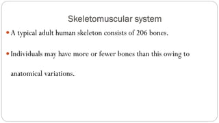
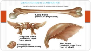
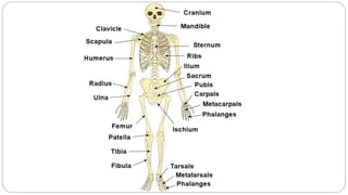
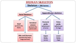
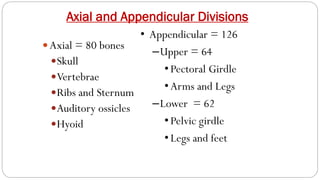
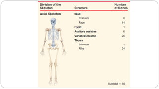
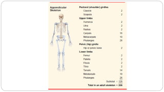
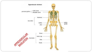
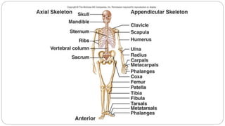
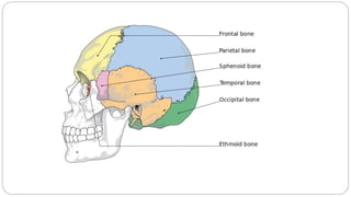
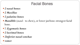
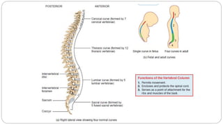
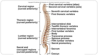
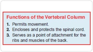
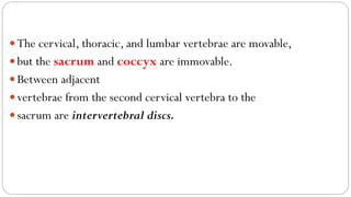
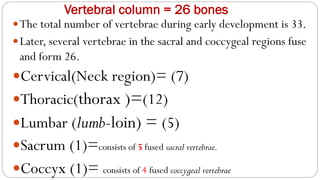
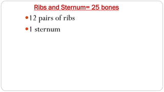
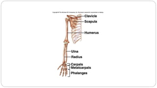
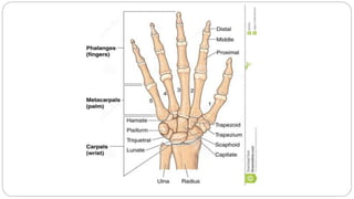
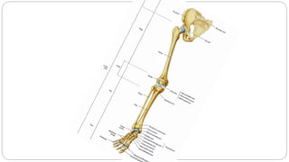
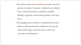
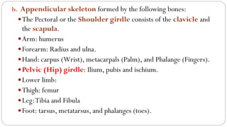
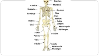
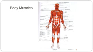
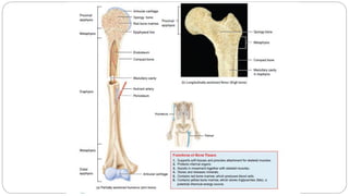
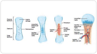
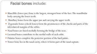
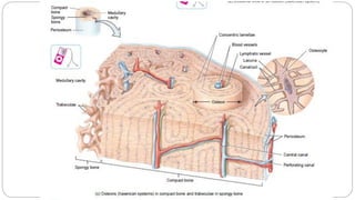
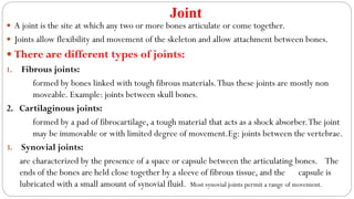
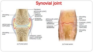
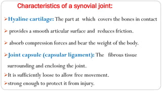
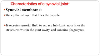
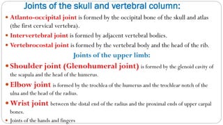
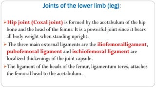
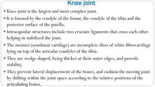
Dr.Jackson Achankunju, MSc,MEd,MPhil,PhD
Department of Science
Chapter 2#
The Skeleto-muscular System
Skeletomuscular system
A typical adult human skeleton consists of 206 bones.
Individuals may have more or fewer bones than this owing to
anatomical variations.
TYPES OF BONE
GROSS ANATOMICAL CLASSIFICATION
Functions of Skeletal System
 The skeleton serves several major functions including:
a. Support:The skeleton provides the framework which supports the body
and maintains its shape.
b. Movement:The joints between bones permit movement.
c. Protection:The skeleton protects many vital organs, such as the skull
protects the brain, the eyes, and the middle and inner ears.The vertebrae
protect the spinal cord.The rib cage, spine, and sternum protect the
human lungs, human heart and major blood vessels.
d. Blood cell production:The skeleton is the site of
haematopoiesis, the development of blood cells that takes place in
the bone marrow.
e. Storage: Bone matrix can store calcium and is involved in
calcium metabolism.
Diet and bone:
Healthy bone tissue requires adequate dietary calcium and vitamins A,
C, and D.
Calcium and smaller amounts of other minerals such as phosphate,
iron and manganese, is essential for adequate mineralization of bone.
VitaminA is needed for osteoblast activity.
Vitamin C is used in collagen synthesis.
Vitamin D is required for calcium and phosphorus absorption from
the intestinal tract.
HUMAN SKELETON
Axial and Appendicular Divisions
Axial = 80 bones
Skull
Vertebrae
Ribs and Sternum
Auditory ossicles
Hyoid
• Appendicular = 126
–Upper = 64
•Pectoral Girdle
•Arms and Legs
–Lower = 62
•Pelvic girdle
•Legs and feet
■ The Skull consists of 8 cranial bones include:
Frontal bone 1 in number anteriorly.
• Temporal bones 2 in number laterally.
• Occipital bone 1 in number posteriorly.
• Parietal bones 2 in number superiorly.
• Sphenoid bone 1 in number, ethmoid bone 1 in number, and parts
• of the frontal, temporal and occipital bones inferiorly
The Skull
The Skull
Facial Bones
2 nasal bones
2 Maxillae
2 palatine bones
Mandible (mand- to chew), or lower jawbone-strongest facial
bone.
2 Zygomatic bones
2 lacrimal bones
2inferior nasal conchae
vomer
The cervical, thoracic, and lumbar vertebrae are movable,
but the sacrum and coccyx are immovable.
Between adjacent
vertebrae from the second cervical vertebra to the
sacrum are intervertebral discs.
Vertebral column = 26 bones
The total number of vertebrae during early development is 33.
Later, several vertebrae in the sacral and coccygeal regions fuse
and form 26.
Cervical(Neck region)= (7)
Thoracic(thorax )=(12)
Lumbar (lumb-loin) = (5)
Sacrum (1)=consists of 5 fused sacral vertebrae.
Coccyx (1)= consists of 4 fused coccygeal vertebrae
Ribs and Sternum= 25 bones
12 pairs of ribs
1 sternum
Bones of the skeleton
 The main bones of the human skeleton are:
a. Axial skeleton formed by the following bones:
 Vertebral column includes 7 cervical vertebrae, 12 thoracic vertebrae, 5 lumbar
vertebrae, 5 fused sacrum vertebrae, and 1coccyx.
 The rib cage (12 pairs of ribs and the sternum).
 The skull consists of 8 cranial bones include 1frontal, 2
parietal, 1occipital, 2 temporal, 1 sphenoid, and 1ethmoid
bones, and the facial bones include the mandible,
Maxillary, zygomatic, nasal, lacrimal, palatine, and vomer
bones.
 The upright posture of humans is maintained by the axial
skeleton, which transmits the weight from the head, the
trunk, and the upper extremities down to the lower
extremities at the hip joints.
b. Appendicular skeleton formed by the following bones:
The Pectoral or the Shoulder girdle consists of the clavicle and
the scapula.
Arm: humerus
Forearm: Radius and ulna.
Hand: carpus (Wrist), metacarpals (Palm), and Phalange (Fingers).
Pelvic (Hip) girdle: Ilium, pubis and ischium.
Lower limb:
Thigh: femur
Leg:Tibia and Fibula
Foot: tarsus, metatarsus, and phalanges (toes).
Body Muscles
Facial bones include:
 Mancfible (lower jaw) bone is the largest, strongest bone of the face.The mandibular
body carrying the lower teeth.
 Maxillary bones form the upper jaw and carrying the upper teeth.
 Zygomatic bones (cheek bones) form the prominences of the cheeks and parts of the
inferolateral margins of the orbits.
 Nasal bones are fused medially forming the bridge of the nose.
 Lacrimal bones contribute to the medial walls of each orbit.
 Palatine bones complete the posterior portion of the hard palate.
 Vomer bone lies in the nasal cavity, where it forms part of the nasal septum.
Nasal bones are fused medially forming the bridge of the
nose.
Lacrimal bones contribute to the medial walls of each orbit.
Palatine bones complete the posterior portion of the hard
palate.
Vomer bone lies in the nasal cavity, where it forms part of
the nasal septum
Joint
 A joint is the site at which any two or more bones articulate or come together.
 Joints allow flexibility and movement of the skeleton and allow attachment between bones.
 There are different types of joints:
1. Fibrous joints:
formed by bones linked with tough fibrous materials.Thus these joints are mostly non
moveable. Example: joints between skull bones.
2. Cartilaginous joints:
formed by a pad of fibrocartilage, a tough material that acts as a shock absorber.The joint
may be immovable or with limited degree of movement.Eg: joints between the vertebrae.
3. Synovial joints:
are characterized by the presence of a space or capsule between the articulating bones. The
ends of the bones are held close together by a sleeve of fibrous tissue, and the capsule is
lubricated with a small amount of synovial fluid. Most synovial joints permit a range of movement.
Synovial joint
Characteristics of a synovial joint:
➢Hyaline cartilage: The part at which covers the bones in contact
➢ provides a smooth articular surface and reduces friction.
➢ absorb compression forces and bear the weight of the body.
➢Joint capsule (capsular ligament): The fibrous tissue
surrounding and enclosing the joint.
➢It is sufficiently loose to allow free movement.
➢strong enough to protect it from injury.
Characteristics of a synovial joint:
Synovial membrane:
➢the epithelial layer that lines the capsule.
➢It secretes synovial fluid to act as a lubricant, nourishes the
structures within the joint cavity, and contains phagocytes.
Joints of the skull and vertebral column:
 Atlanto-occipital joint is formed by the occipital bone of the skull and atlas
(the first cervical vertebra).
 Intervertebral joint is formed by adjacent vertebral bodies.
 Vertebrocostal joint is formed by the vertebral body and the head of the rib.
Joints of the upper limb:
Shoulder joint (Glenohumeral joint) is formed by the glenoid cavity of
the scapula and the head of the humerus.
Elbow joint is formed by the trochlea of the humerus and the trochlear notch of the
ulna and the head of the radius.
Wrist joint between the distal end of the radius and the proximal ends of upper carpal
bones.
 Joints of the hands and fingers
Joints of the lower limb (leg):
➢Hip joint (Coxal joint) is formed by the acetabulum of the hip
bone and the head of the femur. It is a powerful joint since it bears
all body weight when standing upright.
➢The three main external ligaments are the iliofemoralligament,
pubofemoral ligament and ischiofemoral ligament are
localized thickenings of the joint capsule.
➢The ligament of the heads of the femur, ligamentum teres, attaches
the femoral head to the acetabulum.
Knee joint
 Knee joint is the largest and most complex joint.
 It is formed by the condyle of the femur, the condyle of the tibia and the
posterior surface of the patella.
 Intracapsular structures include two cruciate ligaments that cross each other
helping in stabilized the joint.
 The menisci (semilunar cartilage) are incomplete discs of white fibrocartilage
lying on top of the articular condyles of the tibia.
 They are wedge shaped, being thicker at their outer edges, and provide
stability.
 They prevent lateral displacement of the bones, and cushion the moving joint
by shifting within the joint space according to the relative positions of the
articulating bones.
Ankle joint
 Ankle joint is formed by the distal end of the tibia, the distal end of fibula
and the tarsals.
 Joints of the feet and toes are a number of synovial joints between the tarsal
bones, between the tarsal and metatarsal bones, between the metatarsals and
proximal phalanges and between the phalanges.
Body Muscles
 Muscle is a band or bundle of fibrous tissue that has the ability to contract,
producing movement in or maintaining the position of parts of the body.Tendons
and muscles work together to move bones.
 Ligament is a cord or band of connective tissue uniting two structures. Example:
the cruciate ligaments of the knee joint.
 Tendon is a tough band of fibrous connective tissue that usually connects muscle to
bone and is capable of withstanding tension.
 Tendons are similar to ligaments and fasciae as they are all made of collagen except
that ligaments join one bone to another bone, and fasciae connect muscles to other
muscles.
 Fascia is a mixture of connective tissues mostly areolar and collagen fibers that
covers the muscles or units the layers of the skin to the underlying deeper tissues.
Types of Muscle Tissues:
 The three types of muscle tissue are:
 Skeletal, Cardiac, and Smooth muscles.
 Functions of muscles in the body:
1. Produce movement
2. Maintains posture
3. Stabilizes joints
4. Generate heat
Physiology of a muscle fiber
 The skeletal muscle cell contracts in response to stimulation from a nerve
fiber.
 Skelton muscle cells are stimulated by motor neurons of the somatic nervous
system.
 The name given to a synapse between a motor nerve and a skeletal muscle
fiber is the neuromuscular junction.
 When muscle fiber contracts, it obeys the all-or-none law, i.e. the whole
fiber either contracts completely or not at all.
 Muscle fibers need an adequate supply of oxygen and fuel molecules such as
glucose. Fatigue occurs when a muscle works at a level that exceeds those
supplies. In this case the muscle response decreases with fatigue.
Human Anatomy.Skeletal and Muscular system.pdf

Human Anatomy.Skeletal and Muscular system.pdf

  • 1.
    Dr.Jackson Achankunju, MSc,MEd,MPhil,PhD Departmentof Science Chapter 2# The Skeleto-muscular System
  • 2.
    Skeletomuscular system A typicaladult human skeleton consists of 206 bones. Individuals may have more or fewer bones than this owing to anatomical variations.
  • 3.
    TYPES OF BONE GROSSANATOMICAL CLASSIFICATION
  • 5.
    Functions of SkeletalSystem  The skeleton serves several major functions including: a. Support:The skeleton provides the framework which supports the body and maintains its shape. b. Movement:The joints between bones permit movement. c. Protection:The skeleton protects many vital organs, such as the skull protects the brain, the eyes, and the middle and inner ears.The vertebrae protect the spinal cord.The rib cage, spine, and sternum protect the human lungs, human heart and major blood vessels.
  • 6.
    d. Blood cellproduction:The skeleton is the site of haematopoiesis, the development of blood cells that takes place in the bone marrow. e. Storage: Bone matrix can store calcium and is involved in calcium metabolism.
  • 7.
    Diet and bone: Healthybone tissue requires adequate dietary calcium and vitamins A, C, and D. Calcium and smaller amounts of other minerals such as phosphate, iron and manganese, is essential for adequate mineralization of bone. VitaminA is needed for osteoblast activity. Vitamin C is used in collagen synthesis. Vitamin D is required for calcium and phosphorus absorption from the intestinal tract.
  • 8.
  • 9.
    Axial and AppendicularDivisions Axial = 80 bones Skull Vertebrae Ribs and Sternum Auditory ossicles Hyoid • Appendicular = 126 –Upper = 64 •Pectoral Girdle •Arms and Legs –Lower = 62 •Pelvic girdle •Legs and feet
  • 15.
    ■ The Skullconsists of 8 cranial bones include: Frontal bone 1 in number anteriorly. • Temporal bones 2 in number laterally. • Occipital bone 1 in number posteriorly. • Parietal bones 2 in number superiorly. • Sphenoid bone 1 in number, ethmoid bone 1 in number, and parts • of the frontal, temporal and occipital bones inferiorly
  • 16.
  • 17.
  • 19.
    Facial Bones 2 nasalbones 2 Maxillae 2 palatine bones Mandible (mand- to chew), or lower jawbone-strongest facial bone. 2 Zygomatic bones 2 lacrimal bones 2inferior nasal conchae vomer
  • 23.
    The cervical, thoracic,and lumbar vertebrae are movable, but the sacrum and coccyx are immovable. Between adjacent vertebrae from the second cervical vertebra to the sacrum are intervertebral discs.
  • 24.
    Vertebral column =26 bones The total number of vertebrae during early development is 33. Later, several vertebrae in the sacral and coccygeal regions fuse and form 26. Cervical(Neck region)= (7) Thoracic(thorax )=(12) Lumbar (lumb-loin) = (5) Sacrum (1)=consists of 5 fused sacral vertebrae. Coccyx (1)= consists of 4 fused coccygeal vertebrae
  • 25.
    Ribs and Sternum=25 bones 12 pairs of ribs 1 sternum
  • 29.
    Bones of theskeleton  The main bones of the human skeleton are: a. Axial skeleton formed by the following bones:  Vertebral column includes 7 cervical vertebrae, 12 thoracic vertebrae, 5 lumbar vertebrae, 5 fused sacrum vertebrae, and 1coccyx.  The rib cage (12 pairs of ribs and the sternum).
  • 30.
     The skullconsists of 8 cranial bones include 1frontal, 2 parietal, 1occipital, 2 temporal, 1 sphenoid, and 1ethmoid bones, and the facial bones include the mandible, Maxillary, zygomatic, nasal, lacrimal, palatine, and vomer bones.  The upright posture of humans is maintained by the axial skeleton, which transmits the weight from the head, the trunk, and the upper extremities down to the lower extremities at the hip joints.
  • 31.
    b. Appendicular skeletonformed by the following bones: The Pectoral or the Shoulder girdle consists of the clavicle and the scapula. Arm: humerus Forearm: Radius and ulna. Hand: carpus (Wrist), metacarpals (Palm), and Phalange (Fingers). Pelvic (Hip) girdle: Ilium, pubis and ischium. Lower limb: Thigh: femur Leg:Tibia and Fibula Foot: tarsus, metatarsus, and phalanges (toes).
  • 33.
  • 36.
    Facial bones include: Mancfible (lower jaw) bone is the largest, strongest bone of the face.The mandibular body carrying the lower teeth.  Maxillary bones form the upper jaw and carrying the upper teeth.  Zygomatic bones (cheek bones) form the prominences of the cheeks and parts of the inferolateral margins of the orbits.  Nasal bones are fused medially forming the bridge of the nose.  Lacrimal bones contribute to the medial walls of each orbit.  Palatine bones complete the posterior portion of the hard palate.  Vomer bone lies in the nasal cavity, where it forms part of the nasal septum.
  • 37.
    Nasal bones arefused medially forming the bridge of the nose. Lacrimal bones contribute to the medial walls of each orbit. Palatine bones complete the posterior portion of the hard palate. Vomer bone lies in the nasal cavity, where it forms part of the nasal septum
  • 39.
    Joint  A jointis the site at which any two or more bones articulate or come together.  Joints allow flexibility and movement of the skeleton and allow attachment between bones.  There are different types of joints: 1. Fibrous joints: formed by bones linked with tough fibrous materials.Thus these joints are mostly non moveable. Example: joints between skull bones. 2. Cartilaginous joints: formed by a pad of fibrocartilage, a tough material that acts as a shock absorber.The joint may be immovable or with limited degree of movement.Eg: joints between the vertebrae. 3. Synovial joints: are characterized by the presence of a space or capsule between the articulating bones. The ends of the bones are held close together by a sleeve of fibrous tissue, and the capsule is lubricated with a small amount of synovial fluid. Most synovial joints permit a range of movement.
  • 40.
  • 41.
    Characteristics of asynovial joint: ➢Hyaline cartilage: The part at which covers the bones in contact ➢ provides a smooth articular surface and reduces friction. ➢ absorb compression forces and bear the weight of the body. ➢Joint capsule (capsular ligament): The fibrous tissue surrounding and enclosing the joint. ➢It is sufficiently loose to allow free movement. ➢strong enough to protect it from injury.
  • 42.
    Characteristics of asynovial joint: Synovial membrane: ➢the epithelial layer that lines the capsule. ➢It secretes synovial fluid to act as a lubricant, nourishes the structures within the joint cavity, and contains phagocytes.
  • 43.
    Joints of theskull and vertebral column:  Atlanto-occipital joint is formed by the occipital bone of the skull and atlas (the first cervical vertebra).  Intervertebral joint is formed by adjacent vertebral bodies.  Vertebrocostal joint is formed by the vertebral body and the head of the rib. Joints of the upper limb: Shoulder joint (Glenohumeral joint) is formed by the glenoid cavity of the scapula and the head of the humerus. Elbow joint is formed by the trochlea of the humerus and the trochlear notch of the ulna and the head of the radius. Wrist joint between the distal end of the radius and the proximal ends of upper carpal bones.  Joints of the hands and fingers
  • 44.
    Joints of thelower limb (leg): ➢Hip joint (Coxal joint) is formed by the acetabulum of the hip bone and the head of the femur. It is a powerful joint since it bears all body weight when standing upright. ➢The three main external ligaments are the iliofemoralligament, pubofemoral ligament and ischiofemoral ligament are localized thickenings of the joint capsule. ➢The ligament of the heads of the femur, ligamentum teres, attaches the femoral head to the acetabulum.
  • 45.
    Knee joint  Kneejoint is the largest and most complex joint.  It is formed by the condyle of the femur, the condyle of the tibia and the posterior surface of the patella.  Intracapsular structures include two cruciate ligaments that cross each other helping in stabilized the joint.  The menisci (semilunar cartilage) are incomplete discs of white fibrocartilage lying on top of the articular condyles of the tibia.  They are wedge shaped, being thicker at their outer edges, and provide stability.  They prevent lateral displacement of the bones, and cushion the moving joint by shifting within the joint space according to the relative positions of the articulating bones.
  • 46.
    Ankle joint  Anklejoint is formed by the distal end of the tibia, the distal end of fibula and the tarsals.  Joints of the feet and toes are a number of synovial joints between the tarsal bones, between the tarsal and metatarsal bones, between the metatarsals and proximal phalanges and between the phalanges.
  • 47.
    Body Muscles  Muscleis a band or bundle of fibrous tissue that has the ability to contract, producing movement in or maintaining the position of parts of the body.Tendons and muscles work together to move bones.  Ligament is a cord or band of connective tissue uniting two structures. Example: the cruciate ligaments of the knee joint.  Tendon is a tough band of fibrous connective tissue that usually connects muscle to bone and is capable of withstanding tension.  Tendons are similar to ligaments and fasciae as they are all made of collagen except that ligaments join one bone to another bone, and fasciae connect muscles to other muscles.  Fascia is a mixture of connective tissues mostly areolar and collagen fibers that covers the muscles or units the layers of the skin to the underlying deeper tissues.
  • 48.
    Types of MuscleTissues:  The three types of muscle tissue are:  Skeletal, Cardiac, and Smooth muscles.  Functions of muscles in the body: 1. Produce movement 2. Maintains posture 3. Stabilizes joints 4. Generate heat
  • 49.
    Physiology of amuscle fiber  The skeletal muscle cell contracts in response to stimulation from a nerve fiber.  Skelton muscle cells are stimulated by motor neurons of the somatic nervous system.  The name given to a synapse between a motor nerve and a skeletal muscle fiber is the neuromuscular junction.  When muscle fiber contracts, it obeys the all-or-none law, i.e. the whole fiber either contracts completely or not at all.  Muscle fibers need an adequate supply of oxygen and fuel molecules such as glucose. Fatigue occurs when a muscle works at a level that exceeds those supplies. In this case the muscle response decreases with fatigue.