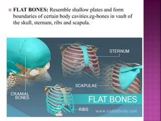
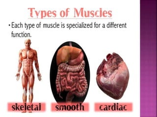
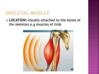
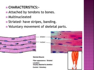
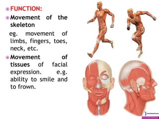
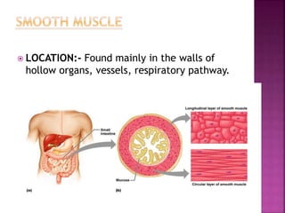
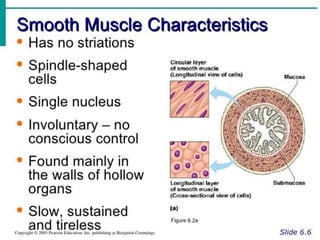
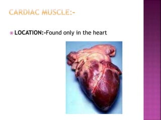
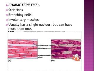
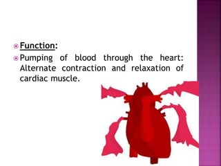
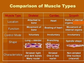
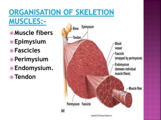
1) There are three main types of muscles in the body - skeletal muscles, which are voluntarily controlled and attached to bones to enable movement; cardiac muscle found only in the heart to enable pumping of blood; and smooth muscle found in organs like the digestive tract and blood vessels to enable involuntary movement.
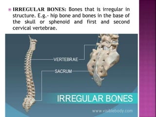
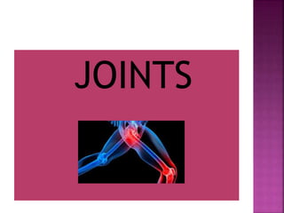
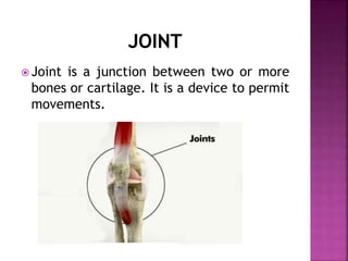
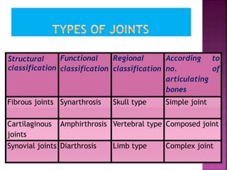
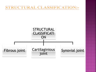
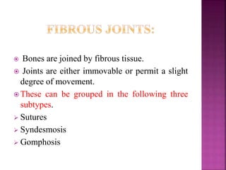
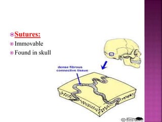
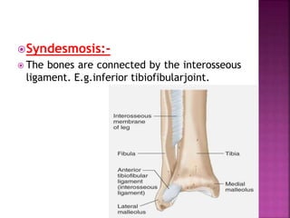
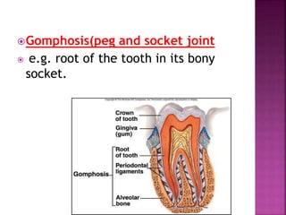
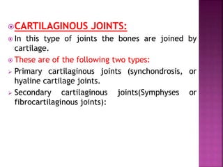
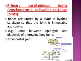
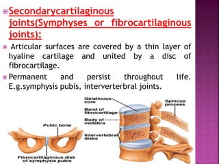
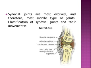
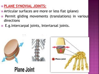
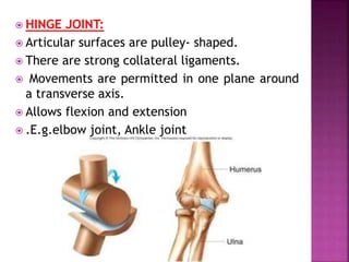
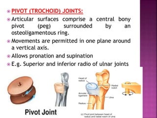
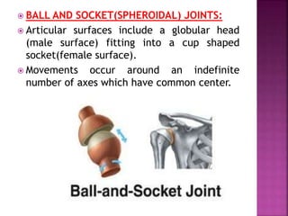
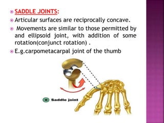
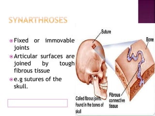
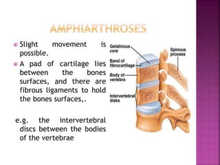
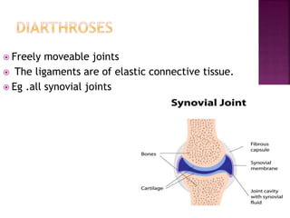
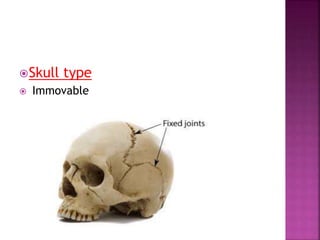
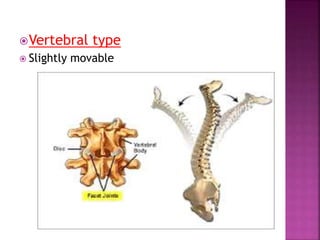
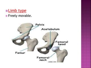
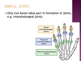
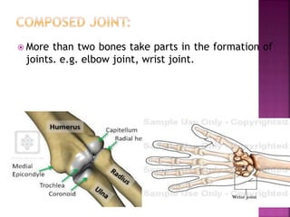
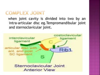
2) Joints can be classified based on their structure, function and region of the body. The main types of joints are fibrous, cartilaginous and synovial joints. Synovial joints which include hinge, pivot, ball-and-socket and saddle joints, allow the most movement.


![Pre-history & Early Man [PDF]](https://cdn.slidesharecdn.com/ss_thumbnails/pre-historyearlymanpowerpointfull-150914164855-lva1-app6892-thumbnail.jpg?width=640&height=640&fit=bounds)


![PERI-PROSTHETIC FRACTURE NAIL-PLATE CONSTRUCT [NPC].pptx](https://cdn.slidesharecdn.com/ss_thumbnails/drarunkumardrmohamedashrafperiprostheticfrasturenail-plateconstructnpc-260209164459-7e9d15a1-thumbnail.jpg?width=640&height=640&fit=bounds)


