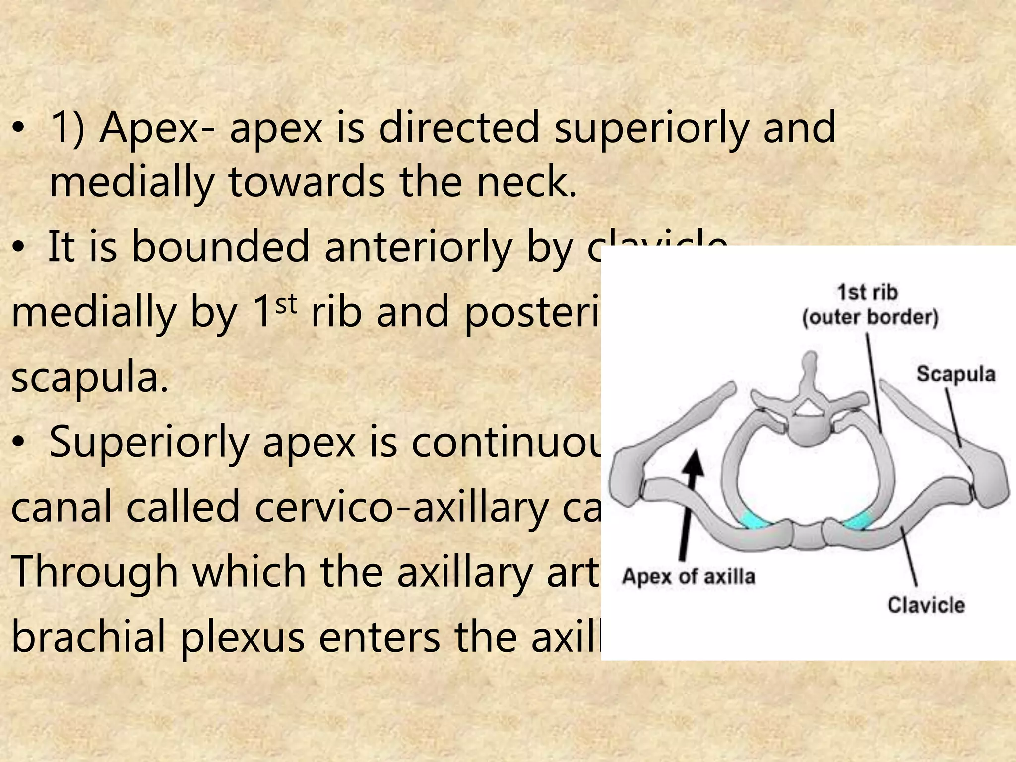
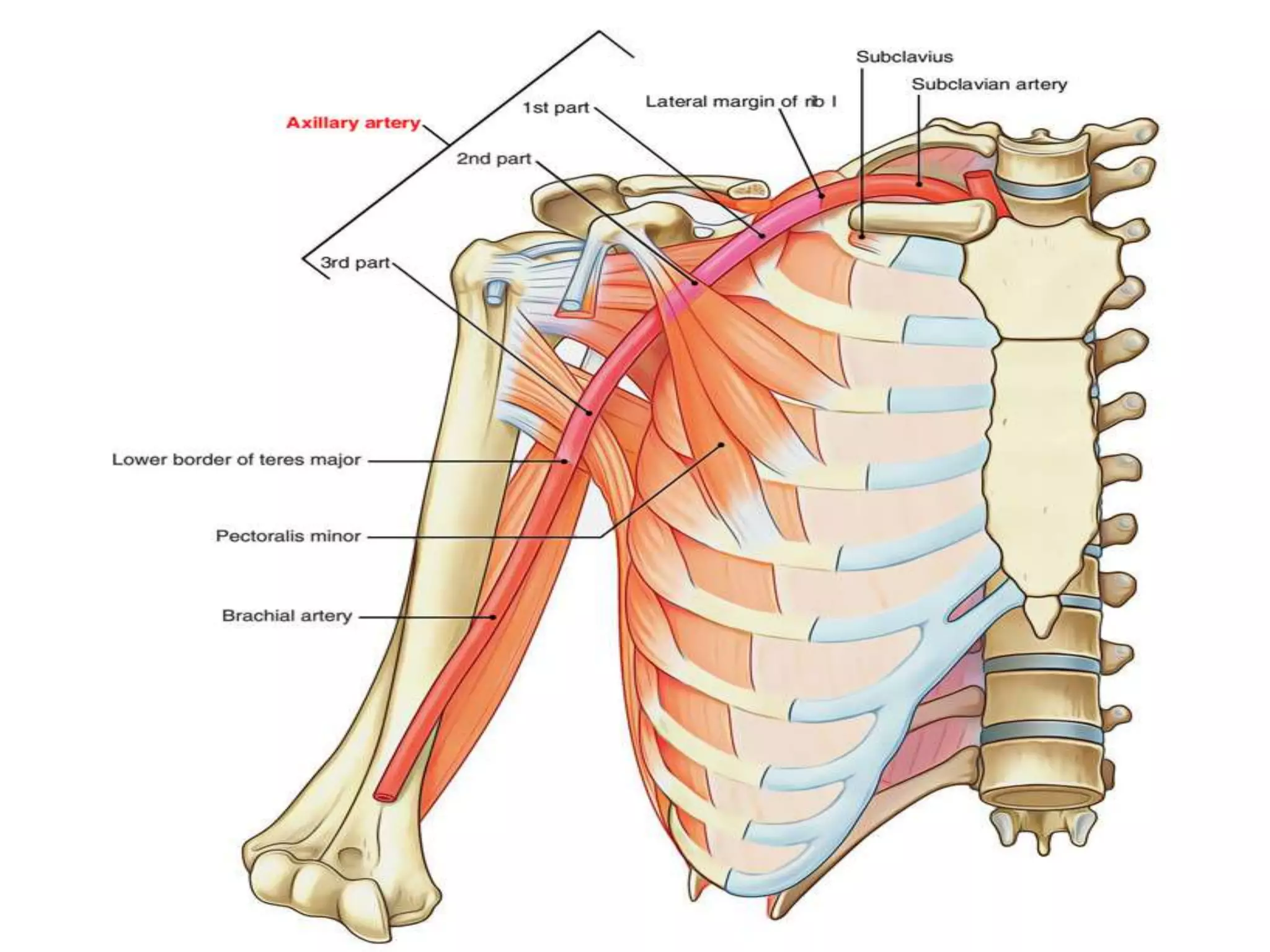
The document describes the anatomy of the axilla, subclavian artery, axillary artery, brachial artery, radial artery, ulnar artery, and intermuscular spaces in the arm. Key points include:
- The axilla is a pyramidal space between the upper arm and chest wall containing lymph nodes, blood vessels and nerves.
- The subclavian artery becomes the axillary artery in the axilla and then the brachial artery in the arm, with named branches along its course.
- The radial and ulnar arteries are terminal branches of the brachial artery, running in the forearm and hand.
- There are three intermuscular spaces in the arm that contain named
brachial plexus, branches of brachial plexus, main nerves of brachial plexus and their innervations, disorders of brachial plexus injury, Erb's palsy, Klumpke's palsy, compression of brachial plexus
rectus sheath, the sheath covering rectus muscle of anterior abdominal wall, formation of the sheath, the muscles involved in ts formation, and the contents the sheath is covering
Seminar presentation on arterial supply of human head & neck - carotid artery, maxillary artery, ophthalmic artery
post-graduate level
MDS- oral & maxillofacial surgery
Anatomy of axilla with Dr- Ameera Al-Humidi .pptxAmeera Al-Humidi
The axilla is the anatomical region under the shoulder joint where the arm connects to the shoulder.
The axilla has five anatomic borders: superior, anterior, posterior, lateral, and medial walls.
The borders of the axilla are composed of muscles, including the serratus anterior, coracobrachialis, and short head of the biceps
The axillary walls are used as landmarks by surgeons to prevent damage to the neurovascular structures within the axilla during surgery
The contents of the axilla include muscles, nerves, vessels, and lymphatics
The axillary artery and vein, brachial plexus, and axillary lymph nodes are some of the neurovascular structures found in the axilla
These slides are based on the notes provided by the K V Sangathan. For the revision of thermodynamics the notes are pretty awseome. I f only want submit the home work they will do so.I am sure they will help.
THANK YOU
It is fully based on the notes provided by the K V Sangathan. For the revision to students they are short but enough to clear the concept of equillibrium.hope you like them.give your reviews.
THANK YOU
These simplified slides by Dr. Sidra Arshad present an overview of the non-respiratory functions of the respiratory tract.
Learning objectives:
1. Enlist the non-respiratory functions of the respiratory tract
2. Briefly explain how these functions are carried out
3. Discuss the significance of dead space
4. Differentiate between minute ventilation and alveolar ventilation
5. Describe the cough and sneeze reflexes
Study Resources:
1. Chapter 39, Guyton and Hall Textbook of Medical Physiology, 14th edition
2. Chapter 34, Ganong’s Review of Medical Physiology, 26th edition
3. Chapter 17, Human Physiology by Lauralee Sherwood, 9th edition
4. Non-respiratory functions of the lungs https://academic.oup.com/bjaed/article/13/3/98/278874
Title: Sense of Taste
Presenter: Dr. Faiza, Assistant Professor of Physiology
Qualifications:
MBBS (Best Graduate, AIMC Lahore)
FCPS Physiology
ICMT, CHPE, DHPE (STMU)
MPH (GC University, Faisalabad)
MBA (Virtual University of Pakistan)
Learning Objectives:
Describe the structure and function of taste buds.
Describe the relationship between the taste threshold and taste index of common substances.
Explain the chemical basis and signal transduction of taste perception for each type of primary taste sensation.
Recognize different abnormalities of taste perception and their causes.
Key Topics:
Significance of Taste Sensation:
Differentiation between pleasant and harmful food
Influence on behavior
Selection of food based on metabolic needs
Receptors of Taste:
Taste buds on the tongue
Influence of sense of smell, texture of food, and pain stimulation (e.g., by pepper)
Primary and Secondary Taste Sensations:
Primary taste sensations: Sweet, Sour, Salty, Bitter, Umami
Chemical basis and signal transduction mechanisms for each taste
Taste Threshold and Index:
Taste threshold values for Sweet (sucrose), Salty (NaCl), Sour (HCl), and Bitter (Quinine)
Taste index relationship: Inversely proportional to taste threshold
Taste Blindness:
Inability to taste certain substances, particularly thiourea compounds
Example: Phenylthiocarbamide
Structure and Function of Taste Buds:
Composition: Epithelial cells, Sustentacular/Supporting cells, Taste cells, Basal cells
Features: Taste pores, Taste hairs/microvilli, and Taste nerve fibers
Location of Taste Buds:
Found in papillae of the tongue (Fungiform, Circumvallate, Foliate)
Also present on the palate, tonsillar pillars, epiglottis, and proximal esophagus
Mechanism of Taste Stimulation:
Interaction of taste substances with receptors on microvilli
Signal transduction pathways for Umami, Sweet, Bitter, Sour, and Salty tastes
Taste Sensitivity and Adaptation:
Decrease in sensitivity with age
Rapid adaptation of taste sensation
Role of Saliva in Taste:
Dissolution of tastants to reach receptors
Washing away the stimulus
Taste Preferences and Aversions:
Mechanisms behind taste preference and aversion
Influence of receptors and neural pathways
Impact of Sensory Nerve Damage:
Degeneration of taste buds if the sensory nerve fiber is cut
Abnormalities of Taste Detection:
Conditions: Ageusia, Hypogeusia, Dysgeusia (parageusia)
Causes: Nerve damage, neurological disorders, infections, poor oral hygiene, adverse drug effects, deficiencies, aging, tobacco use, altered neurotransmitter levels
Neurotransmitters and Taste Threshold:
Effects of serotonin (5-HT) and norepinephrine (NE) on taste sensitivity
Supertasters:
25% of the population with heightened sensitivity to taste, especially bitterness
Increased number of fungiform papillae
Basavarajeeyam is an important text for ayurvedic physician belonging to andhra pradehs. It is a popular compendium in various parts of our country as well as in andhra pradesh. The content of the text was presented in sanskrit and telugu language (Bilingual). One of the most famous book in ayurvedic pharmaceutics and therapeutics. This book contains 25 chapters called as prakaranas. Many rasaoushadis were explained, pioneer of dhatu druti, nadi pareeksha, mutra pareeksha etc. Belongs to the period of 15-16 century. New diseases like upadamsha, phiranga rogas are explained.
New Directions in Targeted Therapeutic Approaches for Older Adults With Mantl...i3 Health
i3 Health is pleased to make the speaker slides from this activity available for use as a non-accredited self-study or teaching resource.
This slide deck presented by Dr. Kami Maddocks, Professor-Clinical in the Division of Hematology and
Associate Division Director for Ambulatory Operations
The Ohio State University Comprehensive Cancer Center, will provide insight into new directions in targeted therapeutic approaches for older adults with mantle cell lymphoma.
STATEMENT OF NEED
Mantle cell lymphoma (MCL) is a rare, aggressive B-cell non-Hodgkin lymphoma (NHL) accounting for 5% to 7% of all lymphomas. Its prognosis ranges from indolent disease that does not require treatment for years to very aggressive disease, which is associated with poor survival (Silkenstedt et al, 2021). Typically, MCL is diagnosed at advanced stage and in older patients who cannot tolerate intensive therapy (NCCN, 2022). Although recent advances have slightly increased remission rates, recurrence and relapse remain very common, leading to a median overall survival between 3 and 6 years (LLS, 2021). Though there are several effective options, progress is still needed towards establishing an accepted frontline approach for MCL (Castellino et al, 2022). Treatment selection and management of MCL are complicated by the heterogeneity of prognosis, advanced age and comorbidities of patients, and lack of an established standard approach for treatment, making it vital that clinicians be familiar with the latest research and advances in this area. In this activity chaired by Michael Wang, MD, Professor in the Department of Lymphoma & Myeloma at MD Anderson Cancer Center, expert faculty will discuss prognostic factors informing treatment, the promising results of recent trials in new therapeutic approaches, and the implications of treatment resistance in therapeutic selection for MCL.
Target Audience
Hematology/oncology fellows, attending faculty, and other health care professionals involved in the treatment of patients with mantle cell lymphoma (MCL).
Learning Objectives
1.) Identify clinical and biological prognostic factors that can guide treatment decision making for older adults with MCL
2.) Evaluate emerging data on targeted therapeutic approaches for treatment-naive and relapsed/refractory MCL and their applicability to older adults
3.) Assess mechanisms of resistance to targeted therapies for MCL and their implications for treatment selection
Flu Vaccine Alert in Bangalore Karnatakaaddon Scans
As flu season approaches, health officials in Bangalore, Karnataka, are urging residents to get their flu vaccinations. The seasonal flu, while common, can lead to severe health complications, particularly for vulnerable populations such as young children, the elderly, and those with underlying health conditions.
Dr. Vidisha Kumari, a leading epidemiologist in Bangalore, emphasizes the importance of getting vaccinated. "The flu vaccine is our best defense against the influenza virus. It not only protects individuals but also helps prevent the spread of the virus in our communities," he says.
This year, the flu season is expected to coincide with a potential increase in other respiratory illnesses. The Karnataka Health Department has launched an awareness campaign highlighting the significance of flu vaccinations. They have set up multiple vaccination centers across Bangalore, making it convenient for residents to receive their shots.
To encourage widespread vaccination, the government is also collaborating with local schools, workplaces, and community centers to facilitate vaccination drives. Special attention is being given to ensuring that the vaccine is accessible to all, including marginalized communities who may have limited access to healthcare.
Residents are reminded that the flu vaccine is safe and effective. Common side effects are mild and may include soreness at the injection site, mild fever, or muscle aches. These side effects are generally short-lived and far less severe than the flu itself.
Healthcare providers are also stressing the importance of continuing COVID-19 precautions. Wearing masks, practicing good hand hygiene, and maintaining social distancing are still crucial, especially in crowded places.
Protect yourself and your loved ones by getting vaccinated. Together, we can help keep Bangalore healthy and safe this flu season. For more information on vaccination centers and schedules, residents can visit the Karnataka Health Department’s official website or follow their social media pages.
Stay informed, stay safe, and get your flu shot today!
263778731218 Abortion Clinic /Pills In Harare ,sisternakatoto
263778731218 Abortion Clinic /Pills In Harare ,ABORTION WOMEN’S CLINIC +27730423979 IN women clinic we believe that every woman should be able to make choices in her pregnancy. Our job is to provide compassionate care, safety,affordable and confidential services. That’s why we have won the trust from all generations of women all over the world. we use non surgical method(Abortion pills) to terminate…Dr.LISA +27730423979women Clinic is committed to providing the highest quality of obstetrical and gynecological care to women of all ages. Our dedicated staff aim to treat each patient and her health concerns with compassion and respect.Our dedicated group ABORTION WOMEN’S CLINIC +27730423979 IN women clinic we believe that every woman should be able to make choices in her pregnancy. Our job is to provide compassionate care, safety,affordable and confidential services. That’s why we have won the trust from all generations of women all over the world. we use non surgical method(Abortion pills) to terminate…Dr.LISA +27730423979women Clinic is committed to providing the highest quality of obstetrical and gynecological care to women of all ages. Our dedicated staff aim to treat each patient and her health concerns with compassion and respect.Our dedicated group of receptionists, nurses, and physicians have worked together as a teamof receptionists, nurses, and physicians have worked together as a team wwww.lisywomensclinic.co.za/
ARTIFICIAL INTELLIGENCE IN HEALTHCARE.pdfAnujkumaranit
Artificial intelligence (AI) refers to the simulation of human intelligence processes by machines, especially computer systems. It encompasses tasks such as learning, reasoning, problem-solving, perception, and language understanding. AI technologies are revolutionizing various fields, from healthcare to finance, by enabling machines to perform tasks that typically require human intelligence.
Lung Cancer: Artificial Intelligence, Synergetics, Complex System Analysis, S...Oleg Kshivets
RESULTS: Overall life span (LS) was 2252.1±1742.5 days and cumulative 5-year survival (5YS) reached 73.2%, 10 years – 64.8%, 20 years – 42.5%. 513 LCP lived more than 5 years (LS=3124.6±1525.6 days), 148 LCP – more than 10 years (LS=5054.4±1504.1 days).199 LCP died because of LC (LS=562.7±374.5 days). 5YS of LCP after bi/lobectomies was significantly superior in comparison with LCP after pneumonectomies (78.1% vs.63.7%, P=0.00001 by log-rank test). AT significantly improved 5YS (66.3% vs. 34.8%) (P=0.00000 by log-rank test) only for LCP with N1-2. Cox modeling displayed that 5YS of LCP significantly depended on: phase transition (PT) early-invasive LC in terms of synergetics, PT N0—N12, cell ratio factors (ratio between cancer cells- CC and blood cells subpopulations), G1-3, histology, glucose, AT, blood cell circuit, prothrombin index, heparin tolerance, recalcification time (P=0.000-0.038). Neural networks, genetic algorithm selection and bootstrap simulation revealed relationships between 5YS and PT early-invasive LC (rank=1), PT N0—N12 (rank=2), thrombocytes/CC (3), erythrocytes/CC (4), eosinophils/CC (5), healthy cells/CC (6), lymphocytes/CC (7), segmented neutrophils/CC (8), stick neutrophils/CC (9), monocytes/CC (10); leucocytes/CC (11). Correct prediction of 5YS was 100% by neural networks computing (area under ROC curve=1.0; error=0.0).
CONCLUSIONS: 5YS of LCP after radical procedures significantly depended on: 1) PT early-invasive cancer; 2) PT N0--N12; 3) cell ratio factors; 4) blood cell circuit; 5) biochemical factors; 6) hemostasis system; 7) AT; 8) LC characteristics; 9) LC cell dynamics; 10) surgery type: lobectomy/pneumonectomy; 11) anthropometric data. Optimal diagnosis and treatment strategies for LC are: 1) screening and early detection of LC; 2) availability of experienced thoracic surgeons because of complexity of radical procedures; 3) aggressive en block surgery and adequate lymph node dissection for completeness; 4) precise prediction; 5) adjuvant chemoimmunoradiotherapy for LCP with unfavorable prognosis.
Lung Cancer: Artificial Intelligence, Synergetics, Complex System Analysis, S...
axilla.pptx
1. Axilla
• Also called armpit.
• It is a pyramidal space situated b/w upper part
of arm and chest wall.
• It resembles four sided pyramid.
• It has-
An apex
A base
And four walls (boundries)-
anterior,posterior, medial, and lateral.
2. • 1) Apex- apex is directed superiorly and
medially towards the neck.
• It is bounded anteriorly by clavicle,
medially by 1st rib and posteriorly by
scapula.
• Superiorly apex is continuous as
canal called cervico-axillary canal.
Through which the axillary artery and
brachial plexus enters the axilla.
3. 2) base- directed downwards, and formed by
skin.
3) anterior wall- formed by pactoralis major,
pactoralis minor, and subclavius muscle.
4) posterior wall- formed by subscapularis,
latissimus dorsi muscles.
5)Medial wall- formed by upper four ribs and
serratus anterior muscle.
6)Lateral wall- formed by upper part of shaft of
humerus and coracobrahiallis and short head
of biceps brachii muscles.
4.
5. • Contents of axilla-
1- axillary artery and its branches.
2- axillary vein and its tributeries.
3- brachial plexus.
4-Axillary lymph nodes.
5-long thoresic nerves.
6. Triangle of auscultation
• It is small triangular interval, in the back. It has
three boundries-
Medial- lateral border of trapezius.
Lateral- medial border of scapula.
Inferior- upper border of latissmus dorsi.
• Respiratory sounds herd through
stethoscope are said to be batter herd
over this triangle.
7. Subclavian artery
• It is a major artery that supply the upper limb.
• Origin-
Right Subclavian artery - rt. Subclavian artery is
branch of brachiocephelic trunk.
left Subclavian artery- lt. Subclavian artery is
direct branch of arch of aorta.
• Course- after its origin it lies behind the
sternoclavicular joint (right and left), then it
takes upward loop into the neck, and leaves
neck by passing into axilla, where it continues
as axillary artery.
8.
9. cont.
• Extent- it extends from its origin to the outer
border of 1st rib.
• Parts of artery- the sclaneus
anterior muscle divides this artery
into three parts-
1st part – it lies medial to the
sclaneus anterior muscle.
2nd part – it lies deep to the
sclaneus anterior muscle.
3rd part – it lies lateral to the sclaneus anterior
10. • Branches of subclavian artery-
V- vertebral artery
I- internal thoracic artery 1st part
T- thyrocervical trunk
C- costo cervical trunk 2nd part
D- dorsal scapular artery 3rd part
• Relations-
1st part-
anterior- Platysma, Sternocleidomastoid, internal
jugular vein, vagus nerve
Below and behind - pleura, apex of the lung, first
12. Axillary artery
• It is continuation of subclavian artery.
• Extent- it extends from outer border of first rib to
lower border of teres major.
• Parts – pactoralis minor muscle crosses the artery
and devides it into three parts-
1st part- superior to the muscle
2nd part- deep to the muscle
3rd part- inferior to the muscle.
13.
14. Continue...
• Relations-
1st part-
Anterior- platysma, medial and lateral pectoral nerve, pacroralis
major muscle.
Posterior- 1st intercostal space, medial cord.
Laterally- lateral and medial cord.
Medial- axillary vein.
2nd part-
Anterior- pacroralis major and pacroralis minor muscle.
Posterior- posterior cord of brachial plexus
Medial- medial cord.
Lateral- lateral cord
15. • 3rd part-
Anterior- pactoralis major muscle.
Posterior- radial and axillary nerve.
Lateral-musculocutaneous nerve, lateral cord.
Medial- axillary vein, medial cutaneous nerve.
BRANCHES OF ARTERY-
(Save The Lions And Protect Species)
1-Superior thoracic artery – arises from 1st part
2-Thoracoacromial artery- arises from 2nd part
3-Lateral thoracic artery -arises from 2nd part
4-Anterior circumflex humeral artery- arises from 3rd part
5-Posterior circumflex humeral artery- arises from 3rd part
6-Subscapular artery - arises from 3rd part (Largest branch)
16. Brachial artery
• It is the continuation of axillary artery.
Extent- it extends from lower border of teres major muscle to
neck of radius/ medial to the tendon of biceps.
Relations-
Anterior- in upper part- medial cuteneous nerve of forearm.
In middle part- median nerve.
In lower part- bicipital aponeurosis.
19. Radial artery
• Radial artery is smaller direct continuation
of brachial artery.
Course and extent-
• it is quite superficial in its course.
• Runs downwards in lateral aspect of
forearm.
• Leaves forearm by turning posteriorly and
entering anatomical snuff box.
• In hand it ends by forming superficial and
deep palmer arch.
20. Cont...
RELATIONS-
Anterior- Brahioradialis muscle and skin.
Posteriorly- Muscles attacahed on ant. Surface of
radius.
Medially- in U 1/3- pronator teres.
in L 2/3 by flexior carpi radialis
Laterally- Brachioradialis and Radial nerve.
22. BRANCHES – in forearm-
1. Radial recurrent branch- runs upward and anastomose
with radial collateral artery.
2. Muscular branches
3. Palmer carpal branch- arises near the lower border of
pronator quadratus, and supplies the wrist joint.
4. Superficial palmer branch- forms Superficial palmer
arch.
In hand-
1. 1st dorsal metacarpal branch
2. Princeps pollicis artery- supplies to thumb.
3. Radial indicis artery- radial side of index finger.
24. Ulnar artery
• Ulnar artery is the larger terminal branch of brachial artery.
It runs downwards in forearm and enters in palm by passing
superficial to the flexor retinaculum.
Relations-
in upper part- muscles of flexor group
Anteriorly-
in lower part- skin and fascia
Posteriorly- flexor digitorum profundus.
Medially- Ulnar nerve.
Lateral- Flexor digitorum superficialis.
25. Cont...
BRANCHES- (in forearm)
1. anterior and posterior ulnar recurrent artery- anastomose with
superior and inferior ulnar collateral artery.
2. Common interosseous artery- runs along the anterior surface of
interosseous membrane.
3. Muscular branch-
4. Palmar and dorsal carpal branch- anastomose with palmar branch of
radial artery.
(in palm)
1. Muscular branch
2. Superficial branch
3. Deep branch
26. Inter muscular spaces in arm
• There are three intermuscular spaces in arm-
1-Superior triangular space
2-Superior quadrangular space
3-Inferior triangular space
27. Boundaries and content
1-Superior triangular space
Boundaries-
Superior- teres minor
Inferior- teres major
Lateral- long head of triceps.
Contents- circumflex scapular vassels. Med. Lat.
2- superior quadrangular space-
Boundaries-
Superior- teres minor and capsule of shoulder joint.
Inferior- teres major
Medial- long head of triceps
Lateral – shaft of humerus.
Contents- axillary nerve and post. Circumflex humeral vessels.