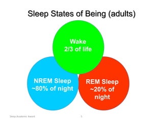
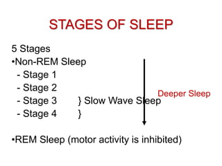
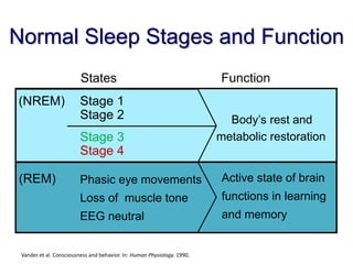
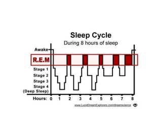
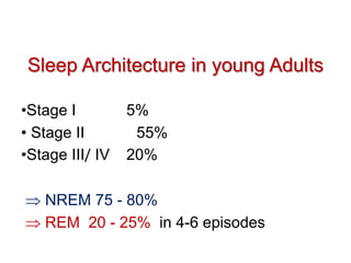
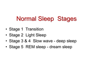
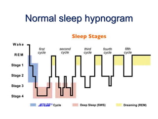
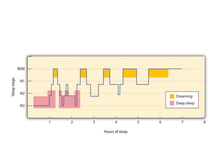
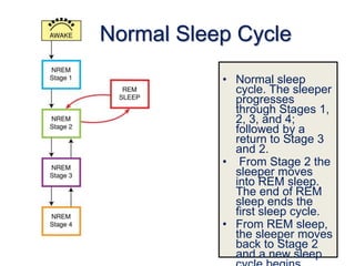
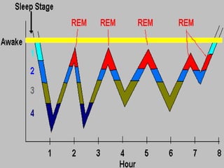
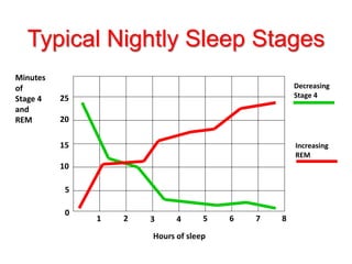
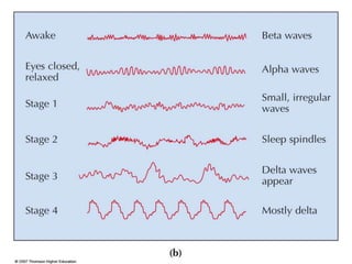
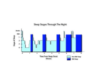
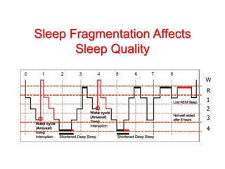
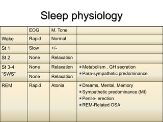
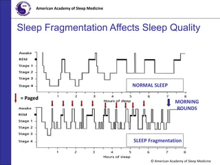
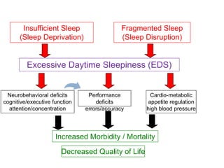
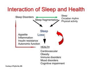
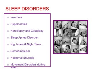
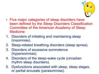
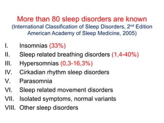
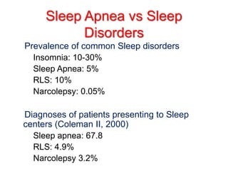
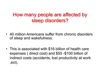
What Happens When you Sleep?
Sleep Academic Award 5
Sleep States of Being (adults)
REM Sleep
~20% of
night
NREM Sleep
~80% of night
Wake
2/3 of life
STAGES OF SLEEP
5 Stages
•Non-REM Sleep
- Stage 1
- Stage 2
- Stage 3 } Slow Wave Sleep
- Stage 4 }
•REM Sleep (motor activity is inhibited)
Deeper Sleep
Normal Sleep Stages and Function
Vander et al. Consciousness and behavior. In: Human Physiology. 1990.
States Function
Active state of brain
functions in learning
and memory
Body’s rest and
metabolic restoration
Phasic eye movements
Loss of muscle tone
EEG neutral
Stage 3
Stage 4
(REM)
Stage 1
Stage 2
(NREM)
Sleep Architecture in young Adults
•Stage I 5%
• Stage II 55%
•Stage III/ IV 20%
 NREM 75 - 80%
 REM 20 - 25% in 4-6 episodes
• Stage 1 Transition
• Stage 2 Light Sleep
• Stage 3 & 4 Slow wave - deep sleep
• Stage 5 REM sleep - dream sleep
Normal Sleep Stages
Normal sleep hypnogram
• Stage 1 is only a transition state from wake to
stage 2 and has no real rest value. A lot of it
means inability to initiate and/or maintain
sleep.
• Stage 3/4 (SWS) decreases as a function of
age, or if you're subject to constant arousals,
as in OSA.
©John Wiley & Sons, Inc. 2007
Huffman: Psychology in Action (8e)
Sleep Over the Life Span
Normal Sleep Cycle
• Normal sleep
cycle. The sleeper
progresses
through Stages 1,
2, 3, and 4;
followed by a
return to Stage 3
and 2.
• From Stage 2 the
sleeper moves
into REM sleep.
The end of REM
sleep ends the
first sleep cycle.
• From REM sleep,
the sleeper moves
back to Stage 2
and a new sleep
Typical Nightly Sleep Stages
Hours of sleep
Minutes
of
Stage 4
and
REM
1 2 3 4 5 6 7 8
0
10
15
20
25
5
Decreasing
Stage 4
Increasing
REM
Sleep Academic Award 19
Awakening
The return to the polysomnographically
defined awake state from any NREM or
REM sleep stages.”
Arousal
“Arousal: An abrupt change from a ‘deeper’ stage
of non-REM (NREM) sleep to a ‘lighter’ stage, or
from REM sleep toward wakefulness, with the
possibility of awakening as the final outcome.
• Arousal may be accompanied by increased tonic
electromyographic activity and heart rate, as well
as by an increased number of body movements.”
• An Arousal is an abrupt change from a
"deeper" stage of non-REM sleep to a "lighter"
stage (Normally to stage 1).
• Micro arousals are “a sudden transient cortical
activation during sleep, but does not
necessarily result in a behavioral awakening.
• An awakening is an arousal that lasts for more
than 15 seconds
• The origin of an arousal is usually "cortical" but it
can also be generated in response to sensory
perturbations, such as respiratory interruption
(apnea or snoring ).
• The criterion for pathology in terms of number of
arousals has not yet been determined, but a
reasonable cut-off point, used in "normal"
sleepers, is approximately 10 to 12 arousals per
hour.”
So it isn’t uncommon to have micro arousals but
when they develop into apneas then it is a concern
• Non-REM and REM sleep alternate throughout the
night in cycles of about 90-110 minutes.
• Relative proportion of REM and non-REM sleep
per cycle changes across the night, such that slow
wave sleep predominates in the first third of the
night and REM sleep in the last third.
• Brief arousals normally followed by a rapid return
to sleep often occur at the end of each sleep cycle
(4-6 times per night).
Spontaneous Arousal Index
• The number of spontaneous arousals (e.g. arousals
not related to respiratory events, limb movements,
snoring, etc) multiplied by the number of hours of
sleep.
• An arousal is a wake or "alpha" pattern for 3 to 15
seconds. You are usually not aware of arousals.
• Micro-arousal - partial awakening from sleep of
which the sleeper is unaware
Spontaneous Arousal
• There are 3 types of arousals reported out on the
sleep studies- those attributed to respiratory
events, periodic limb movements and those that
are spontaneous.
• Spontaneous arousals have no directly
attributable cause, or cannot be linked to the first 2
reasons for arousals.
There are a bunch of things that could create the
scenario for the appearance of a large number of
spontaneous arousals, such as medications that
deter sleep (pseudoephedrine, caffeine, some
antidepressants, too much thyroid medication,
etc.), depression and narcolepsy.”
Spontaneous Arousal
Sleep Fragmentation Affects
Sleep Quality
© American Academy of Sleep Medicine
American Academy of Sleep Medicine
Sleep Fragmentation Affects Sleep Quality
= Paged
NORMAL SLEEP
SLEEP Fragmentation
MORNING
ROUNDS
• When sleep is very fragmented by frequent
interruptions during the night as in OSA.
• As a result, the patient does not obtain an
adequate period of consolidated sleep, spends
very little time in the restorative stages of sleep
(Stages 3 and 4 and REM ).
Delta Sleep
• Delta sleep is the most restorative stage of sleep,
and can be reduced by interruptions in sleep
continuity by stuff like OSA, or simply as a function
of age , Normal is about 20% of the night.
• “Delta Sleep Stage: This stage is indicative of the
stage of sleep in which electroencephalographic
delta waves are prevalent or predominant (sleep
stages 3 and 4, respectively)
Electroencephalogram and the stages of sleep
Sawtooth waves
o Stages 3 and 4 sleep which are otherwise know as
“deep” sleep, slow wave sleep, or delta sleep and
during which the highest arousal threshold (most
difficult to awaken) also occurs.
o Delta sleep is generally considered the most
restorative stage of sleep, and one which tends to
be preserved if the total amount of sleep is
restricted.
o The relative percentage of delta sleep is also
increased during the recovery sleep that follows a
Delta Sleep
Sleep physiology
REM
M. ToneEOG
NormalRapidWake
+/-SlowSt 1
RelaxationNoneSt 2
Metabolism , GH secretion
Para-sympathetic predominance
RelaxationNoneSt 3-4
“SWS”
RelaxationNone
Dreams, Mental, Memory
Sympathetic predominance (MI)
Penile- erection
REM-Related OSA
AtoniaRapidREM
• When we sleep well, we wake up feeling refreshed
and alert for our daily activities. Sleep affects how
we look, feel and perform on a daily basis, and
can have a major impact on our overall quality of
life.
• To get the most out of our sleep, both quantity and
quality are important.
Total Sleep Requirement
Total Sleep Time
• Total sleep time is the actual amount of sleep time
in a sleep period.
• Total sleep time is the total of all REMS and
NREMS in a sleep period.
• This is important because it gives a basic idea as
to whether or not the patient is achieving enough
sleep for the time they are in bed.
Time in bed
• Time in bed is the total number of minutes that a
patient spends in bed.
• This amount varies for different age groups and
can also vary on an individual patient basis.
• This is important because it gives a basic idea as
to whether or not the patient is spending enough
time attempting to sleep.
Sleep Efficiency
• Sleep efficiency is the ratio of time spent asleep
(total Sleep time) to the amount of time spent in
bed.
Examples:
• If a man spends eight hours in bed but only sleeps
four hours, his sleep efficiency would be 50%
Sleep Efficiency
• Normal sleep efficiency is at least 85%
(asleep 85% of the night). It is reduced in
a number of situations, such as insomnia
or simply lab effect.
Sleep Latency
• Sleep Latency: The duration of time from
lights out or bedtime, to the onset of sleep...
refers to how long it takes to fall asleep.
• Normal sleep latency is about 15 minutes
• REM latency is 90 minutes
Sleep Quality
• Sleep Architecture refers to how we progress
through the five stages of sleep throughout the
night. This is not something we have much
control over.
• Restorative stages of sleep (Stages 3 and 4
and REM ).
• Restorative Sleep is sleep that helps us feel
rested and alert during the daytime.
© American Academy of Sleep Medicine
American Academy of Sleep Medicine
Sleep Fragmentation Affects Sleep Quality
= Paged
NORMAL SLEEP
SLEEP Fragmentation
MORNING
ROUNDS
• If sleep is cut short, the body doesn't have time to
complete all of the phases needed for muscle
repair, memory consolidation and release of
hormones regulating growth and appetite.
• Then we wake up less prepared to concentrate,
make decisions, or engage fully in daily and social
activities.
Fragmented Sleep
(Sleep Disruption)
Insufficient Sleep
(Sleep Deprivation)
Excessive Daytime Sleepiness (EDS)
Neurobehavioral deficits
cognitive/executive function
attention/concentration
Cardio-metabolic
appetite regulation
high blood pressure
Increased Morbidity / Mortality
Decreased Quality of Life
Performance
deficits
errors/accuracy
Prevalence of ES
Worldwide ESS Scores >10
n=35,327 survey respondents aged 39 ± 15.3 years.1
ES = excessive sleepiness; ESS = Epworth Sleepiness Scale.
1. Soldatos CR et al. Sleep Med. 2005;6:5-13; 2. Baldwin CM et al. Sleep. 2004;27:305-311; 3. Pallesen S et al. Sleep.
2007;30:619-624.
US women 20.8%,
US men 29.7% of
6,440 (52% women)2
South Africa1
24.5% of 202
Japan1 12.4%
of 10,424
China1 6.2%
of 10,079
Austria1
9.2% of
490
Belgium1
17.5% of
6,832
Brazil1 14.3%
of 1,999
Germany1
7.2% of 2,016
Portugal1
18.3% of 784
Slovakia1
13.7% of 502
Spain1
12.7% of
1,999
Norway3 17.7%
of 2,301
• Excessive daytime drowsiness is a tendency to
sleep at inappropriate times regardless of
activity or circumstances.
• It can manifest itself in mild or severe forms.
• Excessive daytime sleepiness is a common
problem in today's society.
EDS
• It is so common that in some circles people almost
consider it a normal aspect of a productive society.
• The fact is that daytime sleepiness and fatigue are
leading causes of accidents, both on the job and
on the road.
• The cost to society is estimated in billions of
dollars per year..
EDS
56
EDS
• EDS is not a disorder – but a symptom1
• Causes:
– CNS abnormalities, e. g. narcolepsy
– Sleep deficiency, e. g. Sleep apnea
– Circadian imbalances, e. g. jet lag
– Drug side effects, e. g. marijuana
1. Beers MH, et al. Merck Manual of Diagnosis and Therapy; 2006; 2. Roth T & Roehrs TA. Clin
Ther; 1996.
Alternative Names for Excessive
Daytime Sleepiness?
• Somnolence
• Hypersomnia
• Excessive sleepiness
• Dyssomnia
• Sleeplessness
Distinguishing excessive daytime
sleepiness from fatigue
• Excessive sleepiness:
- Describe significant efforts to maintain alertness,
or functioning, expecially when sedentary
- Physical activity often masks symptoms
• Fatigue:
- Physical activity often elicits symptoms
- Sedentary situations do not elicit sleep
Relationship Between Sleep and Health
“Recent research links lack of
sleep to health problems, such
as obesity, diabetes and
cardiovascular disease”
US News & World Report (October 16, 2000)
“Sleep and Health: Everywhere
and in Both Directions”
Zee and Turek. Arch Intern Med. 2006.
Sleep
Loss
Cardiovascular
Obesity
Immune disorders
Mood disorders
Cognitive impairment
HEALTH
Sleep
Circadian rhythm
Physical activity
Sleep fragmentation
Appetite
Inflammation
Insulin resistance
Autonomic function
Interaction of Sleep and Health
Sleep Disorders
Courtesy of Phyllis Zee, MD.
SLEEP DISORDERS
o Insomnia
o Hypersomnia
o Narcolepsy and Cataplexy
o Sleep Apnea Disorder
o Nightmare & Night Terror
o Somnambulism
o Nocturnal Enuresis
o Movement Disorders during
sleep.
How many sleep disorders are there?
• There are 99 sleep disorders listed in the
International Classifications of Sleep
Disorders book ranging from problems with
falling asleep, staying asleep, and how you
feel during the day.
• Five major categories of sleep disorders have
been defined by the Sleep Disorders Classification
Committee of the American Academy of Sleep
Medicine:
1. Disorders of initiating and maintaining sleep
(insomnias).
2. Sleep-related breathing disorders (sleep apnea).
3. Disorders of excessive somnolence
(hypersomnias).
4. Disorders of the sleep-wake cycle (circadian
rhythm sleep disorders).
5. Dysfunctions associated with sleep, sleep stages,
or partial arousals (parasomnias).
More than 80 sleep disorders are known
(International Classification of Sleep Disorders, 2nd Edition
American Academy of Sleep Medicine, 2005)
I. Insomnias (33%)
II. Sleep related breathing disorders (1,4-40%)
III. Hypersomnias (0,3-16,3%)
IV. Cirkadian rhythm sleep disorders
V. Parasomnia
VI. Sleep related movement disorders
VII. Isolated symptoms, normal variants
VIII. Other sleep disorders
Sleep Apnea vs Sleep
Disorders
Prevalence of common Sleep disorders
– Insomnia: 10-30%
– Sleep Apnea: 5%
– RLS: 10%
– Narcolepsy: 0.05%
Diagnoses of patients presenting to Sleep
centers (Coleman II, 2000)
– Sleep apnea: 67.8
– RLS: 4.9%
– Narcolepsy 3.2%
How many people are affected by
sleep disorders?
• 40 million Americans suffer from chronic disorders
of sleep and wakefulness.
• This is associated with $16 billion of health care
expenses ( direct cost) and $50 -$100 billion of
indirect costs (accidents, lost productivity at work
,ect).
 The cumulative effects of sleep loss and sleep
disorder represent an under recognized public
health problem
 95% of these remain unidentified and
undiagnosed.
 The problem is that there are not nearly enough
sleep centers available to help diagnose and treat
them.
Sleep Academic
Award70
What else about sleep?
• Sleep disorders are common
• Sleep disorders are serious
• Sleep disorders are treatable
• Sleep disorders are underdiagnosed
71
Is Sleep the new vital sign?
• Growing evidence shows that sleep is an
important ingredient in good health
• Few MDs address sleep quality in their practices
– <10% of patient charts document sleep history
• Since physicians underestimate the importance of
Sleep , Sleep disorders are underdiagnosed,
undertreated
1. Wilson JF. Am Coll Physicians; 2005; 2. Namen AM, et al. South Med J; 2001.
• Also known as Sleep related breathing disorder ,
is a group of disorders characterized by
breathing difficulties while sleeping
• Obstructive sleep apnea (OSA) is the most
common respiratory disorder of sleep
• It is estimated that more than 18 million American
adults have obstructive sleep apnea
Classification of SDB
• Chronic or habitual Snoring
• UARS-upper airway resistance Syndrome
• SDB characterized by Obstructive hypopneas
• OSAS- obstructive sleep apnea Syndrome
• CSA-central sleep apnea
• OHS-obesity Hypoventilation Syndrome
•
• During sleep, air should move freely and
rhythmically in and out of the lungs via the nose
and/or mouth just as while awake. When this normal
breathing pattern is disrupted during sleep, "sleep-
disordered breathing" is said to occur.
• Sleep-disordered breathing refers to a range of
breathing disturbances, including apneas,
hypopneas, Snoring, and respiratory effort-related
arousals , caused by upper airway obstruction
occurring exclusively during sleep.
• When airflow completely stops during sleep, it's
called an apnea. When airflow slows significantly,
but not completely, it's called an hypopnea.
• In obstructive sleep apnea (OSA), apneas and
hypopneas occur because of complete or near-
complete blockage of the upper airway .
• In central sleep apnea (CSA), apneas and
hypopneas occur because the brain and central
nervous system fails to generate a rhythmic signal to
breathe.
• Snoring occurs when air flows through a narrowed
throat.
• The turbulence created by the air as it passes
relaxed tissues causes them to vibrate, and that
vibration is the sound of snoring.
• Because snoring and OSA are both due to
narrowing of the throat, people who snore are
more likely to have OSA.
• Sometimes the throat narrows enough to cause
loud snoring and hard breathing, but not enough to
produce apneas or hypopneas.
• Breathing in this situation can be difficult enough
to wake the sleeper, an occurrance referred to as
a respiratory effort related arousal (RERA).
Definitions of respiratory events
• Apnea
• Hypopnea
• Respiratory effort–related arousal (RERA)
• Apnea
– Complete Cessation of airflow > 10
seconds (2 respiratory cycles)
• Hypopnea
– Decreased airflow (30% - 50 %) > 10
seconds (2 respiratory cycles) associated
with:
• Arousal
• Oxyhemoglobin desaturation > 4 %
drop
Definitions of respiratory events
Apnea Hypopnea
• The word Apnea , comes from the Greek word
meaning “Without Breathing”
• During sleep, Everyone has brief pauses in their
breathing pattern called Apnea , Usually this is
completely normal
• Apnea is defined by the American Academy of
Sleep Medicine (AASM) as the complete cessation
of airflow for at least 10 seconds.
• Although it's perfectly normal for everyone to
experience occasional pauses in breathing, apnea
can be a problem when breathing stops frequently
or for prolonged periods of time
• Apnea may last for 30 seconds or even longer.
• Abnormal apnea might actually cause decreased
oxygen levels in the body and disrupt sleep
• Under normal conditions, humans cannot store
much oxygen in the body
• Apnea of more than one minute’s duration can lead
to severe lack of oxygen in the circulation system
• Permanent brain damage can occur just a little after
3 minutes and death will occur a few minutes later
unless ventilation is restored.
• Untrained humans can not sustain voluntary apnea
for more than one or two minutes
• Eventually all cells will die if deprived of
oxygen.
Types of Sleep Apnea
• Obstructive (Apnea and Hypopnea)
• Central Sleep Apnea
• Mixed Sleep Apnea
OSA
Somers et al, JACC
Definitions
• Obstructive Sleep apnea
– absence of airflow for at least 10 seconds,in the
presence of continuous respiratory effort
(pharyngeal obstruction)
– absence of airflow despite persistent ventilatory
effort
• Central Sleep apnea
– absence of airflow during which no respiratory
effort is present
—airflow & ventilatory effort are absents
1. Obstructive Sleep Apnea which is correlated
to Snoring
2. Central Sleep Apnea which is correlated to
heart failure.
• Most people with OSA have some
combination of loud snoring, RERAs,
hypopneas, and apneas when they sleep,
so the term OSA is commonly used to
encompass all of these phenomena
Mixed Sleep Apnea
• Mixed apnea has both central and obstructive
components
• Initial absence of both airflow and ventilatory
effort, followed by evidence of a return of effort but
a continued lack of airflow effort…terminates as an
obstructive event
Apnea Patterns
Obstructive Mixed Central
Airflow
Respiratory
effort
Obstructive Apnea
EEG
10 sec
Arousal
Airflow
Effort
(Pes)
SaO2
Effort
(Abdomen)
Effort
(Rib Cage)
Central Apnea
10 sec
Arousal
EEG
Airflow
Effort
(Pes)
SaO2
Effort
(Abdomen)
Effort
(Rib Cage)
Mixed Apnea
EEG
10 sec
Arousal
Airflow
Effort
(Pes)
SaO2
Effort
(Abdomen)
Effort
(Rib Cage)
Obstructive Hypopnea
EEG
10 sec
Arousal
Airflow
Effort
(Pes)
SaO2
Effort
(Abdomen)
Effort
(Rib Cage)
Upper Airway Resistance
Syndrome
EEG
10 sec
Arousal
Airflow
Effort
(Pes)
SaO2
Effort
(Abdomen)
Effort
(Rib Cage)
• Respiratory Effort Related Arousal
(RERA):
Episodes of decreased inspiratory airflow and
increased respiratory effort for 10 seconds or
longer leading to an arousal from sleep , which
culminate in an EEG arousal, but does not fulfill the
criteria for a hypopnea or apnea.
— Respiratory event related arousals during
sleep associated with excessive daytime
sleepiness.
— No apneas or hypopneas
Upper Airway Resistance Syndrome (UARS)
• There is a group of patients who present with
daytime sleepiness due to narrowing of their upper
airway without apneas or hypopneas.
• These patients have repetitive, short arousals
caused by the increased work of breathing required
to overcome the increased resistance of a narrowed
airway.
• With sleep onset, the airway narrows, thereby
increasing resistance to airflow.
Measures of Sleep Apnea Frequency
• Apnea Index
– # apneas per hour of sleep
• Apnea / Hypopnea Index (AHI)
– # apneas + hypopneas per hour of sleep
– Used to grade severity of the Respiratory
disturbance in sleep.
Measures of Sleep Apnea Frequency
• Respiratory Disturbance Index (RDI)
– # apneas + hypopneas + RERAs per hour of
sleep
– Normal value <1-2 per hour of sleep
– Any RDI lower than 5/hr is considered to be
within normal limits.
• RDI Like the apnea-hypopnea index (AHI), it reports
on respiratory events during sleep, but unlike the
AHI, it also includes respiratory-effort related
arousals (RERAs).
• RERAs are arousals from sleep that do not
technically meet the definitions of apneas or
hypopneas, but do disrupt sleep. They are abrupt
transitions from a deeper stage of sleep to a
shallower.
Understanding the Results
• The Apnea Hypopnea Index (AHI) and oxygen
desaturation levels are used to indicate the
severity of obstructive sleep apnea.
Apnea Hypopnea Index (AHI)
Based on the AHI, the severity of OSA is classified
as follows:
—None/Minimal: AHI < 5 per hour
—Mild: AHI ≥ 5, but < 15 per hour
—Moderate: AHI ≥ 15, but < 30 per hour
—Severe: AHI ≥ 30 per hour
An AHI < 5 is considered normal.
Obstructive sleep apnea (OSA) is defined
by an AHI  15 or an AHI  5 with clinical
symptoms including excessive daytime
sleepiness, waking with episodes of
choking, or witnessed apnea.
American Academy of Sleep Medicine. International Classification of Sleep Disorders, 2nd Edition: Diagnostic
and Coding Manual. 2nd ed. Westchester, IL: American Academy of Sleep Medicine; 2005.
• Some sleep clinics use the term respiratory
disturbance index (RDI).
• The RDI may be synonymous with the AHI or
equal to the AHI plus the respiratory-effort-related
arousal index.
• Respiratory-effort-related arousals represent sleep
fragmentation caused by arousals from any form
of obstructive respiratory effort, excluding apneas
and hypopneas.
• Sometimes the Respiratory Disturbance Index
(RDI) is used.
• This can be confusing because the RDI includes
not only apneas and hypopneas, but may also
include other, more subtle, breathing irregularities.
• This means a person's RDI can be higher than his
or her AHI.
Understanding the Results
Oxygen Desaturation
• Reductions in blood oxygen levels (desaturation)
are recorded during polysomnography.
• Although there are no generally accepted
classifications for severity of oxygen desaturation,
reductions to not less than 90% usually are
considered mild.
• Dips into the 80 - 89% range can be considered
moderate, and those below 80% are severe.
Sleep-Disordered Breathing is a
Spectrum
Continuum of Sleep Disordered Breathing
Continuum of Sleep Disordered
Breathing
Normal Non-sleepy
snorer
Sleepy
Snorer
(UARS)
Obstructive
Sleep Apnea
syndrome
Continuum of Sleep Disordered Breathing
• The idea of the SRBD continuum was first described
by Elio Lugaresi: "There is a continuum of
intermediate clinical conditions between trivial
snoring and the most severe forms of OSAS.
• This fact should be taken into consideration for any
meaningful approach to the clinical problems posed
by snoring.
Continuum of Sleep Disordered Breathing
• The continuum ranges from mild snoring at
one end to severe OSA at the other end.
• The goal of initial assessment is to find where
the patient is on the continuum.
Continuum of Sleep Disordered Breathing
• Snoring is quite simply the sound produced during
sleep by the vibration of the soft tissues in the
upper airway, including the nose and throat.
• Snoring is a respiratory sound, typically occurring
during inspiration or expiration.
• Snoring occurs occasionally in nearly everyone,
yet many people snore chronically. In people 30 to
60 years old, 44 percent of men and 28 percent of
women habitually snore.
Snoring
Snoring is caused by vibration
of the soft palate , lateral
pharyngeal wall & base of the
tongue WHY?
1- Hypotonia of the pharyngeal muscles
during sleep
2- partial supralaryngeal obstruction
 Partial collapse of the oropharynx during
inspiration
Vibration of hypotonic
pharyngeal wall
• When snoring is present, it suggests that the nose
or throat may be partially obstructed.
• This obstruction leads to difficulties moving air. As a
result, the air moves turbulently through the airway
and this causes the vibration and sound of snoring.
• If the airway becomes completely blocked the result
is obstructive sleep apnea.
• Even partial obstructions may lead to hypopneas or
upper airway resistance syndrome (UARS).
• Loud disruptive snoring is at least a social
problem that may strain relationships
• Loud snoring may signal a potentially life
threatening disorder: obstructive sleep apnea, or
OSA.
• Snoring is a major symptom of obstructive sleep
apnea, But even though most people who have
sleep apnea snore, not all people who snore have
sleep apnea.
Snoring
•Risk factors include males gender, obesity, alcohol /
sedatives
•In isolation is a poor marker for OSA (<50% of snorers
suspected of OSA have AHI >10)
•Not indication in itself to progress to sleep study
unless other features present (eg witnessed apnoeas,
hypersomnolence)
Snoring
• Simple snoring, usually noted by a patient’s bed
partner, has no clinical sequelae.
• In this scenario, patients do not experience
daytime somnolence and usually present to the
doctor only at the urging of their bed partner or
family member.
• Suggestions to reduce snoring include weight
reduction,positional modification during sleep, or
an oral appliance
SNORING ~ NOT JUST NOISE
Snoring is often a warning of serious health
issues
• Not necessarily benign; associated with
- CAD risk
-Hypertension
- Cerebrovascular risk
• Although snoring has long been regarded as a
social problem of noise pollution, recent evidence
has demonstrated adverse cardiovascular
consequences of both sleep-disordered breathing
and snoring, leading to a viewpoint that sleep is
the “new cardiovascular frontier”.
• Snoring does not always equal OSA; sometimes it
is only a social inconvenience.
• Still, even a social inconvenience can require
treatment, and there are several options available
to chronic snorers.
• Some non-medical treatments that may alleviate
snoring include:
1. Weight loss — as little as 10 pounds may be
enough to make a difference.
2. Change of sleeping position — Because you tend
to snore more when sleeping on your back,
sleeping on your side may be helpful.
3. Avoid alcohol, caffeine and heavy meals —
especially within two hours of bedtime.
4. Avoid sedatives — which can relax your throat
muscles and increase the tendency for airway
obstruction related to snoring.
Other treatment options
Radio Frequency (RF) of the Soft Palate
• Uses radio waves to shrink the tissue in the throat
or tongue, thereby increasing the space in the
throat and making airway obstruction less likely.
• Several treatments may be required, but the long-
term success of this procedure has not yet been
determined.
Laser-Assisted Uvuloplasty (LAUP)
• Is a surgical procedure that removes the uvula
and surrounding tissue to open the airway behind
the palate.
• This procedure is generally used to relieve snoring
and can be performed in the Oral and Maxillofacial
Surgeon's office with local or general anesthesia.
Other treatment options
Not everyone who snores has sleep
apnea, but everyone with sleep apnea
snores.
• SDB comprises a wide spectrum of sleep-related
breathing abnormalities , those related to
increased upper airway resistance include snoring,
upper airway resistance syndrome (UARS), and
obstructive sleep apnea–hypopnea syndrome
(OSAHS).
• This concept suggests that a person who snores
may be exhibiting the first manifestation of SDB
and that snoring should not be viewed as normal.
• The SDB continuum suggests that snoring is the
initial presenting symptom, and it increases in
severity over time and it increases in association
with medical disorders that may serve to
exacerbate the disorder, such as obesity
• As the disease progresses, SDB patients begin to
develop increased UA resistance that results in a
new hallmark symptom ,sleepiness.
• Sleepiness is caused by increased arousals from
sleep , This syndrome has been described as the
UA resistance syndrome (UARS).
Continuum of Sleep Disordered Breathing
UARS
• Upper airway resistance syndrome (UARS) was
first described by researchers at Stanford University
in 1993.
• They described a group of young women and men
who complained of chronic fatigue and excessive
daytime sleepiness.
• They all also underwent a formal sleep study and all
were found not to meet the official criteria for OSA.
• However, by treating them as if they had OSA, most
improved significantly.
• UARS is a condition when the air passageway
narrows so much that chest muscles and diaphragm
have to work very hard to pull air into the lungs.
• Inspiratory negative pressure increase causes a
cortical arousal leading to an abrupt opening of the
airways before an apnea occurs
• The level of negative intrathoracic pressure is the
most likely stimulus for arousal, possibly mediated
by mechanoreceptors in the upper airway
UARS
Air flow gets progressively more and
more restricted (red arrows).
arousal from a deeper level of sleep,
(green arrows)
• In UARS sufferer The air flow gets progressively
more and more restricted (red arrows).
• This increasing restriction takes place as UARS
sufferer progressively goes deeper and deeper into
sleep and the airway slowly collapses with increased
muscle relaxation.
• When the effort of inhalation gets too extreme, this
UARS sufferer arouses from a deeper level of sleep,
takes a few easy, deep breaths (green arrows), and
starts the process all over again.
• UARS manifests as multiple EEG arousals during
the night, leading to daytime fatigue.
• Surprisingly, UARS sufferers are not aware of the
difficulty that they have breathing in their sleep
and often experience daytime sleepiness and high
blood pressure.
• Many UARS patients are NOT obese, and may
never have breathing entirely stop, as happens in
sleep apnea.
• Patients with UARS often present with daytime
somnolence and snoring.
• Airflow is limited, but any subsequent apnea or
hypopnea is minor enough to cause no
concomitant oxygen desaturation.
• Daytime somnolence is a consequence of
disturbed, but not apneic, sleep
• Upper Airway Resistance Syndrome is based on
the presence of a large number of spontaneous
arousals without the presence of scorable
respiratory events such as hypopneas or apneas.
• UARS: No Apneas, But Lots of RERAs
• UARS patients are not hypoxic, and hypoxia does
not explain why they are sleepy
• Unlike sleep apnea where you have obstruction,
apnea, then arousal, UARS patients typically have
mostly obstructions and then arousals.
• As mentioned previously, all UARS patients have
some form of fatigue, almost all state that they are
“light sleepers,” and almost invariably, they don’t
like to sleep on their backs. In some cases, they
actually can’t..
• In deeper levels of sleep, especially during REM
sleep, the normal muscle tone that keeps your
airway open during inspiration diminishes.
• So, if your airway is normal to begin with and you
take a deep breath in, a vacuum-like pressure is set
up and the back of your tongue can fall back
completely.
• There are many reasons for the tongue to cause
obstruction including being too large or being
overweight. But once it occurs, the only thing you
• Regarding tongue collapse , When awake, you’re
fine, but once you start to fall asleep, the tongue
falls back and you wake up, either fully or
subconsciously.
• This is why so many people can’t fall asleep on their
backs and therefore, have unconsciously trained
themselves to roll over to their side or their stomach
where the tongue collapse is less likely, although it
can still happen.
• This can happen 10, 20 or 30 times every hour
preventing you from sustaining deep sleep. You
may realize that you are waking up sometimes,
but the vast majority of arousals are subconscious.
• Poor quality sleep is due to repetitive arousals at
night, especially during the deeper levels of sleep,
one is unable to get the required deep, restorative
sleep that one needs to feel refreshed in the
morning
• If this happens for a few nights in a row and you
return to your normal sleep habits, you’re fine.
But if it occurs continuously for months or years,
then certain events can happen.
• Due to repetitive arousals, your body goes into
almost a chronic state of low-grade stress.
• Physiologically, these multiple arousals also affect
the autonomic nervous system
• In UARS, however, the brain reacts promptly to
difficulties breathing. The brain nudges the sleeper
awake for a moment. The sleeper shifts position,
resumes breathing and goes back to sleep.
• The goods news for UARS sufferers is that UARS
doesn't cause ten-second apneas like sleep apnea
does. Sleeping blood oxygen levels remain normal
all night. That’s good for the sleeper's heart and
cardiovascular system.
• The bad news is that UARS can still ruin sleep.
Every time the sufferer wakes up slightly, he or
she loses the chance to have deep, restorative
sleep.
• In the morning, the sufferer probably won’t
remember most of these small wakings, known as
respiratory effort-related arousals (RERAs). The
body remembers them, though, and they cause
daytime fatigue.
• Aside from daytime somnolence, hypertension is
an important sequela of this disorder, likely
resulting from autonomic and cardiovascular
changes induced by increased negative
intrathoracic pressure.
• Nasal continuous positive airway pressure is the
most efficacious form of therapy, although low
patient compliance may limit its practical
application.
Major differences between those with OSA
and people suffering from UARS
1. While sleep apnea is more common in men,
women are as likely to suffer from UARS as men.
2. While people with sleep apnea are more likely to
be overweight, it is extremely common for UARS
suffers to be normal weight.
3. Chronic insomnia with frequent awakenings and
difficulty resuming sleep are more common in
those with UARS than with sleep apnea.
3. In UARS, the RDI is in the normal range, five per
hour or below. Someone with sleep apnea usually
has an index of 15 or higher.
4. In UARS, blood oxygen levels remain in the normal
range (90% and above) rather than fluctuating.
5. Unfortunately, in UARS, the sleeper experiences a
high number of RERAs, at least 10 per hour.
To identify RERAs, a doctor must look at your brain
wave patterns during the night, as recorded in an
EEG.
• RERAs only appear in a sleep study that includes
an EEG to measure brain activity.
As a result, UARS may not appear in sleep studies
conducted at home with a portable recording
device.
• Home studies can collect data on breathing and
blood oxygen but not brain activity.
• Even though home sleep studies are more
comfortable, the inconvenience of an overnight
sleep study in a lab is worth it for anyone who
PSG vs. HST
The home sleep test measures the same 5 essential channels
for diagnosing OSA as the lab PSG. Other PSG measurements
apply to non-OSA clinical evaluations.
STANDARD IN LAB SLEEP
STUDY
Airflow
Breathing Efforts
Blood Oxygen
Heart Activity
Snoring
Brain Waves
Eye Movements
Chin Movements
Leg Movements
Unusual Behavior
Body Position
Sleep Architecture
IN LAB SLEEP
STUDY
Must record at least
12 channels of
information if a AASM-
accredited center
IN HOME OSA
STUDY
HST captures only
what is necessary
for OSA diagnosis
Airflow
Breathing Efforts
Blood Oxygen
Heart Activity
Snoring
$$$ $
OSAOtherMetrics
> 90% of All Sleep Disorders Diagnosed Are OSA
• The gold standard for measuring RERAs is
esophageal manometry, as recommended by the
American Academy of Sleep Medicine (AASM).
However, esophageal manometry is
uncomfortable for patients and impractical to use
in most sleep centers.
• A reliable and valid way to measure RERAs is with
the use of a nasal cannula and pressure
transducer.
• Treatment for UARS is similar to treatment for
sleep apnea. In fact, some doctors suggest that
UARS and sleep apnea are part of a “spectrum of
diseases,” with UARS possibly leading to sleep
apnea later in life.
• Possible treatments are weight loss, C-PAP, oral
appliances and surgery.
• Because doctors do not yet understand UARS as
well as sleep apnea, sufferers may have to visit
the doctor several times to find the right treatment
A mandibular advancing device is a mouthpiece
which pulls the jaw forward and opens the airways
while sleeping.
• Sleep medicine has undergone an evolution.
UARS was born as part of the efforts to describe a
generally unrecognized patient population that is
nonobese with clinical features not matching those
reported with OSAHS.
• Unfortunately, many sleep breathing abnormalities
are still ignored because of the belief that SDB is
synonymous with OSAHS and that patients must
be obese
• UARS/Upper Airway Resistance Syndrome, type
of sleep disorders, is not sleep apnea
• UARS is at the midpoint of severity in breathing
disorders between snoring and obstructive sleep
apnea.
• As snoring gets worse, it becomes UARS.
Untreated UARS can evolve into obstructive sleep
apnea.
• The SRBD continuum predicts that over time, a
UARS patient develops OSA, if untreated
• OSA has as its hallmark symptoms snoring,
sleepiness, spouse apnea report, and hypoxia.
• Sleep apnea first came to the attention of doctors
in the 1970s, but UARS was unrecognized until
the early 1990s
• Sleep apnea is somewhat easier to diagnose than
UARS.
• In sleep apnea, the sleeper's airflow decreases to
50% of normal (hypopneas) or the sleeper stops
breathing completely (apneas).
Sleep Apnea
• Sleep disorder characterized by pauses in
breathing during sleep ,These episodes, called
apneas, each last long enough so one or more
breaths are missed and occur periodically through
sleep.
• Regardless of the type of apnea (Obstructive,
Central, or Mixed), the individual affected is rarely
aware of having difficulty breathing, even upon
awakening.
Obstructive Sleep Apnea
A Serious Epidemic
Obstructive Sleep Apnea
• Is a chronic respiratory sleep disorder characterized
by recurrent episodes of partial or complete upper
airway obstruction during sleep (apneas,
hypopneas) and are associated with a reduction in
blood oxygen saturation and enhanced autonomic
activity.
• Apneic events may terminate in arousals with
resultant sleep fragmentation leading to excessive
daytime somnolence and other medical co-
morbidities.
• Sleep apnea is the intermittent Recurrent
cessation of airflow at the nose and mouth during
sleep.
• Apneas of at least 10 s duration are important but
in most cases the apneas last 20-30 s and can last
as long as 2-3 min.
• Obstructive sleep apnea (OSA)—also referred to
as obstructive sleep apnea-hypopnea (OSAH)—is
a sleep disorder that involves cessation or
significant decrease in airflow in the presence of
breathing effort.
• It is the most common type of sleep-disordered
breathing (SDB) and is characterized by recurrent
episodes of upper airway (UA) collapse during
sleep.
• These episodes are associated with recurrent
oxyhemoglobin desaturations and arousals from
• During sleep, closure of the upper airway results in
cessation or diminished airflow despite continued
respiratory effort. The termination of the apneic
event is associated with a brief awakening.
• These arousals result in sleep fragmentation
which reduces the amount of slow wave and REM
sleep and causes varying degrees of daytime
sleepiness.
Obstructive Sleep Apnea
Basic Concepts of SDB
• OSA associated with excessive daytime
sleepiness (EDS) is commonly called obstructive
sleep apnea syndrome (OSAS)—also referred to
as obstructive sleep apnea-hypopnea syndrome
(OSAHS).
• Despite being a common disease, OSAS is under
recognized by most primary care physicians in the
United States; an estimated 80% of Americans
with OSAS are not diagnosed
• Often, sleepers are unaware that they have OSA
and may in fact regard themselves as "good
sleepers" because they "can sleep anytime,
anywhere" (eg, in the physician’s waiting room, in
traffic, in class, at his or her office).
• Sleepiness is one of the potentially most morbid
symptoms of sleep apnea, owing to the accidents
that can occur as a result of it
• Sleep apnea is recognized as a problem by others
witnessing the individual during episodes, or is
suspected because of its effects on the body
• Apnea may occur hundreds of times nightly, 1-2
times per minute, in patients with severe OSA, and
it is often accompanied by wide swings in HR , a
precipitous decrease in oxygen saturation, and
brief (EEG) arousals concomitant with stertorous
breathing sounds as a bolus of air is exhaled when
the airway reopens.
Sleep Apnea
• The cardinal symptoms of sleep apnea include the
"3 S ’s": S noring, S leepiness, and S ignificant-
other report of sleep apnea episodes.
• This helpful mnemonic has proven to be valuable in
teaching residents to be sensitive in the
identification and appropriate referral of these
patients for further study.
• Also helpful is if patients’ spouses or others who are
close to them can attend visits
• Snoring may be more than just an annoying habit
– it may be a sign of sleep apnea.
• Persons with sleep apnea characteristically make
periodic gasping or “snorting” noises, during which
their sleep is momentarily interrupted. Those with
sleep apnea may also experience excessive
daytime sleepiness, as their sleep is commonly
interrupted and may not feel restorative
Obstructive Sleep Apnea
• Sleep Apnea is the most common cause of
excessive daytime sleepiness and snoring,
but there are many other disorders that
must be carefully considered.
Alae nasi
Tensor palatini
Genioglossis
Geniohyoid
Thyrohyoid
Sternohyoid
Adv Physiol Educ 32: 196–202, 2008Levitsky – LSU
Normal State
Adv Physiol Educ 32: 196–202, 2008Levitsky – LSU
Sleep Apnea Event
Sites of obstruction during
sleep apnea
Laryngopharynx
With CPAP
Tongue
Tongue
Obstructive Sleep Apnea
Sleep Academic Award 183
Classic Obstructive Apneas
Airflow Airflow Ceases
Chest wall motion continues
Arousal
Sleep Apnea Cycle
Ventilation
Apnea
Arousal
Sleep
Hypoxia
Pleural pressure Δ
Sympathetic
activation
Reoxygenation
Pathophysiology of Sleep
ApneaAwake: Small airway + neuromuscular compensation
Loss of neuromuscular
compensation
+
Decreased pharyngeal
muscle activity
Sleep Onset
Hyperventilate: correct
hypoxia & hypercapnia
Airway opens
Airway collapses
Pharyngeal muscle
activity restored
Apnea Arousal from
sleep
Hypoxia &
Hypercapnia
Increased
ventilatory effort
Clinical Consequences
Cardiovascular
Complications
Morbidity
Mortality
Sleep Fragmentation
Hypoxia/ Hypercapnia
Excessive Daytime
Sleepiness
Sleep Apnea
http:// im.knuh.or.kr
Sequences
Sleep Onset
Apnea
O2 CO2 PH
Arousal from sleep
Resumptiom of Airflow
Return to sleep
Vagal Bradycardaia
Cardiac Ischemia and
Irritability
Pulmonary Vasoconstriction Pulmonary Hypertension
Right heart failure
Stimulation of
Erythropoiesis
Excessive Motor
Activity
SleepFragmentation
loss of deep sleep
Celebral Impairment
or damage
Systemic
Vasoconstriction
Systemic Hypertension
Cardiac Arrhythmias
sudden “unexpected”
death
Polycythemia
Excessive Daytime
Sleepiness Intelletual
Detorioration persnality
changes behavioral
disorders
http:// im.knuh.or.kr
Cardiovascular
Complications
Neuro-cognitive
Complications
Significant Co-morbidities
HTN
CAD
Stroke
CHF
OSA Increases Co-Morbid Health Risks
• OSA is an independent risk factor for HTN & Type II DM
Obesity
Depression
40%
Diabetes
50%
CHF
50%
50%
Stroke
50%
Hypertension
35%
Wolk et al 2003 Javaheri et al 1999,
Somers et al 2007
Einhorn ADA 2005
Sjostrom et al 2004Sandberg et al 2008Smith et al 2002,
Schroder et al 2005
• Left undiagnosed, OSA increases risk of stroke by 2X, risk of fatal
cardiovascular events by 5X, and risk of serious vehicular accidents
%DiseaseCo-morbiditywithOSA
= With OSA
Sources: Yaggi et al, NEJM 2005; Young et al, Sleep 2008; Teran-Santos, NEJM 1999
American Journal of Kidney Diseases
Sleep Disturbances as Nontraditional Risk Factors for Development and
Progression of CKD
Nicolas F. Turek, BA, Ana C. Ricardo, MD, MPH, James P. Lash, MD
Nov 18, 2012 Authors & Disclosures Am J Kidney Dis. 2012;60(5):823-
833. © 2012 The National Kidney Foundation
• The SRBD continuum suggests that over time,
untreated OSA may hasten death through
1. Hypertension, stroke, myocardial infarction, heart
failure, cardiac arrhythmia,
2. Diabetes,
3. Metabolic syndrome, or
4. Vehicular or other accident due to sleepiness
Sleep Apnea is:
• Common
• Dangerous
• Easily recognized
• Treatable
Sleep Disordered Breathing

Sleep Disordered Breathing

  • 4.
  • 5.
    Sleep Academic Award5 Sleep States of Being (adults) REM Sleep ~20% of night NREM Sleep ~80% of night Wake 2/3 of life
  • 6.
    STAGES OF SLEEP 5Stages •Non-REM Sleep - Stage 1 - Stage 2 - Stage 3 } Slow Wave Sleep - Stage 4 } •REM Sleep (motor activity is inhibited) Deeper Sleep
  • 7.
    Normal Sleep Stagesand Function Vander et al. Consciousness and behavior. In: Human Physiology. 1990. States Function Active state of brain functions in learning and memory Body’s rest and metabolic restoration Phasic eye movements Loss of muscle tone EEG neutral Stage 3 Stage 4 (REM) Stage 1 Stage 2 (NREM)
  • 9.
    Sleep Architecture inyoung Adults •Stage I 5% • Stage II 55% •Stage III/ IV 20%  NREM 75 - 80%  REM 20 - 25% in 4-6 episodes
  • 10.
    • Stage 1Transition • Stage 2 Light Sleep • Stage 3 & 4 Slow wave - deep sleep • Stage 5 REM sleep - dream sleep Normal Sleep Stages
  • 11.
  • 13.
    • Stage 1is only a transition state from wake to stage 2 and has no real rest value. A lot of it means inability to initiate and/or maintain sleep. • Stage 3/4 (SWS) decreases as a function of age, or if you're subject to constant arousals, as in OSA.
  • 14.
    ©John Wiley &Sons, Inc. 2007 Huffman: Psychology in Action (8e) Sleep Over the Life Span
  • 16.
    Normal Sleep Cycle •Normal sleep cycle. The sleeper progresses through Stages 1, 2, 3, and 4; followed by a return to Stage 3 and 2. • From Stage 2 the sleeper moves into REM sleep. The end of REM sleep ends the first sleep cycle. • From REM sleep, the sleeper moves back to Stage 2 and a new sleep
  • 18.
    Typical Nightly SleepStages Hours of sleep Minutes of Stage 4 and REM 1 2 3 4 5 6 7 8 0 10 15 20 25 5 Decreasing Stage 4 Increasing REM
  • 19.
  • 22.
    Awakening The return tothe polysomnographically defined awake state from any NREM or REM sleep stages.”
  • 25.
    Arousal “Arousal: An abruptchange from a ‘deeper’ stage of non-REM (NREM) sleep to a ‘lighter’ stage, or from REM sleep toward wakefulness, with the possibility of awakening as the final outcome. • Arousal may be accompanied by increased tonic electromyographic activity and heart rate, as well as by an increased number of body movements.”
  • 26.
    • An Arousalis an abrupt change from a "deeper" stage of non-REM sleep to a "lighter" stage (Normally to stage 1). • Micro arousals are “a sudden transient cortical activation during sleep, but does not necessarily result in a behavioral awakening. • An awakening is an arousal that lasts for more than 15 seconds
  • 28.
    • The originof an arousal is usually "cortical" but it can also be generated in response to sensory perturbations, such as respiratory interruption (apnea or snoring ). • The criterion for pathology in terms of number of arousals has not yet been determined, but a reasonable cut-off point, used in "normal" sleepers, is approximately 10 to 12 arousals per hour.” So it isn’t uncommon to have micro arousals but when they develop into apneas then it is a concern
  • 30.
    • Non-REM andREM sleep alternate throughout the night in cycles of about 90-110 minutes. • Relative proportion of REM and non-REM sleep per cycle changes across the night, such that slow wave sleep predominates in the first third of the night and REM sleep in the last third. • Brief arousals normally followed by a rapid return to sleep often occur at the end of each sleep cycle (4-6 times per night).
  • 31.
    Spontaneous Arousal Index •The number of spontaneous arousals (e.g. arousals not related to respiratory events, limb movements, snoring, etc) multiplied by the number of hours of sleep. • An arousal is a wake or "alpha" pattern for 3 to 15 seconds. You are usually not aware of arousals. • Micro-arousal - partial awakening from sleep of which the sleeper is unaware
  • 32.
    Spontaneous Arousal • Thereare 3 types of arousals reported out on the sleep studies- those attributed to respiratory events, periodic limb movements and those that are spontaneous. • Spontaneous arousals have no directly attributable cause, or cannot be linked to the first 2 reasons for arousals.
  • 33.
    There are abunch of things that could create the scenario for the appearance of a large number of spontaneous arousals, such as medications that deter sleep (pseudoephedrine, caffeine, some antidepressants, too much thyroid medication, etc.), depression and narcolepsy.” Spontaneous Arousal
  • 34.
  • 35.
    © American Academyof Sleep Medicine American Academy of Sleep Medicine Sleep Fragmentation Affects Sleep Quality = Paged NORMAL SLEEP SLEEP Fragmentation MORNING ROUNDS
  • 36.
    • When sleepis very fragmented by frequent interruptions during the night as in OSA. • As a result, the patient does not obtain an adequate period of consolidated sleep, spends very little time in the restorative stages of sleep (Stages 3 and 4 and REM ).
  • 37.
    Delta Sleep • Deltasleep is the most restorative stage of sleep, and can be reduced by interruptions in sleep continuity by stuff like OSA, or simply as a function of age , Normal is about 20% of the night. • “Delta Sleep Stage: This stage is indicative of the stage of sleep in which electroencephalographic delta waves are prevalent or predominant (sleep stages 3 and 4, respectively)
  • 38.
    Electroencephalogram and thestages of sleep Sawtooth waves
  • 39.
    o Stages 3and 4 sleep which are otherwise know as “deep” sleep, slow wave sleep, or delta sleep and during which the highest arousal threshold (most difficult to awaken) also occurs. o Delta sleep is generally considered the most restorative stage of sleep, and one which tends to be preserved if the total amount of sleep is restricted. o The relative percentage of delta sleep is also increased during the recovery sleep that follows a Delta Sleep
  • 40.
    Sleep physiology REM M. ToneEOG NormalRapidWake +/-SlowSt1 RelaxationNoneSt 2 Metabolism , GH secretion Para-sympathetic predominance RelaxationNoneSt 3-4 “SWS” RelaxationNone Dreams, Mental, Memory Sympathetic predominance (MI) Penile- erection REM-Related OSA AtoniaRapidREM
  • 41.
    • When wesleep well, we wake up feeling refreshed and alert for our daily activities. Sleep affects how we look, feel and perform on a daily basis, and can have a major impact on our overall quality of life. • To get the most out of our sleep, both quantity and quality are important.
  • 42.
  • 43.
  • 44.
    Total Sleep Time •Total sleep time is the actual amount of sleep time in a sleep period. • Total sleep time is the total of all REMS and NREMS in a sleep period. • This is important because it gives a basic idea as to whether or not the patient is achieving enough sleep for the time they are in bed.
  • 45.
    Time in bed •Time in bed is the total number of minutes that a patient spends in bed. • This amount varies for different age groups and can also vary on an individual patient basis. • This is important because it gives a basic idea as to whether or not the patient is spending enough time attempting to sleep.
  • 46.
    Sleep Efficiency • Sleepefficiency is the ratio of time spent asleep (total Sleep time) to the amount of time spent in bed. Examples: • If a man spends eight hours in bed but only sleeps four hours, his sleep efficiency would be 50%
  • 47.
    Sleep Efficiency • Normalsleep efficiency is at least 85% (asleep 85% of the night). It is reduced in a number of situations, such as insomnia or simply lab effect.
  • 48.
    Sleep Latency • SleepLatency: The duration of time from lights out or bedtime, to the onset of sleep... refers to how long it takes to fall asleep. • Normal sleep latency is about 15 minutes • REM latency is 90 minutes
  • 49.
    Sleep Quality • SleepArchitecture refers to how we progress through the five stages of sleep throughout the night. This is not something we have much control over. • Restorative stages of sleep (Stages 3 and 4 and REM ). • Restorative Sleep is sleep that helps us feel rested and alert during the daytime.
  • 50.
    © American Academyof Sleep Medicine American Academy of Sleep Medicine Sleep Fragmentation Affects Sleep Quality = Paged NORMAL SLEEP SLEEP Fragmentation MORNING ROUNDS
  • 51.
    • If sleepis cut short, the body doesn't have time to complete all of the phases needed for muscle repair, memory consolidation and release of hormones regulating growth and appetite. • Then we wake up less prepared to concentrate, make decisions, or engage fully in daily and social activities.
  • 52.
    Fragmented Sleep (Sleep Disruption) InsufficientSleep (Sleep Deprivation) Excessive Daytime Sleepiness (EDS) Neurobehavioral deficits cognitive/executive function attention/concentration Cardio-metabolic appetite regulation high blood pressure Increased Morbidity / Mortality Decreased Quality of Life Performance deficits errors/accuracy
  • 53.
    Prevalence of ES WorldwideESS Scores >10 n=35,327 survey respondents aged 39 ± 15.3 years.1 ES = excessive sleepiness; ESS = Epworth Sleepiness Scale. 1. Soldatos CR et al. Sleep Med. 2005;6:5-13; 2. Baldwin CM et al. Sleep. 2004;27:305-311; 3. Pallesen S et al. Sleep. 2007;30:619-624. US women 20.8%, US men 29.7% of 6,440 (52% women)2 South Africa1 24.5% of 202 Japan1 12.4% of 10,424 China1 6.2% of 10,079 Austria1 9.2% of 490 Belgium1 17.5% of 6,832 Brazil1 14.3% of 1,999 Germany1 7.2% of 2,016 Portugal1 18.3% of 784 Slovakia1 13.7% of 502 Spain1 12.7% of 1,999 Norway3 17.7% of 2,301
  • 54.
    • Excessive daytimedrowsiness is a tendency to sleep at inappropriate times regardless of activity or circumstances. • It can manifest itself in mild or severe forms. • Excessive daytime sleepiness is a common problem in today's society. EDS
  • 55.
    • It isso common that in some circles people almost consider it a normal aspect of a productive society. • The fact is that daytime sleepiness and fatigue are leading causes of accidents, both on the job and on the road. • The cost to society is estimated in billions of dollars per year.. EDS
  • 56.
    56 EDS • EDS isnot a disorder – but a symptom1 • Causes: – CNS abnormalities, e. g. narcolepsy – Sleep deficiency, e. g. Sleep apnea – Circadian imbalances, e. g. jet lag – Drug side effects, e. g. marijuana 1. Beers MH, et al. Merck Manual of Diagnosis and Therapy; 2006; 2. Roth T & Roehrs TA. Clin Ther; 1996.
  • 57.
    Alternative Names forExcessive Daytime Sleepiness? • Somnolence • Hypersomnia • Excessive sleepiness • Dyssomnia • Sleeplessness
  • 58.
    Distinguishing excessive daytime sleepinessfrom fatigue • Excessive sleepiness: - Describe significant efforts to maintain alertness, or functioning, expecially when sedentary - Physical activity often masks symptoms • Fatigue: - Physical activity often elicits symptoms - Sedentary situations do not elicit sleep
  • 60.
    Relationship Between Sleepand Health “Recent research links lack of sleep to health problems, such as obesity, diabetes and cardiovascular disease” US News & World Report (October 16, 2000) “Sleep and Health: Everywhere and in Both Directions” Zee and Turek. Arch Intern Med. 2006.
  • 61.
    Sleep Loss Cardiovascular Obesity Immune disorders Mood disorders Cognitiveimpairment HEALTH Sleep Circadian rhythm Physical activity Sleep fragmentation Appetite Inflammation Insulin resistance Autonomic function Interaction of Sleep and Health Sleep Disorders Courtesy of Phyllis Zee, MD.
  • 63.
    SLEEP DISORDERS o Insomnia oHypersomnia o Narcolepsy and Cataplexy o Sleep Apnea Disorder o Nightmare & Night Terror o Somnambulism o Nocturnal Enuresis o Movement Disorders during sleep.
  • 64.
    How many sleepdisorders are there? • There are 99 sleep disorders listed in the International Classifications of Sleep Disorders book ranging from problems with falling asleep, staying asleep, and how you feel during the day.
  • 65.
    • Five majorcategories of sleep disorders have been defined by the Sleep Disorders Classification Committee of the American Academy of Sleep Medicine: 1. Disorders of initiating and maintaining sleep (insomnias). 2. Sleep-related breathing disorders (sleep apnea). 3. Disorders of excessive somnolence (hypersomnias). 4. Disorders of the sleep-wake cycle (circadian rhythm sleep disorders). 5. Dysfunctions associated with sleep, sleep stages, or partial arousals (parasomnias).
  • 66.
    More than 80sleep disorders are known (International Classification of Sleep Disorders, 2nd Edition American Academy of Sleep Medicine, 2005) I. Insomnias (33%) II. Sleep related breathing disorders (1,4-40%) III. Hypersomnias (0,3-16,3%) IV. Cirkadian rhythm sleep disorders V. Parasomnia VI. Sleep related movement disorders VII. Isolated symptoms, normal variants VIII. Other sleep disorders
  • 67.
    Sleep Apnea vsSleep Disorders Prevalence of common Sleep disorders – Insomnia: 10-30% – Sleep Apnea: 5% – RLS: 10% – Narcolepsy: 0.05% Diagnoses of patients presenting to Sleep centers (Coleman II, 2000) – Sleep apnea: 67.8 – RLS: 4.9% – Narcolepsy 3.2%
  • 68.
    How many peopleare affected by sleep disorders? • 40 million Americans suffer from chronic disorders of sleep and wakefulness. • This is associated with $16 billion of health care expenses ( direct cost) and $50 -$100 billion of indirect costs (accidents, lost productivity at work ,ect).
  • 69.
     The cumulativeeffects of sleep loss and sleep disorder represent an under recognized public health problem  95% of these remain unidentified and undiagnosed.  The problem is that there are not nearly enough sleep centers available to help diagnose and treat them.
  • 70.
    Sleep Academic Award70 What elseabout sleep? • Sleep disorders are common • Sleep disorders are serious • Sleep disorders are treatable • Sleep disorders are underdiagnosed
  • 71.
    71 Is Sleep thenew vital sign? • Growing evidence shows that sleep is an important ingredient in good health • Few MDs address sleep quality in their practices – <10% of patient charts document sleep history • Since physicians underestimate the importance of Sleep , Sleep disorders are underdiagnosed, undertreated 1. Wilson JF. Am Coll Physicians; 2005; 2. Namen AM, et al. South Med J; 2001.
  • 73.
    • Also knownas Sleep related breathing disorder , is a group of disorders characterized by breathing difficulties while sleeping • Obstructive sleep apnea (OSA) is the most common respiratory disorder of sleep • It is estimated that more than 18 million American adults have obstructive sleep apnea
  • 74.
    Classification of SDB •Chronic or habitual Snoring • UARS-upper airway resistance Syndrome • SDB characterized by Obstructive hypopneas • OSAS- obstructive sleep apnea Syndrome • CSA-central sleep apnea • OHS-obesity Hypoventilation Syndrome
  • 75.
    • • During sleep,air should move freely and rhythmically in and out of the lungs via the nose and/or mouth just as while awake. When this normal breathing pattern is disrupted during sleep, "sleep- disordered breathing" is said to occur. • Sleep-disordered breathing refers to a range of breathing disturbances, including apneas, hypopneas, Snoring, and respiratory effort-related arousals , caused by upper airway obstruction occurring exclusively during sleep.
  • 76.
    • When airflowcompletely stops during sleep, it's called an apnea. When airflow slows significantly, but not completely, it's called an hypopnea. • In obstructive sleep apnea (OSA), apneas and hypopneas occur because of complete or near- complete blockage of the upper airway . • In central sleep apnea (CSA), apneas and hypopneas occur because the brain and central nervous system fails to generate a rhythmic signal to breathe.
  • 78.
    • Snoring occurswhen air flows through a narrowed throat. • The turbulence created by the air as it passes relaxed tissues causes them to vibrate, and that vibration is the sound of snoring. • Because snoring and OSA are both due to narrowing of the throat, people who snore are more likely to have OSA.
  • 79.
    • Sometimes thethroat narrows enough to cause loud snoring and hard breathing, but not enough to produce apneas or hypopneas. • Breathing in this situation can be difficult enough to wake the sleeper, an occurrance referred to as a respiratory effort related arousal (RERA).
  • 80.
    Definitions of respiratoryevents • Apnea • Hypopnea • Respiratory effort–related arousal (RERA)
  • 81.
    • Apnea – CompleteCessation of airflow > 10 seconds (2 respiratory cycles) • Hypopnea – Decreased airflow (30% - 50 %) > 10 seconds (2 respiratory cycles) associated with: • Arousal • Oxyhemoglobin desaturation > 4 % drop Definitions of respiratory events
  • 82.
  • 83.
    • The wordApnea , comes from the Greek word meaning “Without Breathing” • During sleep, Everyone has brief pauses in their breathing pattern called Apnea , Usually this is completely normal • Apnea is defined by the American Academy of Sleep Medicine (AASM) as the complete cessation of airflow for at least 10 seconds.
  • 84.
    • Although it'sperfectly normal for everyone to experience occasional pauses in breathing, apnea can be a problem when breathing stops frequently or for prolonged periods of time • Apnea may last for 30 seconds or even longer. • Abnormal apnea might actually cause decreased oxygen levels in the body and disrupt sleep
  • 85.
    • Under normalconditions, humans cannot store much oxygen in the body • Apnea of more than one minute’s duration can lead to severe lack of oxygen in the circulation system • Permanent brain damage can occur just a little after 3 minutes and death will occur a few minutes later unless ventilation is restored. • Untrained humans can not sustain voluntary apnea for more than one or two minutes
  • 86.
    • Eventually allcells will die if deprived of oxygen.
  • 87.
    Types of SleepApnea • Obstructive (Apnea and Hypopnea) • Central Sleep Apnea • Mixed Sleep Apnea
  • 89.
  • 90.
    Definitions • Obstructive Sleepapnea – absence of airflow for at least 10 seconds,in the presence of continuous respiratory effort (pharyngeal obstruction) – absence of airflow despite persistent ventilatory effort • Central Sleep apnea – absence of airflow during which no respiratory effort is present —airflow & ventilatory effort are absents
  • 91.
    1. Obstructive SleepApnea which is correlated to Snoring 2. Central Sleep Apnea which is correlated to heart failure.
  • 92.
    • Most peoplewith OSA have some combination of loud snoring, RERAs, hypopneas, and apneas when they sleep, so the term OSA is commonly used to encompass all of these phenomena
  • 93.
    Mixed Sleep Apnea •Mixed apnea has both central and obstructive components • Initial absence of both airflow and ventilatory effort, followed by evidence of a return of effort but a continued lack of airflow effort…terminates as an obstructive event
  • 94.
    Apnea Patterns Obstructive MixedCentral Airflow Respiratory effort
  • 95.
  • 96.
  • 97.
  • 98.
  • 99.
    Upper Airway Resistance Syndrome EEG 10sec Arousal Airflow Effort (Pes) SaO2 Effort (Abdomen) Effort (Rib Cage)
  • 100.
    • Respiratory EffortRelated Arousal (RERA): Episodes of decreased inspiratory airflow and increased respiratory effort for 10 seconds or longer leading to an arousal from sleep , which culminate in an EEG arousal, but does not fulfill the criteria for a hypopnea or apnea. — Respiratory event related arousals during sleep associated with excessive daytime sleepiness. — No apneas or hypopneas
  • 101.
    Upper Airway ResistanceSyndrome (UARS) • There is a group of patients who present with daytime sleepiness due to narrowing of their upper airway without apneas or hypopneas. • These patients have repetitive, short arousals caused by the increased work of breathing required to overcome the increased resistance of a narrowed airway. • With sleep onset, the airway narrows, thereby increasing resistance to airflow.
  • 103.
    Measures of SleepApnea Frequency • Apnea Index – # apneas per hour of sleep • Apnea / Hypopnea Index (AHI) – # apneas + hypopneas per hour of sleep – Used to grade severity of the Respiratory disturbance in sleep.
  • 104.
    Measures of SleepApnea Frequency • Respiratory Disturbance Index (RDI) – # apneas + hypopneas + RERAs per hour of sleep – Normal value <1-2 per hour of sleep – Any RDI lower than 5/hr is considered to be within normal limits.
  • 105.
    • RDI Likethe apnea-hypopnea index (AHI), it reports on respiratory events during sleep, but unlike the AHI, it also includes respiratory-effort related arousals (RERAs). • RERAs are arousals from sleep that do not technically meet the definitions of apneas or hypopneas, but do disrupt sleep. They are abrupt transitions from a deeper stage of sleep to a shallower.
  • 106.
    Understanding the Results •The Apnea Hypopnea Index (AHI) and oxygen desaturation levels are used to indicate the severity of obstructive sleep apnea. Apnea Hypopnea Index (AHI) Based on the AHI, the severity of OSA is classified as follows: —None/Minimal: AHI < 5 per hour —Mild: AHI ≥ 5, but < 15 per hour —Moderate: AHI ≥ 15, but < 30 per hour —Severe: AHI ≥ 30 per hour
  • 107.
    An AHI <5 is considered normal. Obstructive sleep apnea (OSA) is defined by an AHI  15 or an AHI  5 with clinical symptoms including excessive daytime sleepiness, waking with episodes of choking, or witnessed apnea. American Academy of Sleep Medicine. International Classification of Sleep Disorders, 2nd Edition: Diagnostic and Coding Manual. 2nd ed. Westchester, IL: American Academy of Sleep Medicine; 2005.
  • 108.
    • Some sleepclinics use the term respiratory disturbance index (RDI). • The RDI may be synonymous with the AHI or equal to the AHI plus the respiratory-effort-related arousal index. • Respiratory-effort-related arousals represent sleep fragmentation caused by arousals from any form of obstructive respiratory effort, excluding apneas and hypopneas.
  • 109.
    • Sometimes theRespiratory Disturbance Index (RDI) is used. • This can be confusing because the RDI includes not only apneas and hypopneas, but may also include other, more subtle, breathing irregularities. • This means a person's RDI can be higher than his or her AHI. Understanding the Results
  • 110.
    Understanding the Results OxygenDesaturation • Reductions in blood oxygen levels (desaturation) are recorded during polysomnography. • Although there are no generally accepted classifications for severity of oxygen desaturation, reductions to not less than 90% usually are considered mild. • Dips into the 80 - 89% range can be considered moderate, and those below 80% are severe.
  • 112.
  • 113.
    Continuum of SleepDisordered Breathing
  • 114.
    Continuum of SleepDisordered Breathing Normal Non-sleepy snorer Sleepy Snorer (UARS) Obstructive Sleep Apnea syndrome
  • 115.
    Continuum of SleepDisordered Breathing
  • 116.
    • The ideaof the SRBD continuum was first described by Elio Lugaresi: "There is a continuum of intermediate clinical conditions between trivial snoring and the most severe forms of OSAS. • This fact should be taken into consideration for any meaningful approach to the clinical problems posed by snoring. Continuum of Sleep Disordered Breathing
  • 117.
    • The continuumranges from mild snoring at one end to severe OSA at the other end. • The goal of initial assessment is to find where the patient is on the continuum. Continuum of Sleep Disordered Breathing
  • 119.
    • Snoring isquite simply the sound produced during sleep by the vibration of the soft tissues in the upper airway, including the nose and throat. • Snoring is a respiratory sound, typically occurring during inspiration or expiration. • Snoring occurs occasionally in nearly everyone, yet many people snore chronically. In people 30 to 60 years old, 44 percent of men and 28 percent of women habitually snore. Snoring
  • 122.
    Snoring is causedby vibration of the soft palate , lateral pharyngeal wall & base of the tongue WHY? 1- Hypotonia of the pharyngeal muscles during sleep 2- partial supralaryngeal obstruction  Partial collapse of the oropharynx during inspiration Vibration of hypotonic pharyngeal wall
  • 123.
    • When snoringis present, it suggests that the nose or throat may be partially obstructed. • This obstruction leads to difficulties moving air. As a result, the air moves turbulently through the airway and this causes the vibration and sound of snoring. • If the airway becomes completely blocked the result is obstructive sleep apnea. • Even partial obstructions may lead to hypopneas or upper airway resistance syndrome (UARS).
  • 124.
    • Loud disruptivesnoring is at least a social problem that may strain relationships • Loud snoring may signal a potentially life threatening disorder: obstructive sleep apnea, or OSA. • Snoring is a major symptom of obstructive sleep apnea, But even though most people who have sleep apnea snore, not all people who snore have sleep apnea.
  • 125.
    Snoring •Risk factors includemales gender, obesity, alcohol / sedatives •In isolation is a poor marker for OSA (<50% of snorers suspected of OSA have AHI >10) •Not indication in itself to progress to sleep study unless other features present (eg witnessed apnoeas, hypersomnolence)
  • 126.
    Snoring • Simple snoring,usually noted by a patient’s bed partner, has no clinical sequelae. • In this scenario, patients do not experience daytime somnolence and usually present to the doctor only at the urging of their bed partner or family member. • Suggestions to reduce snoring include weight reduction,positional modification during sleep, or an oral appliance
  • 127.
    SNORING ~ NOTJUST NOISE Snoring is often a warning of serious health issues • Not necessarily benign; associated with - CAD risk -Hypertension - Cerebrovascular risk
  • 128.
    • Although snoringhas long been regarded as a social problem of noise pollution, recent evidence has demonstrated adverse cardiovascular consequences of both sleep-disordered breathing and snoring, leading to a viewpoint that sleep is the “new cardiovascular frontier”.
  • 129.
    • Snoring doesnot always equal OSA; sometimes it is only a social inconvenience. • Still, even a social inconvenience can require treatment, and there are several options available to chronic snorers. • Some non-medical treatments that may alleviate snoring include: 1. Weight loss — as little as 10 pounds may be enough to make a difference.
  • 130.
    2. Change ofsleeping position — Because you tend to snore more when sleeping on your back, sleeping on your side may be helpful. 3. Avoid alcohol, caffeine and heavy meals — especially within two hours of bedtime. 4. Avoid sedatives — which can relax your throat muscles and increase the tendency for airway obstruction related to snoring.
  • 131.
    Other treatment options RadioFrequency (RF) of the Soft Palate • Uses radio waves to shrink the tissue in the throat or tongue, thereby increasing the space in the throat and making airway obstruction less likely. • Several treatments may be required, but the long- term success of this procedure has not yet been determined.
  • 132.
    Laser-Assisted Uvuloplasty (LAUP) •Is a surgical procedure that removes the uvula and surrounding tissue to open the airway behind the palate. • This procedure is generally used to relieve snoring and can be performed in the Oral and Maxillofacial Surgeon's office with local or general anesthesia. Other treatment options
  • 133.
    Not everyone whosnores has sleep apnea, but everyone with sleep apnea snores.
  • 134.
    • SDB comprisesa wide spectrum of sleep-related breathing abnormalities , those related to increased upper airway resistance include snoring, upper airway resistance syndrome (UARS), and obstructive sleep apnea–hypopnea syndrome (OSAHS). • This concept suggests that a person who snores may be exhibiting the first manifestation of SDB and that snoring should not be viewed as normal.
  • 135.
    • The SDBcontinuum suggests that snoring is the initial presenting symptom, and it increases in severity over time and it increases in association with medical disorders that may serve to exacerbate the disorder, such as obesity • As the disease progresses, SDB patients begin to develop increased UA resistance that results in a new hallmark symptom ,sleepiness. • Sleepiness is caused by increased arousals from sleep , This syndrome has been described as the UA resistance syndrome (UARS).
  • 136.
    Continuum of SleepDisordered Breathing
  • 137.
    UARS • Upper airwayresistance syndrome (UARS) was first described by researchers at Stanford University in 1993. • They described a group of young women and men who complained of chronic fatigue and excessive daytime sleepiness. • They all also underwent a formal sleep study and all were found not to meet the official criteria for OSA. • However, by treating them as if they had OSA, most improved significantly.
  • 138.
    • UARS isa condition when the air passageway narrows so much that chest muscles and diaphragm have to work very hard to pull air into the lungs. • Inspiratory negative pressure increase causes a cortical arousal leading to an abrupt opening of the airways before an apnea occurs • The level of negative intrathoracic pressure is the most likely stimulus for arousal, possibly mediated by mechanoreceptors in the upper airway
  • 139.
    UARS Air flow getsprogressively more and more restricted (red arrows). arousal from a deeper level of sleep, (green arrows)
  • 140.
    • In UARSsufferer The air flow gets progressively more and more restricted (red arrows). • This increasing restriction takes place as UARS sufferer progressively goes deeper and deeper into sleep and the airway slowly collapses with increased muscle relaxation. • When the effort of inhalation gets too extreme, this UARS sufferer arouses from a deeper level of sleep, takes a few easy, deep breaths (green arrows), and starts the process all over again.
  • 141.
    • UARS manifestsas multiple EEG arousals during the night, leading to daytime fatigue. • Surprisingly, UARS sufferers are not aware of the difficulty that they have breathing in their sleep and often experience daytime sleepiness and high blood pressure. • Many UARS patients are NOT obese, and may never have breathing entirely stop, as happens in sleep apnea.
  • 142.
    • Patients withUARS often present with daytime somnolence and snoring. • Airflow is limited, but any subsequent apnea or hypopnea is minor enough to cause no concomitant oxygen desaturation. • Daytime somnolence is a consequence of disturbed, but not apneic, sleep
  • 143.
    • Upper AirwayResistance Syndrome is based on the presence of a large number of spontaneous arousals without the presence of scorable respiratory events such as hypopneas or apneas. • UARS: No Apneas, But Lots of RERAs • UARS patients are not hypoxic, and hypoxia does not explain why they are sleepy
  • 144.
    • Unlike sleepapnea where you have obstruction, apnea, then arousal, UARS patients typically have mostly obstructions and then arousals. • As mentioned previously, all UARS patients have some form of fatigue, almost all state that they are “light sleepers,” and almost invariably, they don’t like to sleep on their backs. In some cases, they actually can’t..
  • 145.
    • In deeperlevels of sleep, especially during REM sleep, the normal muscle tone that keeps your airway open during inspiration diminishes. • So, if your airway is normal to begin with and you take a deep breath in, a vacuum-like pressure is set up and the back of your tongue can fall back completely. • There are many reasons for the tongue to cause obstruction including being too large or being overweight. But once it occurs, the only thing you
  • 146.
    • Regarding tonguecollapse , When awake, you’re fine, but once you start to fall asleep, the tongue falls back and you wake up, either fully or subconsciously. • This is why so many people can’t fall asleep on their backs and therefore, have unconsciously trained themselves to roll over to their side or their stomach where the tongue collapse is less likely, although it can still happen.
  • 147.
    • This canhappen 10, 20 or 30 times every hour preventing you from sustaining deep sleep. You may realize that you are waking up sometimes, but the vast majority of arousals are subconscious. • Poor quality sleep is due to repetitive arousals at night, especially during the deeper levels of sleep, one is unable to get the required deep, restorative sleep that one needs to feel refreshed in the morning
  • 148.
    • If thishappens for a few nights in a row and you return to your normal sleep habits, you’re fine. But if it occurs continuously for months or years, then certain events can happen. • Due to repetitive arousals, your body goes into almost a chronic state of low-grade stress. • Physiologically, these multiple arousals also affect the autonomic nervous system
  • 149.
    • In UARS,however, the brain reacts promptly to difficulties breathing. The brain nudges the sleeper awake for a moment. The sleeper shifts position, resumes breathing and goes back to sleep. • The goods news for UARS sufferers is that UARS doesn't cause ten-second apneas like sleep apnea does. Sleeping blood oxygen levels remain normal all night. That’s good for the sleeper's heart and cardiovascular system.
  • 150.
    • The badnews is that UARS can still ruin sleep. Every time the sufferer wakes up slightly, he or she loses the chance to have deep, restorative sleep. • In the morning, the sufferer probably won’t remember most of these small wakings, known as respiratory effort-related arousals (RERAs). The body remembers them, though, and they cause daytime fatigue.
  • 151.
    • Aside fromdaytime somnolence, hypertension is an important sequela of this disorder, likely resulting from autonomic and cardiovascular changes induced by increased negative intrathoracic pressure. • Nasal continuous positive airway pressure is the most efficacious form of therapy, although low patient compliance may limit its practical application.
  • 152.
    Major differences betweenthose with OSA and people suffering from UARS 1. While sleep apnea is more common in men, women are as likely to suffer from UARS as men. 2. While people with sleep apnea are more likely to be overweight, it is extremely common for UARS suffers to be normal weight. 3. Chronic insomnia with frequent awakenings and difficulty resuming sleep are more common in those with UARS than with sleep apnea.
  • 153.
    3. In UARS,the RDI is in the normal range, five per hour or below. Someone with sleep apnea usually has an index of 15 or higher. 4. In UARS, blood oxygen levels remain in the normal range (90% and above) rather than fluctuating. 5. Unfortunately, in UARS, the sleeper experiences a high number of RERAs, at least 10 per hour. To identify RERAs, a doctor must look at your brain wave patterns during the night, as recorded in an EEG.
  • 154.
    • RERAs onlyappear in a sleep study that includes an EEG to measure brain activity. As a result, UARS may not appear in sleep studies conducted at home with a portable recording device. • Home studies can collect data on breathing and blood oxygen but not brain activity. • Even though home sleep studies are more comfortable, the inconvenience of an overnight sleep study in a lab is worth it for anyone who
  • 155.
    PSG vs. HST Thehome sleep test measures the same 5 essential channels for diagnosing OSA as the lab PSG. Other PSG measurements apply to non-OSA clinical evaluations. STANDARD IN LAB SLEEP STUDY Airflow Breathing Efforts Blood Oxygen Heart Activity Snoring Brain Waves Eye Movements Chin Movements Leg Movements Unusual Behavior Body Position Sleep Architecture IN LAB SLEEP STUDY Must record at least 12 channels of information if a AASM- accredited center IN HOME OSA STUDY HST captures only what is necessary for OSA diagnosis Airflow Breathing Efforts Blood Oxygen Heart Activity Snoring $$$ $ OSAOtherMetrics > 90% of All Sleep Disorders Diagnosed Are OSA
  • 156.
    • The goldstandard for measuring RERAs is esophageal manometry, as recommended by the American Academy of Sleep Medicine (AASM). However, esophageal manometry is uncomfortable for patients and impractical to use in most sleep centers. • A reliable and valid way to measure RERAs is with the use of a nasal cannula and pressure transducer.
  • 157.
    • Treatment forUARS is similar to treatment for sleep apnea. In fact, some doctors suggest that UARS and sleep apnea are part of a “spectrum of diseases,” with UARS possibly leading to sleep apnea later in life. • Possible treatments are weight loss, C-PAP, oral appliances and surgery. • Because doctors do not yet understand UARS as well as sleep apnea, sufferers may have to visit the doctor several times to find the right treatment
  • 158.
    A mandibular advancingdevice is a mouthpiece which pulls the jaw forward and opens the airways while sleeping.
  • 159.
    • Sleep medicinehas undergone an evolution. UARS was born as part of the efforts to describe a generally unrecognized patient population that is nonobese with clinical features not matching those reported with OSAHS. • Unfortunately, many sleep breathing abnormalities are still ignored because of the belief that SDB is synonymous with OSAHS and that patients must be obese
  • 160.
    • UARS/Upper AirwayResistance Syndrome, type of sleep disorders, is not sleep apnea • UARS is at the midpoint of severity in breathing disorders between snoring and obstructive sleep apnea. • As snoring gets worse, it becomes UARS. Untreated UARS can evolve into obstructive sleep apnea.
  • 161.
    • The SRBDcontinuum predicts that over time, a UARS patient develops OSA, if untreated • OSA has as its hallmark symptoms snoring, sleepiness, spouse apnea report, and hypoxia.
  • 162.
    • Sleep apneafirst came to the attention of doctors in the 1970s, but UARS was unrecognized until the early 1990s • Sleep apnea is somewhat easier to diagnose than UARS. • In sleep apnea, the sleeper's airflow decreases to 50% of normal (hypopneas) or the sleeper stops breathing completely (apneas).
  • 164.
    Sleep Apnea • Sleepdisorder characterized by pauses in breathing during sleep ,These episodes, called apneas, each last long enough so one or more breaths are missed and occur periodically through sleep. • Regardless of the type of apnea (Obstructive, Central, or Mixed), the individual affected is rarely aware of having difficulty breathing, even upon awakening.
  • 165.
    Obstructive Sleep Apnea ASerious Epidemic
  • 167.
    Obstructive Sleep Apnea •Is a chronic respiratory sleep disorder characterized by recurrent episodes of partial or complete upper airway obstruction during sleep (apneas, hypopneas) and are associated with a reduction in blood oxygen saturation and enhanced autonomic activity. • Apneic events may terminate in arousals with resultant sleep fragmentation leading to excessive daytime somnolence and other medical co- morbidities.
  • 168.
    • Sleep apneais the intermittent Recurrent cessation of airflow at the nose and mouth during sleep. • Apneas of at least 10 s duration are important but in most cases the apneas last 20-30 s and can last as long as 2-3 min.
  • 169.
    • Obstructive sleepapnea (OSA)—also referred to as obstructive sleep apnea-hypopnea (OSAH)—is a sleep disorder that involves cessation or significant decrease in airflow in the presence of breathing effort. • It is the most common type of sleep-disordered breathing (SDB) and is characterized by recurrent episodes of upper airway (UA) collapse during sleep. • These episodes are associated with recurrent oxyhemoglobin desaturations and arousals from
  • 170.
    • During sleep,closure of the upper airway results in cessation or diminished airflow despite continued respiratory effort. The termination of the apneic event is associated with a brief awakening. • These arousals result in sleep fragmentation which reduces the amount of slow wave and REM sleep and causes varying degrees of daytime sleepiness. Obstructive Sleep Apnea
  • 171.
  • 172.
    • OSA associatedwith excessive daytime sleepiness (EDS) is commonly called obstructive sleep apnea syndrome (OSAS)—also referred to as obstructive sleep apnea-hypopnea syndrome (OSAHS). • Despite being a common disease, OSAS is under recognized by most primary care physicians in the United States; an estimated 80% of Americans with OSAS are not diagnosed
  • 173.
    • Often, sleepersare unaware that they have OSA and may in fact regard themselves as "good sleepers" because they "can sleep anytime, anywhere" (eg, in the physician’s waiting room, in traffic, in class, at his or her office). • Sleepiness is one of the potentially most morbid symptoms of sleep apnea, owing to the accidents that can occur as a result of it
  • 174.
    • Sleep apneais recognized as a problem by others witnessing the individual during episodes, or is suspected because of its effects on the body • Apnea may occur hundreds of times nightly, 1-2 times per minute, in patients with severe OSA, and it is often accompanied by wide swings in HR , a precipitous decrease in oxygen saturation, and brief (EEG) arousals concomitant with stertorous breathing sounds as a bolus of air is exhaled when the airway reopens. Sleep Apnea
  • 175.
    • The cardinalsymptoms of sleep apnea include the "3 S ’s": S noring, S leepiness, and S ignificant- other report of sleep apnea episodes. • This helpful mnemonic has proven to be valuable in teaching residents to be sensitive in the identification and appropriate referral of these patients for further study. • Also helpful is if patients’ spouses or others who are close to them can attend visits
  • 176.
    • Snoring maybe more than just an annoying habit – it may be a sign of sleep apnea. • Persons with sleep apnea characteristically make periodic gasping or “snorting” noises, during which their sleep is momentarily interrupted. Those with sleep apnea may also experience excessive daytime sleepiness, as their sleep is commonly interrupted and may not feel restorative Obstructive Sleep Apnea
  • 177.
    • Sleep Apneais the most common cause of excessive daytime sleepiness and snoring, but there are many other disorders that must be carefully considered.
  • 178.
  • 179.
    Adv Physiol Educ32: 196–202, 2008Levitsky – LSU Sleep Apnea Event
  • 180.
    Sites of obstructionduring sleep apnea Laryngopharynx With CPAP Tongue Tongue Obstructive Sleep Apnea
  • 182.
    Sleep Academic Award183 Classic Obstructive Apneas Airflow Airflow Ceases Chest wall motion continues Arousal
  • 184.
    Sleep Apnea Cycle Ventilation Apnea Arousal Sleep Hypoxia Pleuralpressure Δ Sympathetic activation Reoxygenation
  • 186.
    Pathophysiology of Sleep ApneaAwake:Small airway + neuromuscular compensation Loss of neuromuscular compensation + Decreased pharyngeal muscle activity Sleep Onset Hyperventilate: correct hypoxia & hypercapnia Airway opens Airway collapses Pharyngeal muscle activity restored Apnea Arousal from sleep Hypoxia & Hypercapnia Increased ventilatory effort
  • 188.
  • 190.
  • 191.
    Sleep Onset Apnea O2 CO2PH Arousal from sleep Resumptiom of Airflow Return to sleep Vagal Bradycardaia Cardiac Ischemia and Irritability Pulmonary Vasoconstriction Pulmonary Hypertension Right heart failure Stimulation of Erythropoiesis Excessive Motor Activity SleepFragmentation loss of deep sleep Celebral Impairment or damage Systemic Vasoconstriction Systemic Hypertension Cardiac Arrhythmias sudden “unexpected” death Polycythemia Excessive Daytime Sleepiness Intelletual Detorioration persnality changes behavioral disorders
  • 192.
  • 201.
    OSA Increases Co-MorbidHealth Risks • OSA is an independent risk factor for HTN & Type II DM Obesity Depression 40% Diabetes 50% CHF 50% 50% Stroke 50% Hypertension 35% Wolk et al 2003 Javaheri et al 1999, Somers et al 2007 Einhorn ADA 2005 Sjostrom et al 2004Sandberg et al 2008Smith et al 2002, Schroder et al 2005 • Left undiagnosed, OSA increases risk of stroke by 2X, risk of fatal cardiovascular events by 5X, and risk of serious vehicular accidents %DiseaseCo-morbiditywithOSA = With OSA Sources: Yaggi et al, NEJM 2005; Young et al, Sleep 2008; Teran-Santos, NEJM 1999
  • 202.
    American Journal ofKidney Diseases Sleep Disturbances as Nontraditional Risk Factors for Development and Progression of CKD Nicolas F. Turek, BA, Ana C. Ricardo, MD, MPH, James P. Lash, MD Nov 18, 2012 Authors & Disclosures Am J Kidney Dis. 2012;60(5):823- 833. © 2012 The National Kidney Foundation
  • 203.
    • The SRBDcontinuum suggests that over time, untreated OSA may hasten death through 1. Hypertension, stroke, myocardial infarction, heart failure, cardiac arrhythmia, 2. Diabetes, 3. Metabolic syndrome, or 4. Vehicular or other accident due to sleepiness
  • 204.
    Sleep Apnea is: •Common • Dangerous • Easily recognized • Treatable