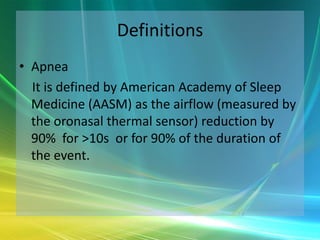
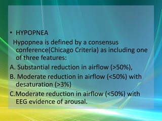
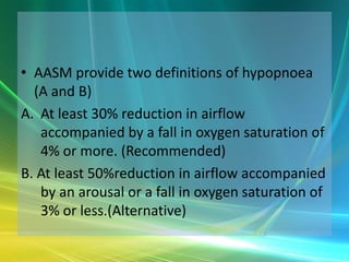
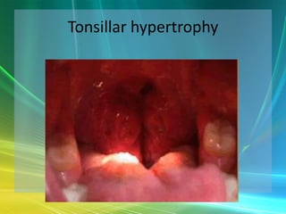
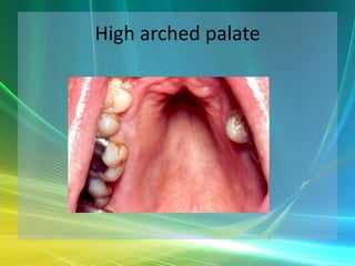
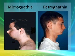
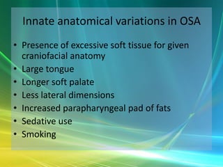
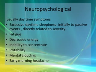
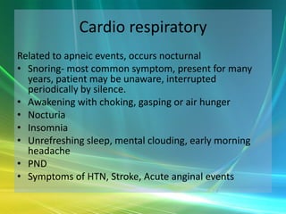
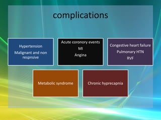
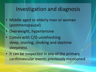
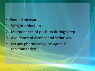
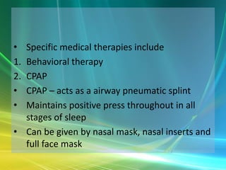
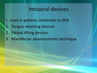
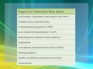
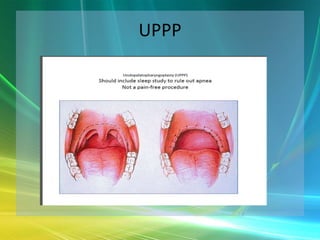
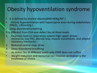
SLEEP APNEA


       Dr.Sushrut Ganpule
       Dr. K.K. Dang
Definitions
• Apnea
  It is defined by American Academy of Sleep
  Medicine (AASM) as the airflow (measured by
  the oronasal thermal sensor) reduction by
  90% for >10s or for 90% of the duration of
  the event.
• HYPOPNEA
 Hypopnea is defined by a consensus
  conference(Chicago Criteria) as including one
  of three features:
A. Substantial reduction in airflow (>50%),
B. Moderate reduction in airflow (<50%) with
  desaturation (>3%)
C.Moderate reduction in airflow (<50%) with
  EEG evidence of arousal.
• AASM provide two definitions of hypopnoea
  (A and B)
A. At least 30% reduction in airflow
   accompanied by a fall in oxygen saturation of
   4% or more. (Recommended)
B. At least 50%reduction in airflow accompanied
   by an arousal or a fall in oxygen saturation of
   3% or less.(Alternative)
• RERA ( Respiratory Effort Related Arousal)
  It is defined as increasing respiratory effort for 10
  seconds or longer leading to an arousal from sleep but
  one that does not fulfill the criteria for a hypopnea or
  apnea.
• AROUSAL RESPONSE
   Arousal response has an important role in termination
  of sleep, inducer of cough reflex, life saving
  gestures, termination of apnea episode etc. BUT
  associated with
1. Fragmentation of sleep
2. Sympathetic surge that is associated with various
    cardiovascular events
• AHI (Apnea Hypopnea Index)
   Apnea and Hypopnea episodes are measured by
   an index known as Apnea Hypopnea Index (AHI)-
   No. of Apnea or Hypopnea episodes per Hr of
   sleep.
• Up to 5 AHI’s can occur during sleep in normal
   individuals and is considered irrelevant.
• > 5 AHI are clinically relevant and they are sub
   classified as mild (5-15) moderate
  (15-30)and severe (>30).
SLEEP
Sleep predominantly occurs in two stages
1. REM
2. NREM
• Both have different effects on blood pressure
   heart rate and respiration
• Adults usually spend 70-80% sleep in NREM
   while children and aged usually spend 70% of
   sleep in REM.Physiological effects of these
   stages are as follows
STAGES OF SLEEP
• It is characterized by two stages which alternates during whole of
  sleep
1. NREM ( Non Rapid Eye Movement ):-
• 90-120 mnts. (each cycle)
• 75% of total duration.
• It is further classified into 4 stages as I, II, III,IV. Stage IV is deepest
    sleep ( slow wave sleep)
• In general NREM is characterized by reduction in sympathetic
    outflow and increase in parasympathetic outflow. (↑ Vagal tone)
• Leads to ↓ B.P. , ↓H.R. , ↓C.O. ↓ Tidal ventilation and minute
    ventilation. Control of ventilation is majorly by chemical means
    (compared with values when person is awake)
Stages of sleep cont…
2. REM ( rapid eye movement sleep)
• 10-20 mnts duration. alternates with NREM
  sleep .
• Makes 15-20% of total sleep duration
• It is characterized by loss of all sensory inputs
  and motor outputs of brain but brain is
  metabolically active, rapid rolling type eye
  movements, dreaming, awakening, paralysis, c
  onsolidation of memory, satisfaction of sleep.
Continued..
• Return of sympathetic tone causes ↑ in
  H.R., B.P. and C.O. to the values similar to
  wakefulness
• Phasic activity that occurs in bursts
• Causes both ↑ and ↓ in H.R. and predisposes
  to MI and Asystole resp.
• Ventilation is reduced than NREM. v High
  propensity of apnea,
SLEEP APNEA
• It is further classified as
1. OBSTRUCTIVE

2. CENTRAL

3. MIXED
Types contd..
• OBSTRUCTIVE
  It is most common type, occurs because of collapse of
  upper airways at intervals during sleep
• CENTRAL
  It occurs because of problem in brain or its neural
  pathways stimulating and controlling breathing without
  any change in the physical status of the airways.
• MIXED
   Absence of thoracoabdominal movements during
   initial part of airflow cessation but appear gradualy as
   the episode terminates.
Collapse of airways

    Apnea (↑PCO2,↓PO2 )

 Sympathetic surge

    Awakening

Openning of airways

Termination of episode
Structural factors
Structural factors related to craniofacial bony
  anatomy that predispose patients with OSA
  to pharyngeal collapse during sleep include
  the following:
• Innate anatomic variations
• Retrognathia and micrognathia
• Mandibular hypoplasia
• Inferior displacement of the hyoid
• Adenotonsillar hypertrophy, particularly in
  children and young adults
• Pierre Robin syndrome
• Down syndrome
• Marfan syndrome
• Prader-Willi syndrome
• High, arched palate (particularly in women)
• Brachycephalic head form - Associated with an
  increased AHI in whites but not in African
  Americans.
Tonsillar hypertrophy
High arched palate
Micrognathia   Retrognathia
Innate anatomical variations in OSA
• Presence of excessive soft tissue for given
  craniofacial anatomy
• Large tongue
• Longer soft palate
• Less lateral dimensions
• Increased parapharyngeal pad of fats
• Sedative use
• Smoking
Nonstructural risk factors
    Nonstructural risk factors for OSA include the
    following:
•   Obesity
•   Central fat distribution
•   Male sex
•   Age
•   Postmenopausal state
•   Alcohol use
Nonstructural factors contd…
•   Sedative use
•   Smoking
•   Habitual snoring with daytime somnolence
•   Supine sleep position
Etiology contd..
 Other conditions associated with the
  development of OSA are as follows:
• Hypothyroidism
• Neurologic syndromes
• Stroke
• Acromegaly
• Environmental exposures
Clinical manifestation
                  Symptoms


Neuropsychological           Cardio respiratory
(repeated arousals)          (recurrent apnea)
Neuropsychological
 usually day time symptoms
• Excessive daytime sleepiness- initially to passive
  events , directly related to severity
• Fatigue
• Decreased energy
• Inability to concentrate
• Irritability
• Mental clouding
• Early morning headache
Cardio respiratory
Related to apneic events, occurs nocturnal
• Snoring- most common symptom, present for many
  years, patient may be unaware, interrupted
  periodically by silence.
• Awakening with choking, gasping or air hunger
• Nocturia
• Insomnia
• Unrefreshing sleep, mental clouding, early morning
  headache
• PND
• Symptoms of HTN, Stroke, Acute anginal events
complications

                       Acute coronory events
  Hypertension                                       Congestive heart failure
                                  MI
Malignant and non                                        Pulmonary HTN
                                 Angina
    respnsive                                                   RVF


            Metabolic syndrome            Chronic hyprecapnia
Investigation and diagnosis
• Middle aged or elderly man or woman
  (postmenopausal)
• Overweight, hypertensive
• Comes with C/O unrefreshing
  sleep, snoring, choking and daytime
  sleepiness
• It can be suspected in any of the primary
  cardiovascular events previously mentioned
Physical examination
• Age
• Neck circumference
• BMI
• Visualization of pharynx to assess crowding
• Soft tissue dimensions and craniofacial
  abnormalities
• Blood pressure
Evaluation of daytime sleepiness
PSG
• AASM guidelines for the indications and performance of
  PSG include the following:
• Sleep stages are recorded via an EEG, electrooculogram,
  and chin electromyogram (EMG).
• Heart rhythm is monitored with a single-lead ECG.
• Leg movements are recorded via an anterior tibialis EMG.
• Breathing is monitored, including airflow at the nose and
  mouth (using both a thermal sensors and a nasal pressure
  transducer), effort (using inductance plethysmography),
  and oxygen saturation.
• The breathing pattern is analyzed for the presence of
  apneas and hypopneas, determined according to
  definitions standardized by the AASM.
• PSG can be done in home by portable PSG devices -
  unattended, cheap. Sleep stage, sleep position, RERA
  cant be measured and negative study cant exclude
  OSA.
• In lab or attended PSG – gold slandered
1. Standard PSG- evaluation and severity of OSA on
   night 1 pt again comes on next day for CPAP
   monitoring.
2. Split night study-both can be done in single
   night,     cost effective ,user friendly , OSA evaluation
   in 1st half and CPAP monitoring in 2nd half.
Treatment
•   General measures
•   Specific medical therapies
•   Intraoral devices
•   Surgical intervention
• General measures
1. Weight reduction
2. Maintenance of position during sleep
3. Avoidance of alcohol and sedatives
• No any pharmacological agent is
   recommended
• Specific medical therapies include
1.Behavioral therapy
2.CPAP
• CPAP – acts as a airway pneumatic splint
• Maintains positive press throughout in all
  stages of sleep
• Can be given by nasal mask, nasal inserts and
  full face mask
Intraoral devices
• Used in patients intolerant to OSA
1. Tongue retaining devices
2. Palatal lifting devices
3. Mandibular advancements technique
UPPP
Obesity hypoventilation syndrome
• it is defined by morbid obesity(BMI>40Kg/M2 )
• chronic hypoventilation with hypercapnia even during wakefulness
    (PACO2 >45mmHg )
• Sleep disordered breathing
It is different from OSA and defect lies at three levels
1. Excessive load on respiratory system-high upper airway
      resistance, low FRC, altered resp. muscle movements ,and altered
      resiratory mechanics
2. Reduced central resp. drive
3. Sleep disordered breathing
• As defect lies at different levels only CPAP does not suffice
• Weight reduction and nocturnal non invasive ventilation is the
      treatment of choice
Sleep apnea

Sleep apnea

  • 1.
    SLEEP APNEA Dr.Sushrut Ganpule Dr. K.K. Dang
  • 2.
    Definitions • Apnea It is defined by American Academy of Sleep Medicine (AASM) as the airflow (measured by the oronasal thermal sensor) reduction by 90% for >10s or for 90% of the duration of the event.
  • 3.
    • HYPOPNEA Hypopneais defined by a consensus conference(Chicago Criteria) as including one of three features: A. Substantial reduction in airflow (>50%), B. Moderate reduction in airflow (<50%) with desaturation (>3%) C.Moderate reduction in airflow (<50%) with EEG evidence of arousal.
  • 4.
    • AASM providetwo definitions of hypopnoea (A and B) A. At least 30% reduction in airflow accompanied by a fall in oxygen saturation of 4% or more. (Recommended) B. At least 50%reduction in airflow accompanied by an arousal or a fall in oxygen saturation of 3% or less.(Alternative)
  • 5.
    • RERA (Respiratory Effort Related Arousal) It is defined as increasing respiratory effort for 10 seconds or longer leading to an arousal from sleep but one that does not fulfill the criteria for a hypopnea or apnea. • AROUSAL RESPONSE Arousal response has an important role in termination of sleep, inducer of cough reflex, life saving gestures, termination of apnea episode etc. BUT associated with 1. Fragmentation of sleep 2. Sympathetic surge that is associated with various cardiovascular events
  • 6.
    • AHI (ApneaHypopnea Index) Apnea and Hypopnea episodes are measured by an index known as Apnea Hypopnea Index (AHI)- No. of Apnea or Hypopnea episodes per Hr of sleep. • Up to 5 AHI’s can occur during sleep in normal individuals and is considered irrelevant. • > 5 AHI are clinically relevant and they are sub classified as mild (5-15) moderate (15-30)and severe (>30).
  • 7.
    SLEEP Sleep predominantly occursin two stages 1. REM 2. NREM • Both have different effects on blood pressure heart rate and respiration • Adults usually spend 70-80% sleep in NREM while children and aged usually spend 70% of sleep in REM.Physiological effects of these stages are as follows
  • 8.
    STAGES OF SLEEP •It is characterized by two stages which alternates during whole of sleep 1. NREM ( Non Rapid Eye Movement ):- • 90-120 mnts. (each cycle) • 75% of total duration. • It is further classified into 4 stages as I, II, III,IV. Stage IV is deepest sleep ( slow wave sleep) • In general NREM is characterized by reduction in sympathetic outflow and increase in parasympathetic outflow. (↑ Vagal tone) • Leads to ↓ B.P. , ↓H.R. , ↓C.O. ↓ Tidal ventilation and minute ventilation. Control of ventilation is majorly by chemical means (compared with values when person is awake)
  • 9.
    Stages of sleepcont… 2. REM ( rapid eye movement sleep) • 10-20 mnts duration. alternates with NREM sleep . • Makes 15-20% of total sleep duration • It is characterized by loss of all sensory inputs and motor outputs of brain but brain is metabolically active, rapid rolling type eye movements, dreaming, awakening, paralysis, c onsolidation of memory, satisfaction of sleep.
  • 10.
    Continued.. • Return ofsympathetic tone causes ↑ in H.R., B.P. and C.O. to the values similar to wakefulness • Phasic activity that occurs in bursts • Causes both ↑ and ↓ in H.R. and predisposes to MI and Asystole resp. • Ventilation is reduced than NREM. v High propensity of apnea,
  • 11.
    SLEEP APNEA • Itis further classified as 1. OBSTRUCTIVE 2. CENTRAL 3. MIXED
  • 12.
    Types contd.. • OBSTRUCTIVE It is most common type, occurs because of collapse of upper airways at intervals during sleep • CENTRAL It occurs because of problem in brain or its neural pathways stimulating and controlling breathing without any change in the physical status of the airways. • MIXED Absence of thoracoabdominal movements during initial part of airflow cessation but appear gradualy as the episode terminates.
  • 14.
    Collapse of airways Apnea (↑PCO2,↓PO2 ) Sympathetic surge Awakening Openning of airways Termination of episode
  • 15.
    Structural factors Structural factorsrelated to craniofacial bony anatomy that predispose patients with OSA to pharyngeal collapse during sleep include the following: • Innate anatomic variations • Retrognathia and micrognathia • Mandibular hypoplasia • Inferior displacement of the hyoid
  • 16.
    • Adenotonsillar hypertrophy,particularly in children and young adults • Pierre Robin syndrome • Down syndrome • Marfan syndrome • Prader-Willi syndrome • High, arched palate (particularly in women) • Brachycephalic head form - Associated with an increased AHI in whites but not in African Americans.
  • 17.
  • 18.
  • 19.
    Micrognathia Retrognathia
  • 20.
    Innate anatomical variationsin OSA • Presence of excessive soft tissue for given craniofacial anatomy • Large tongue • Longer soft palate • Less lateral dimensions • Increased parapharyngeal pad of fats • Sedative use • Smoking
  • 21.
    Nonstructural risk factors Nonstructural risk factors for OSA include the following: • Obesity • Central fat distribution • Male sex • Age • Postmenopausal state • Alcohol use
  • 22.
    Nonstructural factors contd… • Sedative use • Smoking • Habitual snoring with daytime somnolence • Supine sleep position
  • 23.
    Etiology contd.. Otherconditions associated with the development of OSA are as follows: • Hypothyroidism • Neurologic syndromes • Stroke • Acromegaly • Environmental exposures
  • 24.
    Clinical manifestation Symptoms Neuropsychological Cardio respiratory (repeated arousals) (recurrent apnea)
  • 25.
    Neuropsychological usually daytime symptoms • Excessive daytime sleepiness- initially to passive events , directly related to severity • Fatigue • Decreased energy • Inability to concentrate • Irritability • Mental clouding • Early morning headache
  • 26.
    Cardio respiratory Related toapneic events, occurs nocturnal • Snoring- most common symptom, present for many years, patient may be unaware, interrupted periodically by silence. • Awakening with choking, gasping or air hunger • Nocturia • Insomnia • Unrefreshing sleep, mental clouding, early morning headache • PND • Symptoms of HTN, Stroke, Acute anginal events
  • 27.
    complications Acute coronory events Hypertension Congestive heart failure MI Malignant and non Pulmonary HTN Angina respnsive RVF Metabolic syndrome Chronic hyprecapnia
  • 28.
    Investigation and diagnosis •Middle aged or elderly man or woman (postmenopausal) • Overweight, hypertensive • Comes with C/O unrefreshing sleep, snoring, choking and daytime sleepiness • It can be suspected in any of the primary cardiovascular events previously mentioned
  • 29.
    Physical examination • Age •Neck circumference • BMI • Visualization of pharynx to assess crowding • Soft tissue dimensions and craniofacial abnormalities • Blood pressure
  • 30.
  • 31.
    PSG • AASM guidelinesfor the indications and performance of PSG include the following: • Sleep stages are recorded via an EEG, electrooculogram, and chin electromyogram (EMG). • Heart rhythm is monitored with a single-lead ECG. • Leg movements are recorded via an anterior tibialis EMG. • Breathing is monitored, including airflow at the nose and mouth (using both a thermal sensors and a nasal pressure transducer), effort (using inductance plethysmography), and oxygen saturation. • The breathing pattern is analyzed for the presence of apneas and hypopneas, determined according to definitions standardized by the AASM.
  • 32.
    • PSG canbe done in home by portable PSG devices - unattended, cheap. Sleep stage, sleep position, RERA cant be measured and negative study cant exclude OSA. • In lab or attended PSG – gold slandered 1. Standard PSG- evaluation and severity of OSA on night 1 pt again comes on next day for CPAP monitoring. 2. Split night study-both can be done in single night, cost effective ,user friendly , OSA evaluation in 1st half and CPAP monitoring in 2nd half.
  • 33.
    Treatment • General measures • Specific medical therapies • Intraoral devices • Surgical intervention
  • 34.
    • General measures 1.Weight reduction 2. Maintenance of position during sleep 3. Avoidance of alcohol and sedatives • No any pharmacological agent is recommended
  • 35.
    • Specific medicaltherapies include 1.Behavioral therapy 2.CPAP • CPAP – acts as a airway pneumatic splint • Maintains positive press throughout in all stages of sleep • Can be given by nasal mask, nasal inserts and full face mask
  • 36.
    Intraoral devices • Usedin patients intolerant to OSA 1. Tongue retaining devices 2. Palatal lifting devices 3. Mandibular advancements technique
  • 38.
  • 39.
    Obesity hypoventilation syndrome •it is defined by morbid obesity(BMI>40Kg/M2 ) • chronic hypoventilation with hypercapnia even during wakefulness (PACO2 >45mmHg ) • Sleep disordered breathing It is different from OSA and defect lies at three levels 1. Excessive load on respiratory system-high upper airway resistance, low FRC, altered resp. muscle movements ,and altered resiratory mechanics 2. Reduced central resp. drive 3. Sleep disordered breathing • As defect lies at different levels only CPAP does not suffice • Weight reduction and nocturnal non invasive ventilation is the treatment of choice