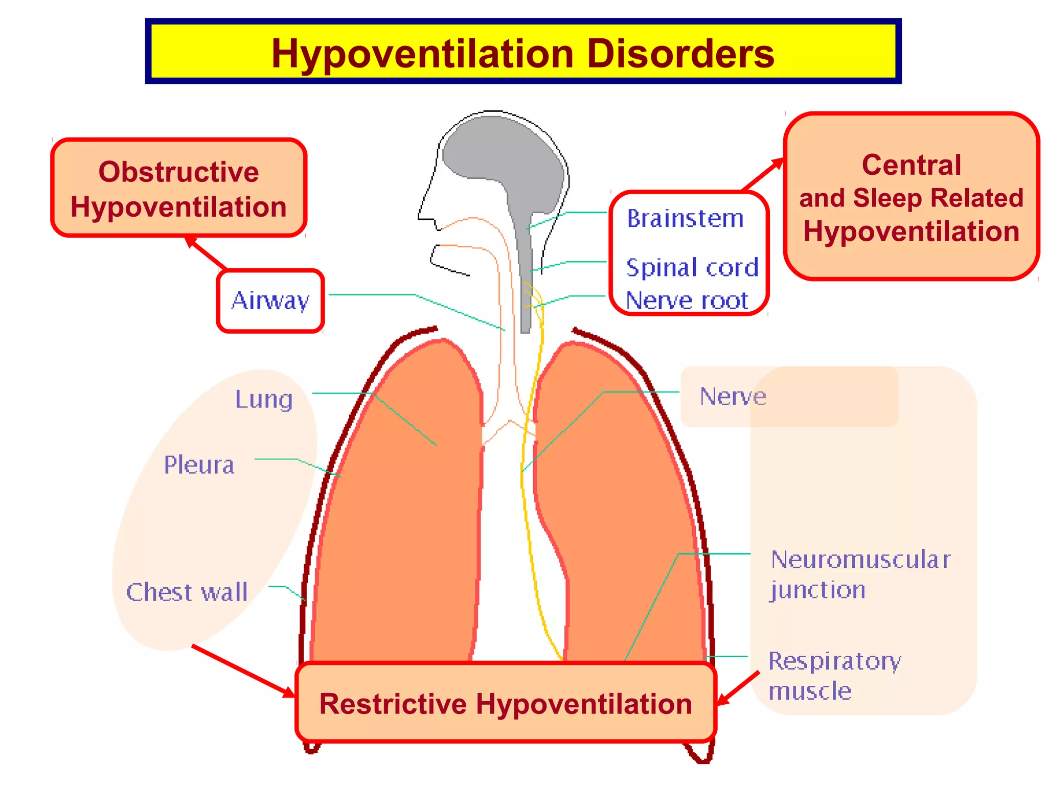
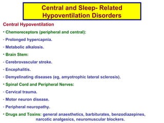
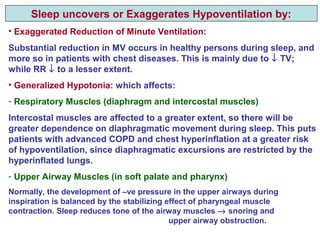
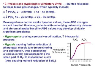
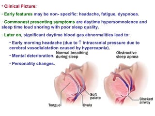
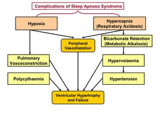
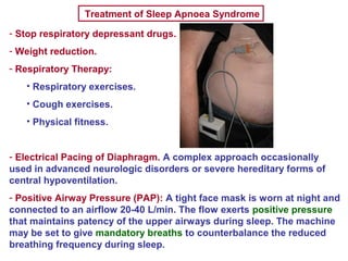
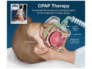
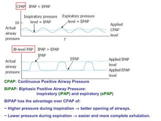
Hypoventilation Disorders
Obstructive
Hypoventilation
Central
and Sleep Related
Hypoventilation
Restrictive Hypoventilation
Central and Sleep- Related
Hypoventilation Disorders
Central Hypoventilation
• Chemoreceptors (peripheral and central):
- Prolonged hypercapnia.
- Metabolic alkalosis.
• Brain Stem:
- Cerebrovascular stroke.
- Encephalitis.
- Demyelinating diseases (eg, amyotrophic lateral sclerosis).
• Spinal Cord and Peripheral Nerves:
- Cervical trauma.
- Motor neuron disease.
- Peripheral neuropathy.
• Drugs and Toxins: general anaesthetics, barbiturates, benzodiazepines,
narcotic analgesics, neuromuscular blockers.
Sleep- Related Hypoventilation
• Hereditary (rare):
- Congenital central alveolar hypoventilation.
- Late onset central alveolar hypoventilation with hypothalamic dysfunction.
- Idiopathic central alveolar hypoventilation.
• Acquired (common):
- Sleep apnoea syndrome (SAS).
- Sleep- related hypoventilation due to a medical disorder.
- Sleep related hypoventilation due to a medication or substance.
Central and sleep related hypoventilation are closely related.
Ventilation is normally reduced during sleep, more so in patients
with central hypoventilation.
Therefore, when hypoventilation occurs only during sleep, this can
be considered as an early phase of central hypoventilation.
Respiratory Failure
Since the main function of the respiratory system is to maintain normal
levels of arterial PaO2 and PaCO2, respiratory failure is defined by the
presence of abnormally low PaO2 (< 60 mmHg) and/or abnormally high
PaCO2 (> 46 mmHg) (ie, presence of some defined degree of
hypoxaemia and/or hypercapnia).
Hypoxaemia almost always reflects failure of gas exchange in the lungs
(occasionally it results from low oxygen fraction of inspired air).
Hypercapnia usually reflects failure of ventilatory control or ventilatory
mechanics.
There are thus 2 main types of respiratory failure (1 and 2), in addition
to a 3rd
combined or mixed type.
Types 2 and 3 may be considered one entity because hypercapnia is
almost always accompanied by some degree of hypoxaema.
Type 1 Failure
Hypoxic Failure
Oxygenation Failure
Lung failure
Type 2 Failure
Hypercapnic Failure
Ventilatory Failure
Pump Failure
Types of Respiratory Failure
- Pulmonary oedema: cardiogenic
and non- Cardiogenic (ARDS).
- IPF.
- Pneumonia.
- Pulmonary embolism
- Rt to Lt shunt
Central and sleep- related
hypoventilation
Typical Examples
Common Features of Central Hypoventilation
And Hypercapnic Respiratory Failure
• Reduction of Minute Ventilation: (TV X RR)
Either TV or RR or both are reduced.
Hypoventilation is NOT, however, defined by reduced minute ventilation.
It is defined by the occurrence of CO2 retention (hypercapnia):
• Abnormal ABG:
- PaCO2 > 45 mmHg.
- pH < 7.4.
- SaO2 < 95%. Some degree of hypoxaemia accompanies hypercapnia
because increased alveolar PCO2 encroaches upon alveolar PO2.
- ↑ Plasma bicarbonate (> 30 mEq/L): metabolic alkalosis to compensate
for respiratory acidosis. This finding may the only indicator of the
underlying hypoventilation if other ABG changes are not persistent.
• Hypoventilation is more pronounced (or even only confined) to sleep:
Sleep uncovers or Exaggerates Hypoventilation by:
• Exaggerated Reduction of Minute Ventilation:
Substantial reduction in MV occurs in healthy persons during sleep, and
more so in patients with chest diseases. This is mainly due to ↓ TV;
while RR ↓ to a lesser extent.
• Generalized Hypotonia: which affects:
- Respiratory Muscles (diaphragm and intercostal muscles)
Intercostal muscles are affected to a greater extent, so there will be
greater dependence on diaphragmatic movement during sleep. This puts
patients with advanced COPD and chest hyperinflation at a greater risk
of hypoventilation, since diaphragmatic excursions are restricted by the
hyperinflated lungs.
- Upper Airway Muscles (in soft palate and pharynx)
Normally, the development of –ve pressure in the upper airways during
inspiration is balanced by the stabilizing effect of pharyngeal muscle
contraction. Sleep reduces tone of the airway muscles → snoring and
upper airway obstruction.
• ↓ Hypoxic and Hypercapnic Ventilatory Drive: → blunted response
to these blood gas changes, which typically include:
o ↑ PaCO2 2 – 3 mmHg → 42 – 43 mmHg.
o ↓ PaO2 15 – 25 mmHg → 75 – 85 mmHg.
Developed on a normal awake baseline values, these ABG changes
are not harmful. However, patients with underlying pulmonary disease
and abnormal awake baseline ABG values may develop clinically
significant problems:
- Hypercapnia causing cerebral vasodilatation, ↑ intracranial
pressure.
- Hypoxia causing further reduction of
pharyngeal muscle tone (more snoring
and obstruction, thus establishing
a vicious circle) and approaching the
steep part of O2-Hb dissociation curve
(thus causing marked reduction of SaO2)
Sleep Apnoea Syndrome
• A relatively common type of sleep- related breathing disorder characterized by
frequent and prolonged apnoeic periods during sleep.
• Apnoeic Period or Spell: complete cessation of airflow at mouth and nose for
> 10 seconds. Such periods may occur infrequently during normal sleep.
In SAS, they occur more frequently (> 40 apnoeic periods during 7h sleep) and
each period lasts much longer (occasionally up to 90 seconds).
• Apnoeic periods have a very characteristic pattern that comprises the
following consecutive phases:
Gradually increasing loud
snoring
Vibration and partial obstruction of floppy
narrow upper airways
Apnoea and silence Exhaustion and complete upper airway
obstruction coupled with low respiratory
drive
Gradually increasing
ineffective respiratory efforts
Gradual build up of severe hypercapnia,
hypoxia
Restoration of breathing with
an explosive snore
Intense respiratory centre stimulation
ultimately overcomes the upper airway
obstruction
• Typical Patient:
- Middle aged male.
- May have underlying chronic respiratory disease, most commonly COPD
(overlap syndrome).
- May have predisposing factors affecting:
• Nose:
o Allergic rhinitis
o Deviated nasal septum
• Pharynx:
o Obesity: Commonest factor → Obesity Hypoventilaton Syndrome
(OHS)
o Endocrinal: myxoedema, acromegaly
o Anatomical: micrognathia, macroglossia, large tonsils, adenoids
• Brain Stem:
o Respiratory depressant drugs
o Hypoxaemia → floppy upper airway muscles → vicious circle.
Obesity Hypoventilation Syndrome (OHS)
• Diagnostic Features:
- Obesity: BMI > 30 Kg/m2
.
- Chronic Hypoventilation: PaCO2 > 45 mmHg.
- Absence of Other Causes of Hypercapnia: as obstructive, restrictive disorders.
- Sleep- Related Breathing Disorder:
• Obtructive sleep apnoea (OSA) (90 %).
• Non- obstructive sleep apnoea (10 %).
• Possible Mechanisms by which Obesity Leads to Chronic Hypoventilation
−↑ Upper airway resistance → OSA.
- ↓ Chest wall compliance.
- ↑ Work of breathing.
- Inspiratory muscle weakness.
- Blunted respiratory drive to hypoxia, hypercapnia.
• Clinical Picture:
- Early features may be non- specific: headache, fatigue, dyspnoea.
- Commonest presenting symptoms are daytime hypersomnolence and
sleep time loud snoring with poor sleep quality.
- Later on, significant daytime blood gas abnormalities lead to:
• Early morning headache (due to ↑ intracranial pressure due to
cerebral vasodialatation caused by hypercapnia).
• Mental deterioration.
• Personality changes.
Complications of Sleep Apnoea Syndrome
Hypoxia
Hypercapnia
(Respiratory Acidosis)
Peripheral
Vasodilatation
Ventricular Hypertrophy
and Failure
Pulmonary
Vascoconstriction
Polycythaemia
Bicarbonate Retention
(Metabolic Alkalosis)
Hypervolaemia
Hypertension
Investigations of Sleep- Related Hypoventilation
• Daytime:
- Spirometry and ABG may be normal, with abnormalities being
detectable only during sleep.
- In overlap syndrome, it may be difficult to attribute spirometry and
ABG changes to the underlying sleep- related problem.
- Exaggerated FVC decline in supine as compared to upright position:
The decline exceeds 25%; normally it is 8 – 10%.
- Increased serum bicarbonate: indirect indicator of hypercapnia,
even if PaCO2 was normal during wakefulness.
• Sleep Time:
- ↑ PaCO2 > 55 mmHg for > 10 mins.
- ↑ PaCO2 > 10 mmHg as compared to awake supine value for > 10 mins.
- ↓ SaO2 to < 90% for > 5 mins: indirect indicator of hypercapnia.
- Polysomnography (Sleep Study): gold standard: Parameters recorded
during sleep include oxymetry, EEG, EMG, oral and nasal air flow.
Treatment of Sleep Apnoea Syndrome
- Stop respiratory depressant drugs.
- Weight reduction.
- Respiratory Therapy:
• Respiratory exercises.
• Cough exercises.
• Physical fitness.
- Electrical Pacing of Diaphragm. A complex approach occasionally
used in advanced neurologic disorders or severe hereditary forms of
central hypoventilation.
- Positive Airway Pressure (PAP): A tight face mask is worn at night and
connected to an airflow 20-40 L/min. The flow exerts positive pressure
that maintains patency of the upper airways during sleep. The machine
may be set to give mandatory breaths to counterbalance the reduced
breathing frequency during sleep.
CPAP: Continuous Positive Airway Pressure
BiPAP: Biphasic Positive Airway Pressure:
inspiratory (iPAP) and expiratory (ePAP)
BiPAP has the advantage over CPAP of:
~ Higher pressure during inspiration → better opening of airways.
~ Lower pressure during expiration → easier and more complete exhalation.
- Nocturnal Oxygen Therapy: controversial:
It may improve hypoxaemia, but the ventilatory drive may be
suppressed further → exaggeration of hypercapnia.
Therefore, PaCO2 should be closely monitored if supplemental oxygen
is given.
- ENT Management of OSA, eg, Uvulo-Palato-Pharyngo-Plasty (UPPP).
Respiratory Stimulants
Doxapram
Amp 100 mg/5mL (Dopram)Indications:
It is the only respiratory stimulant used with any frequency for acute
ventilatory failure. It should be reserved for cases that are potentially
shortly reversible in an attempt to avoid mechanical ventilation, or it
may be used as a temporarily measure until mechanical ventilation is
started:
- Post- operative delayed recovery from general anaesthesia.
- Acute exacerbation of COPD.
- Excessive sedation and respiratory depression caused by opiates or
benzodiazepines if specific antidotes not available.
- To limit respiratory depression during oxygen therapy.
Patients with problems unlikely to resolve within few hours are usually
better managed by mechanical ventilation.
Not Useful in:
• Mechanical causes of hypoventilation (obstructive / restrictive).
• Paralytic / musculoskeletal disorders, eg, prolonged effect of muscle
relaxants.
• Absence of significant hypercapnia. This excludes the presence of
significant central hypoventilation, for which the drug is specifically
used.
• During mechanical ventilation.
Mechanism of Action
• Stim. of peripheral chemoreceptors.
• Direct stim. of respiratory centre at higher doses.
It provides only a small additive drive to ventilation →
↑ minute ventilation, mainly due to ↑ TV, while respiratory rate
is increased to a lesser extent.
Onset of action after IV injection: 20 – 40 seconds
Peak: 1 – 2 minutes
Duration: 5 – 10 minute
Dose:
IV infusion started at 5 mg/min, reduced according to response to
1 - 3 mg/min to a maximum of 600 mg over a period 24 – 36 hh.
Side Effects:
• Generalized CNS Stimulation and Increased Catecholamine Release:
o Agitation, tremors, convulsions.
o Stimulation of vasomotor centre → ↑ heart rate, BP, arrhythmias
including ventricular tachycardia and fibrillation (especially if the patient
was given a general anaesthetic that sensitizes the myocardium to effect of
catecholamines, as halothane).
o Flushing, pyrexia, sweating, pruritus.
• Recurrence of Respiratory Depression: Pt should be kept under close
observation till 1 hour after full recovery.
• Respiratory System: laryngospasm, bronchospasm.
• GIT: nausea, vomiting, diarrhoea.
Airway should be protected to prevent aspiration.
• Extravasation → thrombophlebitis, skin irritation
• Haemolysis: with rapid infusion.
Contraindications:
• Severe hypertension.
• Severe coronary heart disease.
• Decompensated heart failure.
• Epilepsy or other convulsive disorders.
• Head injury.
• Cerebral stroke.
• Hyperthyroidism.
• Phaeochromocytoma.
• General anaesthesia with a volatile agent that sensitizes the
myocardium to catecholamines, eg halothane. It may be given after the
anaesthetic has been excreted.
• Severe hepatic or renal impairment.
Precautions:
• Monitor heart rate, BP, deep tendon reflexes and modify dose
accordingly, to prevent overdose.
• Patients on sympathomimetics and MAOIs may suffer additive pressor
effect.
• Monitor ABG in case of COPD exacerbation so that mechanical
ventilation is not unduly delayed if required.
• Concomitant administration with methylxanthines ↑ risk of agitation
and convulsions.
• The vial contains benzyl alcohol as a preservative. This may produce
serious adverse events and even death in infants and children (gasping
syndrome). So, the combined daily metabolic load of benzyl alcohol
from all sources should be considered.
Naloxone
Amp 0.4 and 0.04 mg/mL (Narcan)
Indications:
It is not a respiratory stimulant, but a competitive opiate receptor
antagonist. It has no significant agonist activity.
• Reversal of respiratory depression (and other effects) of opioid drugs
(narcotic analgesics), eg, post- operative, post- partum.
It has a shorter t ½ than most opioids. Onset of action after IV infusion:
2 minutes; duration of action: 20 – 90 minutes.
It may need to be continued for several days in patients who received
an overdose of a long acting opioid.
Patient should be monitored for a minimum of 2 – 3 hours following the
last dose, to ensure full recovery.
No further opioids should be given for a minimum of 2 – 3 hours
following the last dose of naloxone
• Diagnosis of opioid toxicity in patients with coma of unknown origin.
Most Characteristic Features of Opioid Overdose / Addiction
• Respiratory depression: A respiratory rate < 10 – 12/min is the best
clinical predictor of opioid intoxication.
• Extreme somnolence / disturbed level of consciousness.
• Miosis (PPP: pin point pupil).
• ↓ heart rate, BP.
• Constipation.
• Cold clammy skin.
Ongoing assessment of opioid intoxication should largely be based on
respiratory rate and mental status.
Dose:
IV 0.1 – 0.2 mg. If no adequate response after 2 mins,
repeat to a maximum of 0.4 mg.
If no adequate response, give IV infusion 0.1 – 0.4 mg/h, adjusted
according to response.
If IV route is not readily available, naloxone may be given IM or SC.
Side Effects:
• Withdrawal Symptoms: may be precipitated in opioid addicts and in neonates
exposed to maternal opioid use. Features of opioid withdrawal:
o Agitation, tremors.
ο↑ heart rate, BP, arrhythmias including ventricular tachycardia and
fibrillation.
o Flushing, pyrexia, sweating.
o Nausea, vomiting, diarrhoea
o Body aches.
Opioid withdrawal in neonates may be life threatening. Features include
excessive crying, hyperreflexia, convulsions.
• Recurrence of Respiratory and CNS Depression: Pt should be kept under close
observation till 2 - 3 hours after full recovery. Additional doses or more
prolonged administration may be required if the patient is not adequately
responding, or responds and then relapses back into respiratory depression.
Reversal of respiratory depression caused by partial agonists or mixed
agonist/antagonist (as pentazocine) may be incomplete.
So, larger or repeated doses may be required.
Flumazenil
Amp 0.1 mg/mL (Anexate)
Indications:
It is not a respiratory stimulant, but a competitive benzodiazepine (BD)
receptor antagonist. It has no significant agonist activity.
• Reversal of respiratory depression and sedative effects of BD after
anaesthesia or drug overdose.
• Reversal of respiratory depression and sedative effects of non-BD
drugs that act on BD receptors, eg, Zopiclone.
Onset after IV injection: 1 – 2 mins.
Peak: 5 – 10 mins
The t ½ of flumazenil is shorter than most BDs, so it may need to be
given as continuous IV infusion.
• Hepatic encephalopathy (thought to antagonise the effect of BD- like
substances assumed to stimulate GABAA receptors and cause impaired
level of consciousness.
Mechanism of Action
Competitive inhibition of BD at GABA/BD receptor complex.
GABA (gamma amino butyric acid) is the major inhibitory
neurotransmitter in CNS.
It activates 3 different classes of receptors: GABAA, GABAB, GABAC.
GABAA receptors play a pivotal role in regulation of brain excitability.
Many CNS depressant drugs exert their action through these GABAA
receptors, as BD, barbiturates, anticonvulsants and general
anaesthetics.
Flumazenil does not antagonize the CNS effects of drugs affecting the
GABA-ergic neurons by means other than the BD receptors,
eg, barbiturates.
Flumazenil effectively reverses the respiratory depression, sedation and
psychomotor effects of BD, but amnesia is less completely reversed.
So, patient instructions need to be directed to family members or be
repeated to the patient after full recovery.
Dose:
0.2 mg given IV over 15 seconds, followed if needed by additional doses of 0.2
mg every minute up to a total of 1 mg.
This dose may be repeated after 20 minutes, up to a maximum of 3 mg/hour.
Patients not responding to doses of 3 – 5 mg are unlikely to respond to further
doses.
Side Effects:
• Withdrawal Symptoms: may be precipitated in BD addicts and those who
received long acting or large doses of short acting BDs. Patients may acquire
BD dependence after a few days of being in ICU. Features of withdrawal
include:
o Agitation, panic attacks, tremors, convulsions (most serious).
Cyclic antidepressant overdose ↑ risk of convulsions.
ο↑ heart rate, BP, arrhythmias including ventricular tachycardia.
o Nausea, vomiting, diarrhoea.
o Body aches.
It may be difficult to distinguish SE attributable to the drug itself from
withdrawal symptoms.
Contraindications:
• Patients given BDs for potentially life threatening indications,
eg, status epilepticus.
• Serious cyclic antidepressant overdose
(high risk of withdrawal seizures).

5 central and sleep related hypoventilation

  • 1.
    Hypoventilation Disorders Obstructive Hypoventilation Central and SleepRelated Hypoventilation Restrictive Hypoventilation
  • 2.
    Central and Sleep-Related Hypoventilation Disorders Central Hypoventilation • Chemoreceptors (peripheral and central): - Prolonged hypercapnia. - Metabolic alkalosis. • Brain Stem: - Cerebrovascular stroke. - Encephalitis. - Demyelinating diseases (eg, amyotrophic lateral sclerosis). • Spinal Cord and Peripheral Nerves: - Cervical trauma. - Motor neuron disease. - Peripheral neuropathy. • Drugs and Toxins: general anaesthetics, barbiturates, benzodiazepines, narcotic analgesics, neuromuscular blockers.
  • 3.
    Sleep- Related Hypoventilation •Hereditary (rare): - Congenital central alveolar hypoventilation. - Late onset central alveolar hypoventilation with hypothalamic dysfunction. - Idiopathic central alveolar hypoventilation. • Acquired (common): - Sleep apnoea syndrome (SAS). - Sleep- related hypoventilation due to a medical disorder. - Sleep related hypoventilation due to a medication or substance. Central and sleep related hypoventilation are closely related. Ventilation is normally reduced during sleep, more so in patients with central hypoventilation. Therefore, when hypoventilation occurs only during sleep, this can be considered as an early phase of central hypoventilation.
  • 4.
    Respiratory Failure Since themain function of the respiratory system is to maintain normal levels of arterial PaO2 and PaCO2, respiratory failure is defined by the presence of abnormally low PaO2 (< 60 mmHg) and/or abnormally high PaCO2 (> 46 mmHg) (ie, presence of some defined degree of hypoxaemia and/or hypercapnia). Hypoxaemia almost always reflects failure of gas exchange in the lungs (occasionally it results from low oxygen fraction of inspired air). Hypercapnia usually reflects failure of ventilatory control or ventilatory mechanics. There are thus 2 main types of respiratory failure (1 and 2), in addition to a 3rd combined or mixed type. Types 2 and 3 may be considered one entity because hypercapnia is almost always accompanied by some degree of hypoxaema.
  • 5.
    Type 1 Failure HypoxicFailure Oxygenation Failure Lung failure Type 2 Failure Hypercapnic Failure Ventilatory Failure Pump Failure Types of Respiratory Failure - Pulmonary oedema: cardiogenic and non- Cardiogenic (ARDS). - IPF. - Pneumonia. - Pulmonary embolism - Rt to Lt shunt Central and sleep- related hypoventilation Typical Examples
  • 6.
    Common Features ofCentral Hypoventilation And Hypercapnic Respiratory Failure • Reduction of Minute Ventilation: (TV X RR) Either TV or RR or both are reduced. Hypoventilation is NOT, however, defined by reduced minute ventilation. It is defined by the occurrence of CO2 retention (hypercapnia): • Abnormal ABG: - PaCO2 > 45 mmHg. - pH < 7.4. - SaO2 < 95%. Some degree of hypoxaemia accompanies hypercapnia because increased alveolar PCO2 encroaches upon alveolar PO2. - ↑ Plasma bicarbonate (> 30 mEq/L): metabolic alkalosis to compensate for respiratory acidosis. This finding may the only indicator of the underlying hypoventilation if other ABG changes are not persistent. • Hypoventilation is more pronounced (or even only confined) to sleep:
  • 7.
    Sleep uncovers orExaggerates Hypoventilation by: • Exaggerated Reduction of Minute Ventilation: Substantial reduction in MV occurs in healthy persons during sleep, and more so in patients with chest diseases. This is mainly due to ↓ TV; while RR ↓ to a lesser extent. • Generalized Hypotonia: which affects: - Respiratory Muscles (diaphragm and intercostal muscles) Intercostal muscles are affected to a greater extent, so there will be greater dependence on diaphragmatic movement during sleep. This puts patients with advanced COPD and chest hyperinflation at a greater risk of hypoventilation, since diaphragmatic excursions are restricted by the hyperinflated lungs. - Upper Airway Muscles (in soft palate and pharynx) Normally, the development of –ve pressure in the upper airways during inspiration is balanced by the stabilizing effect of pharyngeal muscle contraction. Sleep reduces tone of the airway muscles → snoring and upper airway obstruction.
  • 8.
    • ↓ Hypoxicand Hypercapnic Ventilatory Drive: → blunted response to these blood gas changes, which typically include: o ↑ PaCO2 2 – 3 mmHg → 42 – 43 mmHg. o ↓ PaO2 15 – 25 mmHg → 75 – 85 mmHg. Developed on a normal awake baseline values, these ABG changes are not harmful. However, patients with underlying pulmonary disease and abnormal awake baseline ABG values may develop clinically significant problems: - Hypercapnia causing cerebral vasodilatation, ↑ intracranial pressure. - Hypoxia causing further reduction of pharyngeal muscle tone (more snoring and obstruction, thus establishing a vicious circle) and approaching the steep part of O2-Hb dissociation curve (thus causing marked reduction of SaO2)
  • 9.
    Sleep Apnoea Syndrome •A relatively common type of sleep- related breathing disorder characterized by frequent and prolonged apnoeic periods during sleep. • Apnoeic Period or Spell: complete cessation of airflow at mouth and nose for > 10 seconds. Such periods may occur infrequently during normal sleep. In SAS, they occur more frequently (> 40 apnoeic periods during 7h sleep) and each period lasts much longer (occasionally up to 90 seconds). • Apnoeic periods have a very characteristic pattern that comprises the following consecutive phases: Gradually increasing loud snoring Vibration and partial obstruction of floppy narrow upper airways Apnoea and silence Exhaustion and complete upper airway obstruction coupled with low respiratory drive Gradually increasing ineffective respiratory efforts Gradual build up of severe hypercapnia, hypoxia Restoration of breathing with an explosive snore Intense respiratory centre stimulation ultimately overcomes the upper airway obstruction
  • 10.
    • Typical Patient: -Middle aged male. - May have underlying chronic respiratory disease, most commonly COPD (overlap syndrome). - May have predisposing factors affecting: • Nose: o Allergic rhinitis o Deviated nasal septum • Pharynx: o Obesity: Commonest factor → Obesity Hypoventilaton Syndrome (OHS) o Endocrinal: myxoedema, acromegaly o Anatomical: micrognathia, macroglossia, large tonsils, adenoids • Brain Stem: o Respiratory depressant drugs o Hypoxaemia → floppy upper airway muscles → vicious circle.
  • 11.
    Obesity Hypoventilation Syndrome(OHS) • Diagnostic Features: - Obesity: BMI > 30 Kg/m2 . - Chronic Hypoventilation: PaCO2 > 45 mmHg. - Absence of Other Causes of Hypercapnia: as obstructive, restrictive disorders. - Sleep- Related Breathing Disorder: • Obtructive sleep apnoea (OSA) (90 %). • Non- obstructive sleep apnoea (10 %). • Possible Mechanisms by which Obesity Leads to Chronic Hypoventilation −↑ Upper airway resistance → OSA. - ↓ Chest wall compliance. - ↑ Work of breathing. - Inspiratory muscle weakness. - Blunted respiratory drive to hypoxia, hypercapnia.
  • 12.
    • Clinical Picture: -Early features may be non- specific: headache, fatigue, dyspnoea. - Commonest presenting symptoms are daytime hypersomnolence and sleep time loud snoring with poor sleep quality. - Later on, significant daytime blood gas abnormalities lead to: • Early morning headache (due to ↑ intracranial pressure due to cerebral vasodialatation caused by hypercapnia). • Mental deterioration. • Personality changes.
  • 13.
    Complications of SleepApnoea Syndrome Hypoxia Hypercapnia (Respiratory Acidosis) Peripheral Vasodilatation Ventricular Hypertrophy and Failure Pulmonary Vascoconstriction Polycythaemia Bicarbonate Retention (Metabolic Alkalosis) Hypervolaemia Hypertension
  • 14.
    Investigations of Sleep-Related Hypoventilation • Daytime: - Spirometry and ABG may be normal, with abnormalities being detectable only during sleep. - In overlap syndrome, it may be difficult to attribute spirometry and ABG changes to the underlying sleep- related problem. - Exaggerated FVC decline in supine as compared to upright position: The decline exceeds 25%; normally it is 8 – 10%. - Increased serum bicarbonate: indirect indicator of hypercapnia, even if PaCO2 was normal during wakefulness. • Sleep Time: - ↑ PaCO2 > 55 mmHg for > 10 mins. - ↑ PaCO2 > 10 mmHg as compared to awake supine value for > 10 mins. - ↓ SaO2 to < 90% for > 5 mins: indirect indicator of hypercapnia. - Polysomnography (Sleep Study): gold standard: Parameters recorded during sleep include oxymetry, EEG, EMG, oral and nasal air flow.
  • 15.
    Treatment of SleepApnoea Syndrome - Stop respiratory depressant drugs. - Weight reduction. - Respiratory Therapy: • Respiratory exercises. • Cough exercises. • Physical fitness. - Electrical Pacing of Diaphragm. A complex approach occasionally used in advanced neurologic disorders or severe hereditary forms of central hypoventilation. - Positive Airway Pressure (PAP): A tight face mask is worn at night and connected to an airflow 20-40 L/min. The flow exerts positive pressure that maintains patency of the upper airways during sleep. The machine may be set to give mandatory breaths to counterbalance the reduced breathing frequency during sleep.
  • 17.
    CPAP: Continuous PositiveAirway Pressure BiPAP: Biphasic Positive Airway Pressure: inspiratory (iPAP) and expiratory (ePAP) BiPAP has the advantage over CPAP of: ~ Higher pressure during inspiration → better opening of airways. ~ Lower pressure during expiration → easier and more complete exhalation.
  • 18.
    - Nocturnal OxygenTherapy: controversial: It may improve hypoxaemia, but the ventilatory drive may be suppressed further → exaggeration of hypercapnia. Therefore, PaCO2 should be closely monitored if supplemental oxygen is given. - ENT Management of OSA, eg, Uvulo-Palato-Pharyngo-Plasty (UPPP).
  • 19.
    Respiratory Stimulants Doxapram Amp 100mg/5mL (Dopram)Indications: It is the only respiratory stimulant used with any frequency for acute ventilatory failure. It should be reserved for cases that are potentially shortly reversible in an attempt to avoid mechanical ventilation, or it may be used as a temporarily measure until mechanical ventilation is started: - Post- operative delayed recovery from general anaesthesia. - Acute exacerbation of COPD. - Excessive sedation and respiratory depression caused by opiates or benzodiazepines if specific antidotes not available. - To limit respiratory depression during oxygen therapy. Patients with problems unlikely to resolve within few hours are usually better managed by mechanical ventilation.
  • 20.
    Not Useful in: •Mechanical causes of hypoventilation (obstructive / restrictive). • Paralytic / musculoskeletal disorders, eg, prolonged effect of muscle relaxants. • Absence of significant hypercapnia. This excludes the presence of significant central hypoventilation, for which the drug is specifically used. • During mechanical ventilation. Mechanism of Action • Stim. of peripheral chemoreceptors. • Direct stim. of respiratory centre at higher doses. It provides only a small additive drive to ventilation → ↑ minute ventilation, mainly due to ↑ TV, while respiratory rate is increased to a lesser extent. Onset of action after IV injection: 20 – 40 seconds Peak: 1 – 2 minutes Duration: 5 – 10 minute
  • 21.
    Dose: IV infusion startedat 5 mg/min, reduced according to response to 1 - 3 mg/min to a maximum of 600 mg over a period 24 – 36 hh. Side Effects: • Generalized CNS Stimulation and Increased Catecholamine Release: o Agitation, tremors, convulsions. o Stimulation of vasomotor centre → ↑ heart rate, BP, arrhythmias including ventricular tachycardia and fibrillation (especially if the patient was given a general anaesthetic that sensitizes the myocardium to effect of catecholamines, as halothane). o Flushing, pyrexia, sweating, pruritus. • Recurrence of Respiratory Depression: Pt should be kept under close observation till 1 hour after full recovery. • Respiratory System: laryngospasm, bronchospasm. • GIT: nausea, vomiting, diarrhoea. Airway should be protected to prevent aspiration. • Extravasation → thrombophlebitis, skin irritation • Haemolysis: with rapid infusion.
  • 22.
    Contraindications: • Severe hypertension. •Severe coronary heart disease. • Decompensated heart failure. • Epilepsy or other convulsive disorders. • Head injury. • Cerebral stroke. • Hyperthyroidism. • Phaeochromocytoma. • General anaesthesia with a volatile agent that sensitizes the myocardium to catecholamines, eg halothane. It may be given after the anaesthetic has been excreted. • Severe hepatic or renal impairment.
  • 23.
    Precautions: • Monitor heartrate, BP, deep tendon reflexes and modify dose accordingly, to prevent overdose. • Patients on sympathomimetics and MAOIs may suffer additive pressor effect. • Monitor ABG in case of COPD exacerbation so that mechanical ventilation is not unduly delayed if required. • Concomitant administration with methylxanthines ↑ risk of agitation and convulsions. • The vial contains benzyl alcohol as a preservative. This may produce serious adverse events and even death in infants and children (gasping syndrome). So, the combined daily metabolic load of benzyl alcohol from all sources should be considered.
  • 24.
    Naloxone Amp 0.4 and0.04 mg/mL (Narcan) Indications: It is not a respiratory stimulant, but a competitive opiate receptor antagonist. It has no significant agonist activity. • Reversal of respiratory depression (and other effects) of opioid drugs (narcotic analgesics), eg, post- operative, post- partum. It has a shorter t ½ than most opioids. Onset of action after IV infusion: 2 minutes; duration of action: 20 – 90 minutes. It may need to be continued for several days in patients who received an overdose of a long acting opioid. Patient should be monitored for a minimum of 2 – 3 hours following the last dose, to ensure full recovery. No further opioids should be given for a minimum of 2 – 3 hours following the last dose of naloxone • Diagnosis of opioid toxicity in patients with coma of unknown origin.
  • 25.
    Most Characteristic Featuresof Opioid Overdose / Addiction • Respiratory depression: A respiratory rate < 10 – 12/min is the best clinical predictor of opioid intoxication. • Extreme somnolence / disturbed level of consciousness. • Miosis (PPP: pin point pupil). • ↓ heart rate, BP. • Constipation. • Cold clammy skin. Ongoing assessment of opioid intoxication should largely be based on respiratory rate and mental status. Dose: IV 0.1 – 0.2 mg. If no adequate response after 2 mins, repeat to a maximum of 0.4 mg. If no adequate response, give IV infusion 0.1 – 0.4 mg/h, adjusted according to response. If IV route is not readily available, naloxone may be given IM or SC.
  • 26.
    Side Effects: • WithdrawalSymptoms: may be precipitated in opioid addicts and in neonates exposed to maternal opioid use. Features of opioid withdrawal: o Agitation, tremors. ο↑ heart rate, BP, arrhythmias including ventricular tachycardia and fibrillation. o Flushing, pyrexia, sweating. o Nausea, vomiting, diarrhoea o Body aches. Opioid withdrawal in neonates may be life threatening. Features include excessive crying, hyperreflexia, convulsions. • Recurrence of Respiratory and CNS Depression: Pt should be kept under close observation till 2 - 3 hours after full recovery. Additional doses or more prolonged administration may be required if the patient is not adequately responding, or responds and then relapses back into respiratory depression. Reversal of respiratory depression caused by partial agonists or mixed agonist/antagonist (as pentazocine) may be incomplete. So, larger or repeated doses may be required.
  • 27.
    Flumazenil Amp 0.1 mg/mL(Anexate) Indications: It is not a respiratory stimulant, but a competitive benzodiazepine (BD) receptor antagonist. It has no significant agonist activity. • Reversal of respiratory depression and sedative effects of BD after anaesthesia or drug overdose. • Reversal of respiratory depression and sedative effects of non-BD drugs that act on BD receptors, eg, Zopiclone. Onset after IV injection: 1 – 2 mins. Peak: 5 – 10 mins The t ½ of flumazenil is shorter than most BDs, so it may need to be given as continuous IV infusion. • Hepatic encephalopathy (thought to antagonise the effect of BD- like substances assumed to stimulate GABAA receptors and cause impaired level of consciousness.
  • 28.
    Mechanism of Action Competitiveinhibition of BD at GABA/BD receptor complex. GABA (gamma amino butyric acid) is the major inhibitory neurotransmitter in CNS. It activates 3 different classes of receptors: GABAA, GABAB, GABAC. GABAA receptors play a pivotal role in regulation of brain excitability. Many CNS depressant drugs exert their action through these GABAA receptors, as BD, barbiturates, anticonvulsants and general anaesthetics. Flumazenil does not antagonize the CNS effects of drugs affecting the GABA-ergic neurons by means other than the BD receptors, eg, barbiturates. Flumazenil effectively reverses the respiratory depression, sedation and psychomotor effects of BD, but amnesia is less completely reversed. So, patient instructions need to be directed to family members or be repeated to the patient after full recovery.
  • 29.
    Dose: 0.2 mg givenIV over 15 seconds, followed if needed by additional doses of 0.2 mg every minute up to a total of 1 mg. This dose may be repeated after 20 minutes, up to a maximum of 3 mg/hour. Patients not responding to doses of 3 – 5 mg are unlikely to respond to further doses. Side Effects: • Withdrawal Symptoms: may be precipitated in BD addicts and those who received long acting or large doses of short acting BDs. Patients may acquire BD dependence after a few days of being in ICU. Features of withdrawal include: o Agitation, panic attacks, tremors, convulsions (most serious). Cyclic antidepressant overdose ↑ risk of convulsions. ο↑ heart rate, BP, arrhythmias including ventricular tachycardia. o Nausea, vomiting, diarrhoea. o Body aches. It may be difficult to distinguish SE attributable to the drug itself from withdrawal symptoms.
  • 30.
    Contraindications: • Patients givenBDs for potentially life threatening indications, eg, status epilepticus. • Serious cyclic antidepressant overdose (high risk of withdrawal seizures).

Editor's Notes

  • #2 Obstructive and restrictive disorders are considered mechanical causes of hypoventilation.
  • #3 The list provides some examples and is not all inclusive. Prolonged post- operative action of neuromuscular blockers given during anaesthesia is now the commonest neuromuscular cause of ventilatory failure.
  • #7 Normal Serum bicarb: 21 – 27 mEq/L
  • #14 The blood gas abnormalities initiate the complications Both hypoxia and hypercapnia lead to peripheral VD Hypoxia leads to pulmonary VC  pulmonary hypertension The kidneys compensate for respiratory acidosis by bicarbonate retention, generating a state of metabolic alkalosis, but sodium retention leads to hypervolaemia and hypertension. These factors collectively increase both pressure and volume load on both right and left ventricles, leading to ensuing biventricular failure which further increases salt and water retention. The persistent hypoxia stimulates marrow production of RBCs  polycythaemia  increased blood viscosity  hypercaogulability and tendency for thromboembolic disease.
  • #20 Respiratory stimulants have only a limited role in management of central hypoventilation. Because there is as yet no really effective and specific respiratory stimulant. Agents in use have some limited specific indications and narrow safety margin