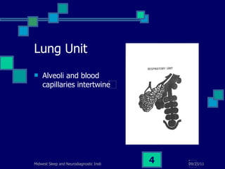
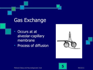
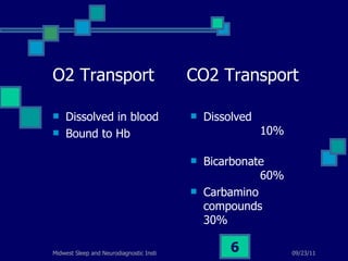
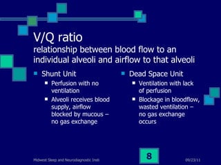
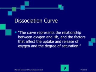
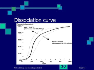
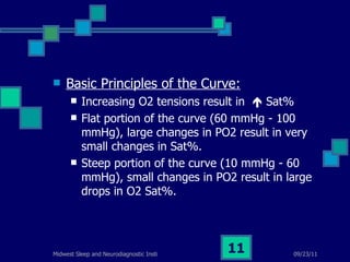
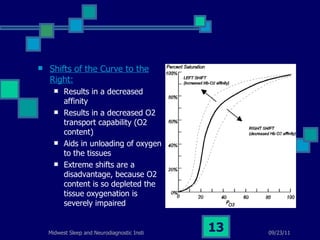
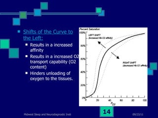
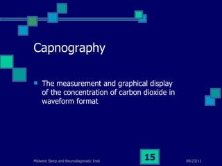
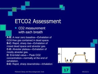
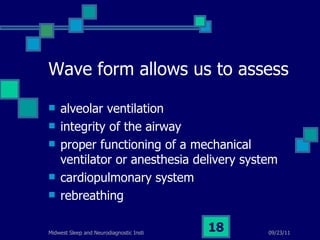
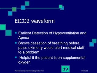
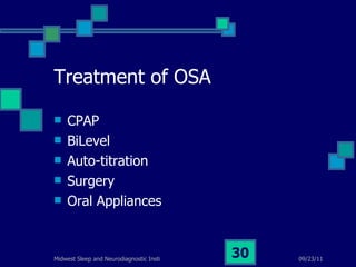
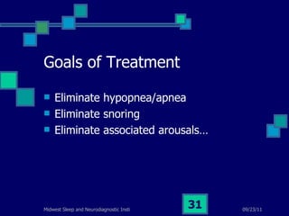
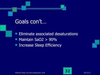
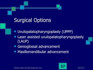
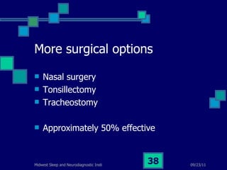
Sleep Disordered Breathing
Objectives Understand CO2, O2 relationships in the lungs Describe types of Sleep Disordered Breathing Treatment options for OSA
Spontaneous Breathing Diaphragm flattens Thoracic cage increases in volume, intrathoracic pressure decreases Negative pressure causes intake of air
Lung Unit Alveoli and blood capillaries intertwine
Gas Exchange Occurs at at alveolar-capillary membrane Process of diffusion
O2 Transport  CO2 Transport Dissolved in blood Bound to Hb Dissolved 10%   Bicarbonate 60% Carbamino compounds 30%
Diffusion affected by: Thickness of membrane Inflammation Fibrosis Surface area diminished Emphysema
V/Q ratio relationship between blood flow to an individual alveoli and airflow to that alveoli Shunt Unit Perfusion with no ventilation Alveoli receives blood supply, airflow blocked by mucous – no gas exchange Dead Space Unit Ventilation with lack of perfusion Blockage in bloodflow, wasted ventilation – no gas exchange occurs
Dissociation Curve “ The curve represents the relationship between oxygen and Hb, and the factors that affect the uptake and release of oxygen and the degree of saturation.”
Dissociation curve
Basic Principles of the Curve: Increasing O2 tensions result in    Sat% Flat portion of the curve (60 mmHg - 100 mmHg), large changes in PO2 result in very small changes in Sat%. Steep portion of the curve (10 mmHg - 60 mmHg), small changes in PO2 result in large drops in O2 Sat%.
Factors Altering HB Affinity for O2: Ph Temperature PaCO2 Hemoglobin Variants
Shifts of the Curve to the Right: Results in a decreased affinity Results in a decreased O2 transport capability (O2 content) Aids in unloading of oxygen to the tissues Extreme shifts are a disadvantage, because O2 content is so depleted the tissue oxygenation is severely impaired
Shifts of the Curve to the Left: Results in a increased affinity Results in a increased O2 transport capability (O2 content) Hinders unloading of oxygen to the tissues.
Capnography The measurement and graphical display of the concentration of carbon dioxide in waveform format
EtCO2 Refers to the measurment of carbon dioxide concentration at the end of expiration
ETCO2 Assessment CO2 measurement with each breath A-B:  A near zero baseline—Exhalation of CO2-free gas contained in dead space. B-C:  Rapid, sharp rise—Exhalation of mixed dead space and alveolar gas. C-D:  Alveolar plateau—Exhalation of mostly alveolar gas. D:  End-tidal value— Peak CO2 concentration—normally at the end of exhalation. D-E:  Rapid, sharp downstroke—Inhalation
Wave form allows us to assess alveolar ventilation integrity of the airway proper functioning of a mechanical ventilator or anesthesia delivery system cardiopulmonary system rebreathing
EtCO2 waveform Earliest Detection of Hypoventilation and Apnea   Shows cessation of breathing before pulse oximetry would alert medical staff to a problem Helpful if the patient is on supplemental oxygen
Why are dissociation curve and EtCO2 important? Windows to ventilation and perfusion
Abnormal capnograms Sudden loss of EtCO2 to zero or near zero   Possible causes: Airway disconnection Dislodged ET tube/esophageal intubation Totally obstructed/kinked ET tube Complete ventilator malfunction
Abnormal capnograms Sustained  low  EtCO2 with good alveolar plateau Possible causes: Hyperventilation Hypothermia Sedation, anesthesia Dead space ventilation                                                          
Abnormal Capnography Elevated  EtCO2 with good alveolar plateau Possible causes:  Hypoventilation Respiratory-depressant drugs Hyperthermia, pain, shivering                                                          
ABG Values 28 88 50 60 7.38 COPD 22-26 94-99 35-45 80-100 7.40 Normal HCO3 SaO2 PCO2 PO2 pH
Cyanosis (bluish coloring) occurs with a PaO2 reduction of 5 gm %
Hypoventilation Causes retention of CO2 pH drop
Hyperventilation Causes decrease of CO2 pH to increase
Hypoxic Drive Normal drive- CO2 build up COPD- low O2
Characteristics of Respiratory Events Not required Usually mild cyclic desats Crescendo-Decrescendo pattern Waz-wane May last 15-30 min or more Cheyne-Stoles Respiration Mandatory Not required   Slight increase, may crescendo to end of event   Slight decrease from baseline Respiratory Event Related Arousal Not required 4% required   Yes    30% from baseline 10-120 sec, Longer  hypo-ventilation Hypopnea Not required Common; not mandatory NO Absent Minimum 10 sec Central Apnea Not required Not required   Yes Absent Minimum 10 sec   Obstructive Apnea   Arousal Desaturation Effort Airflow Duration
Treatment of OSA CPAP BiLevel Auto-titration Surgery Oral Appliances
Goals of Treatment Eliminate hypopnea/apnea Eliminate snoring Eliminate associated arousals…
Goals con’t… Eliminate associated desaturations Maintain SaO2 > 90% Increase Sleep Efficiency
Titration Techniques Initiate at 4-5 cm Increase 1-2 cm increments Record each pressure for a minimum of 15 minutes Record REM sleep while in supine position
BiPAP Varying pressures; insp 4 cm > expiration Initiate if: Pt intolerant to CPAP Optimal CPAP pressure > 15 Optimal CPAP pressure is associated with the occurrence of central apneas
Lung Volumes
The use of CPAP / BiPAP increases FRC which in turn improves oxygenation
Surgical Options Uvulopalatopharyngoplasty (UPPP) Laser assisted uvulopalatopharyngoplasty (LAUP) Genioglossal advancement Maxillomandibular advancement
More surgical options Nasal surgery Tonsillectomy Tracheostomy Approximately 50% effective
Oral Appliances Mandibular repostitioners Tongue retaining devices Palatal lifting devices
 
 
Removable Fitted by dentist Treatment of patients with: Snoring Mild OSA
Identified Risks Intraoral gingival, palatal, or dental soreness TMJ Syndrome Obstruction of oral breathing Loosening or flaring of lower anterior teeth Excessive salivation
Oral appliances, contraindications Central sleep apnea Severe respiratory disorders Loose teeth or advanced periodontal disease < 18 yrs of age

Sleep disordered breathing and sleep apnea

  • 1.
  • 2.
    Objectives Understand CO2,O2 relationships in the lungs Describe types of Sleep Disordered Breathing Treatment options for OSA
  • 3.
    Spontaneous Breathing Diaphragmflattens Thoracic cage increases in volume, intrathoracic pressure decreases Negative pressure causes intake of air
  • 4.
    Lung Unit Alveoliand blood capillaries intertwine
  • 5.
    Gas Exchange Occursat at alveolar-capillary membrane Process of diffusion
  • 6.
    O2 Transport CO2 Transport Dissolved in blood Bound to Hb Dissolved 10% Bicarbonate 60% Carbamino compounds 30%
  • 7.
    Diffusion affected by:Thickness of membrane Inflammation Fibrosis Surface area diminished Emphysema
  • 8.
    V/Q ratio relationshipbetween blood flow to an individual alveoli and airflow to that alveoli Shunt Unit Perfusion with no ventilation Alveoli receives blood supply, airflow blocked by mucous – no gas exchange Dead Space Unit Ventilation with lack of perfusion Blockage in bloodflow, wasted ventilation – no gas exchange occurs
  • 9.
    Dissociation Curve “The curve represents the relationship between oxygen and Hb, and the factors that affect the uptake and release of oxygen and the degree of saturation.”
  • 10.
  • 11.
    Basic Principles ofthe Curve: Increasing O2 tensions result in  Sat% Flat portion of the curve (60 mmHg - 100 mmHg), large changes in PO2 result in very small changes in Sat%. Steep portion of the curve (10 mmHg - 60 mmHg), small changes in PO2 result in large drops in O2 Sat%.
  • 12.
    Factors Altering HBAffinity for O2: Ph Temperature PaCO2 Hemoglobin Variants
  • 13.
    Shifts of theCurve to the Right: Results in a decreased affinity Results in a decreased O2 transport capability (O2 content) Aids in unloading of oxygen to the tissues Extreme shifts are a disadvantage, because O2 content is so depleted the tissue oxygenation is severely impaired
  • 14.
    Shifts of theCurve to the Left: Results in a increased affinity Results in a increased O2 transport capability (O2 content) Hinders unloading of oxygen to the tissues.
  • 15.
    Capnography The measurementand graphical display of the concentration of carbon dioxide in waveform format
  • 16.
    EtCO2 Refers tothe measurment of carbon dioxide concentration at the end of expiration
  • 17.
    ETCO2 Assessment CO2measurement with each breath A-B: A near zero baseline—Exhalation of CO2-free gas contained in dead space. B-C: Rapid, sharp rise—Exhalation of mixed dead space and alveolar gas. C-D: Alveolar plateau—Exhalation of mostly alveolar gas. D: End-tidal value— Peak CO2 concentration—normally at the end of exhalation. D-E: Rapid, sharp downstroke—Inhalation
  • 18.
    Wave form allowsus to assess alveolar ventilation integrity of the airway proper functioning of a mechanical ventilator or anesthesia delivery system cardiopulmonary system rebreathing
  • 19.
    EtCO2 waveform EarliestDetection of Hypoventilation and Apnea Shows cessation of breathing before pulse oximetry would alert medical staff to a problem Helpful if the patient is on supplemental oxygen
  • 20.
    Why are dissociationcurve and EtCO2 important? Windows to ventilation and perfusion
  • 21.
    Abnormal capnograms Suddenloss of EtCO2 to zero or near zero Possible causes: Airway disconnection Dislodged ET tube/esophageal intubation Totally obstructed/kinked ET tube Complete ventilator malfunction
  • 22.
    Abnormal capnograms Sustained low EtCO2 with good alveolar plateau Possible causes: Hyperventilation Hypothermia Sedation, anesthesia Dead space ventilation                                                          
  • 23.
    Abnormal Capnography Elevated EtCO2 with good alveolar plateau Possible causes: Hypoventilation Respiratory-depressant drugs Hyperthermia, pain, shivering                                                          
  • 24.
    ABG Values 2888 50 60 7.38 COPD 22-26 94-99 35-45 80-100 7.40 Normal HCO3 SaO2 PCO2 PO2 pH
  • 25.
    Cyanosis (bluish coloring)occurs with a PaO2 reduction of 5 gm %
  • 26.
  • 27.
    Hyperventilation Causes decreaseof CO2 pH to increase
  • 28.
    Hypoxic Drive Normaldrive- CO2 build up COPD- low O2
  • 29.
    Characteristics of RespiratoryEvents Not required Usually mild cyclic desats Crescendo-Decrescendo pattern Waz-wane May last 15-30 min or more Cheyne-Stoles Respiration Mandatory Not required Slight increase, may crescendo to end of event Slight decrease from baseline Respiratory Event Related Arousal Not required 4% required Yes  30% from baseline 10-120 sec, Longer  hypo-ventilation Hypopnea Not required Common; not mandatory NO Absent Minimum 10 sec Central Apnea Not required Not required Yes Absent Minimum 10 sec Obstructive Apnea Arousal Desaturation Effort Airflow Duration
  • 30.
    Treatment of OSACPAP BiLevel Auto-titration Surgery Oral Appliances
  • 31.
    Goals of TreatmentEliminate hypopnea/apnea Eliminate snoring Eliminate associated arousals…
  • 32.
    Goals con’t… Eliminateassociated desaturations Maintain SaO2 > 90% Increase Sleep Efficiency
  • 33.
    Titration Techniques Initiateat 4-5 cm Increase 1-2 cm increments Record each pressure for a minimum of 15 minutes Record REM sleep while in supine position
  • 34.
    BiPAP Varying pressures;insp 4 cm > expiration Initiate if: Pt intolerant to CPAP Optimal CPAP pressure > 15 Optimal CPAP pressure is associated with the occurrence of central apneas
  • 35.
  • 36.
    The use ofCPAP / BiPAP increases FRC which in turn improves oxygenation
  • 37.
    Surgical Options Uvulopalatopharyngoplasty(UPPP) Laser assisted uvulopalatopharyngoplasty (LAUP) Genioglossal advancement Maxillomandibular advancement
  • 38.
    More surgical optionsNasal surgery Tonsillectomy Tracheostomy Approximately 50% effective
  • 39.
    Oral Appliances Mandibularrepostitioners Tongue retaining devices Palatal lifting devices
  • 40.
  • 41.
  • 42.
    Removable Fitted bydentist Treatment of patients with: Snoring Mild OSA
  • 43.
    Identified Risks Intraoralgingival, palatal, or dental soreness TMJ Syndrome Obstruction of oral breathing Loosening or flaring of lower anterior teeth Excessive salivation
  • 44.
    Oral appliances, contraindicationsCentral sleep apnea Severe respiratory disorders Loose teeth or advanced periodontal disease < 18 yrs of age

Editor's Notes