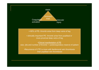
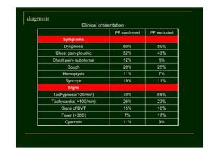
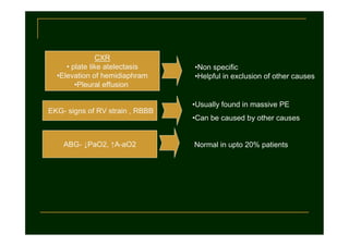
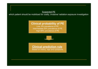
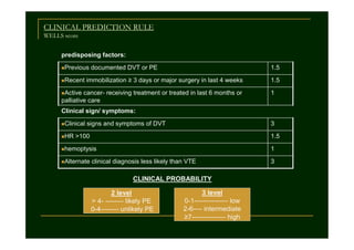
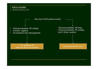
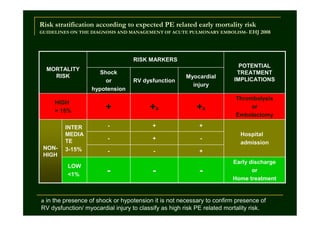
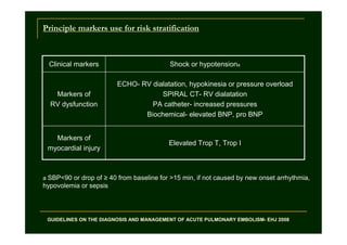
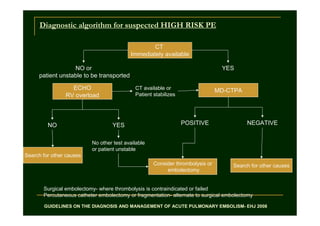
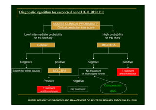
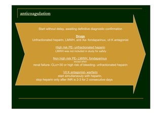
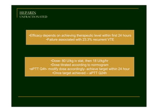
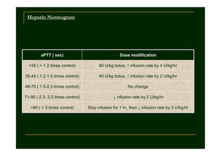
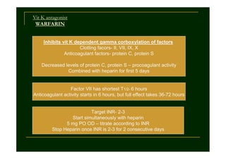
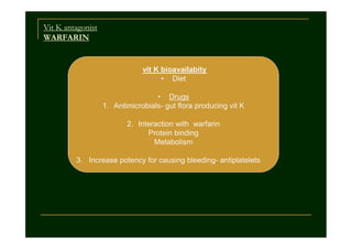
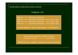
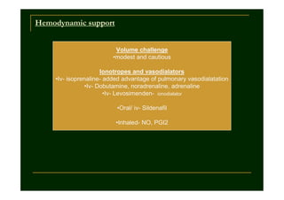
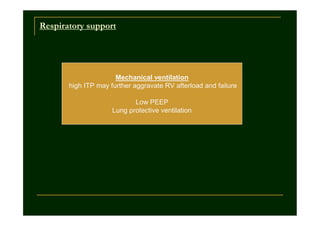
VENOUS THROMBOEMBOLISM
Ubaidur Rahaman
Senior Resident, CCM, SGPGIMS
Lucknow, India
stasis

Coagulation
activation

Virchow’s
triad

Vascular
injury

•>90% of PE- thrombi arise from deep veins of leg
• clinically important PE- thrombi arise from popliteal or
more proximal deep veins of leg
•Clinical manifestation of PE
size, site and number of thrombi + cardiorespiratory reserve of patient
•Recurrence of VTE is more with ileofemoral vein thrombosis
than popliteal vein thrombosis
diagnosis
Clinical presentation
PE confirmed

PE excluded

Dyspnoea

80%

59%

Chest pain-pleuritic

52%

43%

Chest pain- substernal

12%

8%

Cough

20%

25%

Hemoptysis

11%

7%

Syncope

19%

11%

Tachypnoea(>20/min)

70%

68%

Tachycardia( >100/min)

26%

23%

Signs of DVT

15%

10%

Fever (>38C)

7%

17%

Cyanosis

11%

9%

Symptoms

Signs
CXR
• plate like atelectasis
•Elevation of hemidiaphram
•Pleural effusion

EKG- signs of RV strain , RBBB

•Non specific
•Helpful in exclusion of other causes

•Usually found in massive PE
•Can be caused by other causes

ABG- ↓PaO2, ↑A-aO2

Normal in upto 20% patients
D- dimer
degradation product of cross linked fibrin
Elevated in presence of acute clot formation
simultaneous activation of coagulation and fibrinolysis
But fibrin is also produced in
inflammation, necrosis, malignancy, dissection of aorta, aging
high negative predictive value, low positive predictive value
DVT
Detection of DVT in proven PE
venography – 70%
compression USG – 50%

Compression USG
•Sensitivity-90%, specificity-95% for proximal DVT
•Not sensitive for isolated calf vein thrombosis
•Negative result-Should be repeated after 1 week

COMPRESSION USG **
•back up procedure to avoid false positive results with SDCT
•Patients with contraindication to dye or irradiation

**GUIDELINES ON THE DIAGNOSIS AND MANAGEMENT OF ACUTE PULMONARY EMBOLISM- EHJ 2008
Objectively documented DVT
50% suffer PE, many are asymptomatic
Angiographically documented PE
50-70% have detectable DVT
clinically suspected PE
>50%-diagnosis not confirmed by investigation

Objective test for diagnosis of PE
•V/Q scan
•Pulmonary angiography
•Spiral CT- chest
•MR angiography

•costly
•Invasive
•Radiation
•Mobilization of patient
•≥40% of vascular bed obtstruction
to produce detectable features of RV overload

•2 D echocardiography

•TEE more valuable than TTE
•Coexistent cardiorespiratory disease
•Not useful in hemodaynamically stable patients
•Clinical signs, symptoms and routine investigation do not help in
confirmation or exclusion of PE

•Help in increasing the index of suspicion
Suspected PE
which patient should be mobilized for costly, invasive/ radiation exposure investigation

Clinical probability of PE
Low- 9% prevalence of PE
Intermediate-30% prevalence of PE
High-68% prevalence of PE

Clinical prediction rule
Based on history, sign and symptoms
CLINICAL PREDICTION RULE
WELLS score
predisposing factors:
nPrevious
nRecent

documented DVT or PE

1.5

immobilization ≥ 3 days or major surgery in last 4 weeks

nActive

cancer- receiving treatment or treated in last 6 months or
palliative care

1.5
1

Clinical sign/ symptoms:
nClinical
nHR

signs and symptoms of DVT

3

>100

1.5

nhemoptysis

1

nAlternate

3

clinical diagnosis less likely than VTE

CLINICAL PROBABILITY
2 level
> 4- -------- likely PE
0-4-------- unlikely PE

3 level
0-1--------------- low
2-6---- intermediate
≥7--------------- high
WELLS SCORE
clinical prediction rule

More than10,000 patients studied

•
•
•

Clinical probability- PE unlikely
D-dimer- negative
No treatment with anticoagulants

<1% develop VTE
within 90 days of evaluation

•Clinical probability- PE likely
•Clinical probability- PE unlikely
but D- dimer- positive

Prevalence of PE- 20%
Risk stratification according to expected PE related early mortality risk
GUIDELINES ON THE DIAGNOSIS AND MANAGEMENT OF ACUTE PULMONARY EMBOLISM- EHJ 2008

RISK MARKERS
MORTALITY
RISK

HIGH
> 15%

NONHIGH

INTER
MEDIA
TE
3-15%
LOW
<1%

Shock
or
hypotension

RV dysfunction

Myocardial
injury

+

+a

+a

-

+
+

-

-

-

Thrombolysis
or
Embolectomy

+

-

POTENTIAL
TREATMENT
IMPLICATIONS

+

-

-

-

Hospital
admission
Early discharge
or
Home treatment

a in the presence of shock or hypotension it is not necessary to confirm presence of

RV dysfunction/ myocardial injury to classify as high risk PE related mortality risk.
Principle markers use for risk stratification

Clinical markers

Shock or hypotensiona

Markers of
RV dysfunction

ECHO- RV dialatation, hypokinesia or pressure overload
SPIRAL CT- RV dialatation
PA catheter- increased pressures
Biochemical- elevated BNP, pro BNP

Markers of
myocardial injury

Elevated Trop T, Trop I

a SBP<90 or drop of ≥ 40 from baseline for >15 min, if not caused by new onset arrhythmia,

hypovolemia or sepsis

GUIDELINES ON THE DIAGNOSIS AND MANAGEMENT OF ACUTE PULMONARY EMBOLISM- EHJ 2008
Diagnostic algorithm for suspected HIGH RISK PE
CT
Immediately available
NO or
patient unstable to be transported
ECHO
RV overload

NO

YES
CT available or
Patient stabilizes

YES

MD-CTPA

POSITIVE

NEGATIVE

No other test available
or patient unstable
Search for other causes
Consider thrombolysis or
embolectomy

Search for other causes

Surgical embolectomy- where thrombolysis is contraindicated or failed
Percutaneous catheter embolectomy or fragmentation- alternate to surgical embolectomy
GUIDELINES ON THE DIAGNOSIS AND MANAGEMENT OF ACUTE PULMONARY EMBOLISM- EHJ 2008
Diagnostic algorithm for suspected non-HIGH RISK PE

ASSESS CLINICAL PROBABILITY
Clinical prediction rule score
Low/ intermediate probability
or PE unlikely

High probability
or PE likely

D-dimer

MD-CTPA

Negative

positive

negative

positive

Search for other causes

MD-CTPA

No treatment
or investigate further

Treatment
antithrombosis

Positive

negative
Compression

Treatment
antithrombosis

No treatment

USG

GUIDELINES ON THE DIAGNOSIS AND MANAGEMENT OF ACUTE PULMONARY EMBOLISM- EHJ 2008
anticoagulation

Start without delay, awaiting definitive diagnostic confirmation
Drugs
Unfractionated heparin, LMWH, anti Xa- fondaparinux, vit K antagonist
High risk PE- unfractionated heparin
LMWH was not included in study for safety

Non high risk PE- LMWH, fondaparinux
except when

renal failure- CLcr<30 or high risk of bleeding- unfractionated heparin
Vit K antagonist- warfarin
start simultaneouly with heparin,
stop heparin only after INR is 2-3 for 2 consecutive days
HEPARIN
UNFRACTIONATED

•Efficacy depends on achieving therapeutic level within first 24 hours
•Failure associated with 23.3% recurrent VTE

•Dose- 80 U/kg iv stat, then 18 U/kg/hr
•Dose titrated according to normogram
•aPTT Q4h- modify dose accordingly- achieve target within 24 hour
•Once target achieved – aPTT Q24h
Heparin Normogram

aPTT ( sec)

Dose modification

<35 ( < 1.2 times control)

80 U/kg bolus, ↑ infusion rate by 4 U/kg/hr

35-45 ( 1.2-1.5 times control)

40 U/kg bolus, ↑ infusion rate by 2 U/kg/hr

46-70 ( 1.5-2.3 times control)

No change

71-90 ( 2.3- 3.0 times control)

↓ infusion rate by 2 U/kg/hr

>90 ( > 3 times control)

Stop infusion for 1 hr, then ↓ infusion rate by 3 U/kg/hr
Vit K antagonist
WARFARIN
Inhibits vit K dependent gamma corboxylation of factors
Clotting facors- II, VII, IX, X
Anticoagulant factors- protein C, protein S
Decreased levels of protein C, protein S – procoagulant activity
Combined with heparin for first 5 days

Factor VII has shortest T1/2- 6 hours
Anticoagulant activity starts in 6 hours, but full effect takes 36-72 hours

Target INR- 2-3
Start simultaneously with heparin
5 mg PO OD – titrate according to INR
Stop Heparin once INR is 2-3 for 2 consecutive days
Vit K antagonist
WARFARIN

vit K bioavailabity
• Diet
• Drugs
1. Antimicrobials- gut flora producing vit K
2. Interaction with warfarin
Protein binding
Metabolism
3. Increase potency for causing bleeding- antiplatelets
WARFARIN OVERANTICOAGULATION
Antagonist- vit K
•INR 3-5 -------- hold dose of that day
•INR ≥5 – 7.5-- hold dose of that day + vit K 1 mg ivi stat
•INR ≥7.5-10----hold dose of that day + vit K 2 mg ivi stat
•INR ≥10 --------hold dose of that day + vit K 3 mg ivi stat
•If active bleeding – fresh frozen plasma- 10-15ml/kg bw

demonstrable reduction in INR- 6-8 hours
correction on INR-----------------12-24 hours
Half life of vit K < warfarin– repeat dose may be required
Hemodynamic support
Volume challenge
•modest and cautious
Ionotropes and vasodialators
•Iv- isoprenaline- added advantage of pulmonary vasodialatation
•Iv- Dobutamine, noradrenaline, adrenaline
•Iv- Levosimenden- ionodialator
•Oral/ iv- Sildenafil
•Inhaled- NO, PGI2
Respiratory support

Mechanical ventilation
high ITP may further aggravate RV afterload and failure
Low PEEP
Lung protective ventilation
Not everything that counts can be counted. And not everything
that can be counted counts.
--Albert Einstein

Venous thromboembolism

  • 1.
    VENOUS THROMBOEMBOLISM Ubaidur Rahaman SeniorResident, CCM, SGPGIMS Lucknow, India
  • 2.
    stasis Coagulation activation Virchow’s triad Vascular injury •>90% of PE-thrombi arise from deep veins of leg • clinically important PE- thrombi arise from popliteal or more proximal deep veins of leg •Clinical manifestation of PE size, site and number of thrombi + cardiorespiratory reserve of patient •Recurrence of VTE is more with ileofemoral vein thrombosis than popliteal vein thrombosis
  • 3.
    diagnosis Clinical presentation PE confirmed PEexcluded Dyspnoea 80% 59% Chest pain-pleuritic 52% 43% Chest pain- substernal 12% 8% Cough 20% 25% Hemoptysis 11% 7% Syncope 19% 11% Tachypnoea(>20/min) 70% 68% Tachycardia( >100/min) 26% 23% Signs of DVT 15% 10% Fever (>38C) 7% 17% Cyanosis 11% 9% Symptoms Signs
  • 4.
    CXR • plate likeatelectasis •Elevation of hemidiaphram •Pleural effusion EKG- signs of RV strain , RBBB •Non specific •Helpful in exclusion of other causes •Usually found in massive PE •Can be caused by other causes ABG- ↓PaO2, ↑A-aO2 Normal in upto 20% patients
  • 5.
    D- dimer degradation productof cross linked fibrin Elevated in presence of acute clot formation simultaneous activation of coagulation and fibrinolysis But fibrin is also produced in inflammation, necrosis, malignancy, dissection of aorta, aging high negative predictive value, low positive predictive value
  • 6.
    DVT Detection of DVTin proven PE venography – 70% compression USG – 50% Compression USG •Sensitivity-90%, specificity-95% for proximal DVT •Not sensitive for isolated calf vein thrombosis •Negative result-Should be repeated after 1 week COMPRESSION USG ** •back up procedure to avoid false positive results with SDCT •Patients with contraindication to dye or irradiation **GUIDELINES ON THE DIAGNOSIS AND MANAGEMENT OF ACUTE PULMONARY EMBOLISM- EHJ 2008
  • 7.
    Objectively documented DVT 50%suffer PE, many are asymptomatic Angiographically documented PE 50-70% have detectable DVT clinically suspected PE >50%-diagnosis not confirmed by investigation Objective test for diagnosis of PE •V/Q scan •Pulmonary angiography •Spiral CT- chest •MR angiography •costly •Invasive •Radiation •Mobilization of patient •≥40% of vascular bed obtstruction to produce detectable features of RV overload •2 D echocardiography •TEE more valuable than TTE •Coexistent cardiorespiratory disease •Not useful in hemodaynamically stable patients
  • 8.
    •Clinical signs, symptomsand routine investigation do not help in confirmation or exclusion of PE •Help in increasing the index of suspicion
  • 9.
    Suspected PE which patientshould be mobilized for costly, invasive/ radiation exposure investigation Clinical probability of PE Low- 9% prevalence of PE Intermediate-30% prevalence of PE High-68% prevalence of PE Clinical prediction rule Based on history, sign and symptoms
  • 10.
    CLINICAL PREDICTION RULE WELLSscore predisposing factors: nPrevious nRecent documented DVT or PE 1.5 immobilization ≥ 3 days or major surgery in last 4 weeks nActive cancer- receiving treatment or treated in last 6 months or palliative care 1.5 1 Clinical sign/ symptoms: nClinical nHR signs and symptoms of DVT 3 >100 1.5 nhemoptysis 1 nAlternate 3 clinical diagnosis less likely than VTE CLINICAL PROBABILITY 2 level > 4- -------- likely PE 0-4-------- unlikely PE 3 level 0-1--------------- low 2-6---- intermediate ≥7--------------- high
  • 11.
    WELLS SCORE clinical predictionrule More than10,000 patients studied • • • Clinical probability- PE unlikely D-dimer- negative No treatment with anticoagulants <1% develop VTE within 90 days of evaluation •Clinical probability- PE likely •Clinical probability- PE unlikely but D- dimer- positive Prevalence of PE- 20%
  • 12.
    Risk stratification accordingto expected PE related early mortality risk GUIDELINES ON THE DIAGNOSIS AND MANAGEMENT OF ACUTE PULMONARY EMBOLISM- EHJ 2008 RISK MARKERS MORTALITY RISK HIGH > 15% NONHIGH INTER MEDIA TE 3-15% LOW <1% Shock or hypotension RV dysfunction Myocardial injury + +a +a - + + - - - Thrombolysis or Embolectomy + - POTENTIAL TREATMENT IMPLICATIONS + - - - Hospital admission Early discharge or Home treatment a in the presence of shock or hypotension it is not necessary to confirm presence of RV dysfunction/ myocardial injury to classify as high risk PE related mortality risk.
  • 13.
    Principle markers usefor risk stratification Clinical markers Shock or hypotensiona Markers of RV dysfunction ECHO- RV dialatation, hypokinesia or pressure overload SPIRAL CT- RV dialatation PA catheter- increased pressures Biochemical- elevated BNP, pro BNP Markers of myocardial injury Elevated Trop T, Trop I a SBP<90 or drop of ≥ 40 from baseline for >15 min, if not caused by new onset arrhythmia, hypovolemia or sepsis GUIDELINES ON THE DIAGNOSIS AND MANAGEMENT OF ACUTE PULMONARY EMBOLISM- EHJ 2008
  • 14.
    Diagnostic algorithm forsuspected HIGH RISK PE CT Immediately available NO or patient unstable to be transported ECHO RV overload NO YES CT available or Patient stabilizes YES MD-CTPA POSITIVE NEGATIVE No other test available or patient unstable Search for other causes Consider thrombolysis or embolectomy Search for other causes Surgical embolectomy- where thrombolysis is contraindicated or failed Percutaneous catheter embolectomy or fragmentation- alternate to surgical embolectomy GUIDELINES ON THE DIAGNOSIS AND MANAGEMENT OF ACUTE PULMONARY EMBOLISM- EHJ 2008
  • 15.
    Diagnostic algorithm forsuspected non-HIGH RISK PE ASSESS CLINICAL PROBABILITY Clinical prediction rule score Low/ intermediate probability or PE unlikely High probability or PE likely D-dimer MD-CTPA Negative positive negative positive Search for other causes MD-CTPA No treatment or investigate further Treatment antithrombosis Positive negative Compression Treatment antithrombosis No treatment USG GUIDELINES ON THE DIAGNOSIS AND MANAGEMENT OF ACUTE PULMONARY EMBOLISM- EHJ 2008
  • 16.
    anticoagulation Start without delay,awaiting definitive diagnostic confirmation Drugs Unfractionated heparin, LMWH, anti Xa- fondaparinux, vit K antagonist High risk PE- unfractionated heparin LMWH was not included in study for safety Non high risk PE- LMWH, fondaparinux except when renal failure- CLcr<30 or high risk of bleeding- unfractionated heparin Vit K antagonist- warfarin start simultaneouly with heparin, stop heparin only after INR is 2-3 for 2 consecutive days
  • 17.
    HEPARIN UNFRACTIONATED •Efficacy depends onachieving therapeutic level within first 24 hours •Failure associated with 23.3% recurrent VTE •Dose- 80 U/kg iv stat, then 18 U/kg/hr •Dose titrated according to normogram •aPTT Q4h- modify dose accordingly- achieve target within 24 hour •Once target achieved – aPTT Q24h
  • 18.
    Heparin Normogram aPTT (sec) Dose modification <35 ( < 1.2 times control) 80 U/kg bolus, ↑ infusion rate by 4 U/kg/hr 35-45 ( 1.2-1.5 times control) 40 U/kg bolus, ↑ infusion rate by 2 U/kg/hr 46-70 ( 1.5-2.3 times control) No change 71-90 ( 2.3- 3.0 times control) ↓ infusion rate by 2 U/kg/hr >90 ( > 3 times control) Stop infusion for 1 hr, then ↓ infusion rate by 3 U/kg/hr
  • 19.
    Vit K antagonist WARFARIN Inhibitsvit K dependent gamma corboxylation of factors Clotting facors- II, VII, IX, X Anticoagulant factors- protein C, protein S Decreased levels of protein C, protein S – procoagulant activity Combined with heparin for first 5 days Factor VII has shortest T1/2- 6 hours Anticoagulant activity starts in 6 hours, but full effect takes 36-72 hours Target INR- 2-3 Start simultaneously with heparin 5 mg PO OD – titrate according to INR Stop Heparin once INR is 2-3 for 2 consecutive days
  • 20.
    Vit K antagonist WARFARIN vitK bioavailabity • Diet • Drugs 1. Antimicrobials- gut flora producing vit K 2. Interaction with warfarin Protein binding Metabolism 3. Increase potency for causing bleeding- antiplatelets
  • 21.
    WARFARIN OVERANTICOAGULATION Antagonist- vitK •INR 3-5 -------- hold dose of that day •INR ≥5 – 7.5-- hold dose of that day + vit K 1 mg ivi stat •INR ≥7.5-10----hold dose of that day + vit K 2 mg ivi stat •INR ≥10 --------hold dose of that day + vit K 3 mg ivi stat •If active bleeding – fresh frozen plasma- 10-15ml/kg bw demonstrable reduction in INR- 6-8 hours correction on INR-----------------12-24 hours Half life of vit K < warfarin– repeat dose may be required
  • 22.
    Hemodynamic support Volume challenge •modestand cautious Ionotropes and vasodialators •Iv- isoprenaline- added advantage of pulmonary vasodialatation •Iv- Dobutamine, noradrenaline, adrenaline •Iv- Levosimenden- ionodialator •Oral/ iv- Sildenafil •Inhaled- NO, PGI2
  • 23.
    Respiratory support Mechanical ventilation highITP may further aggravate RV afterload and failure Low PEEP Lung protective ventilation
  • 24.
    Not everything thatcounts can be counted. And not everything that can be counted counts. --Albert Einstein