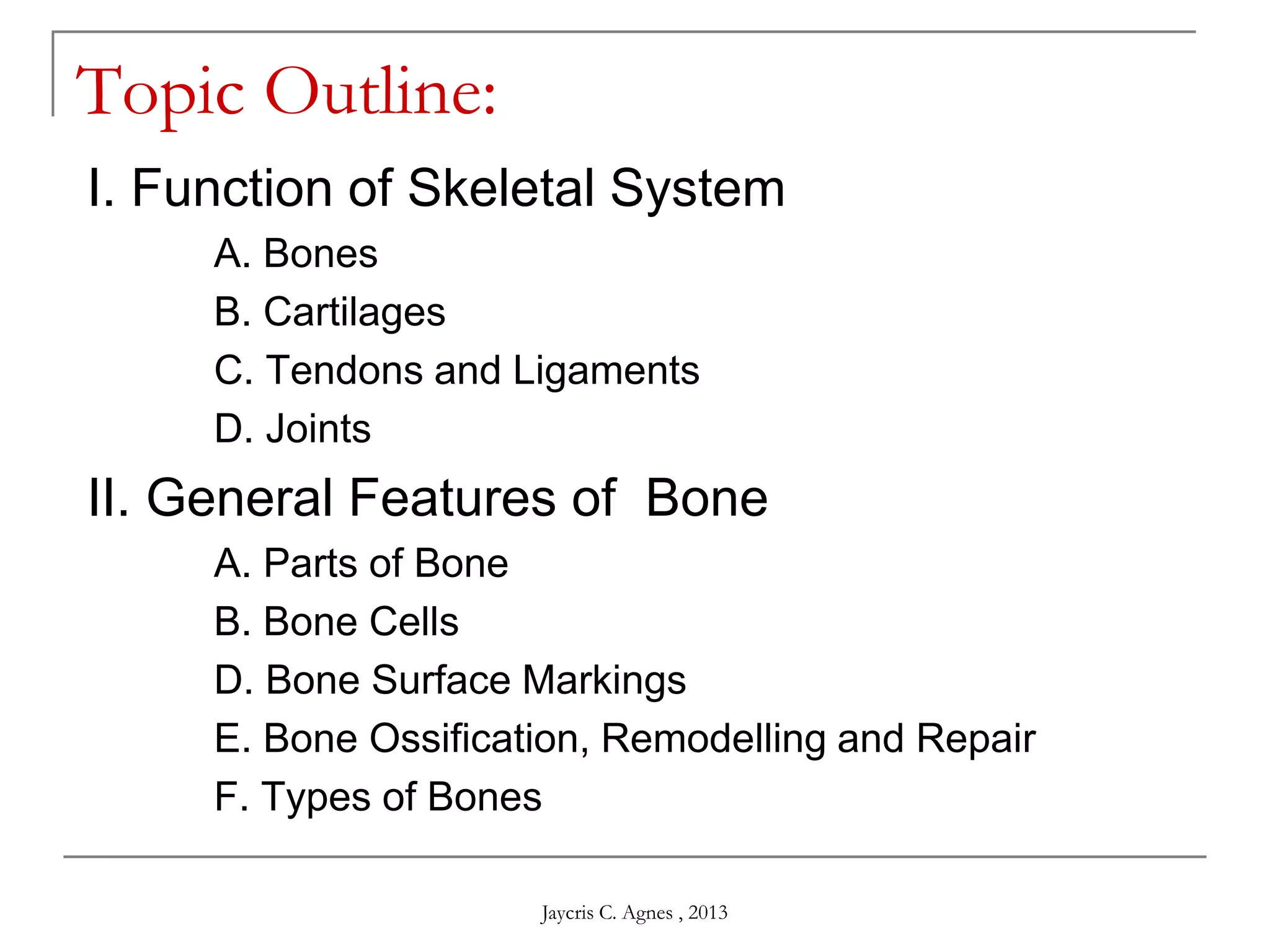
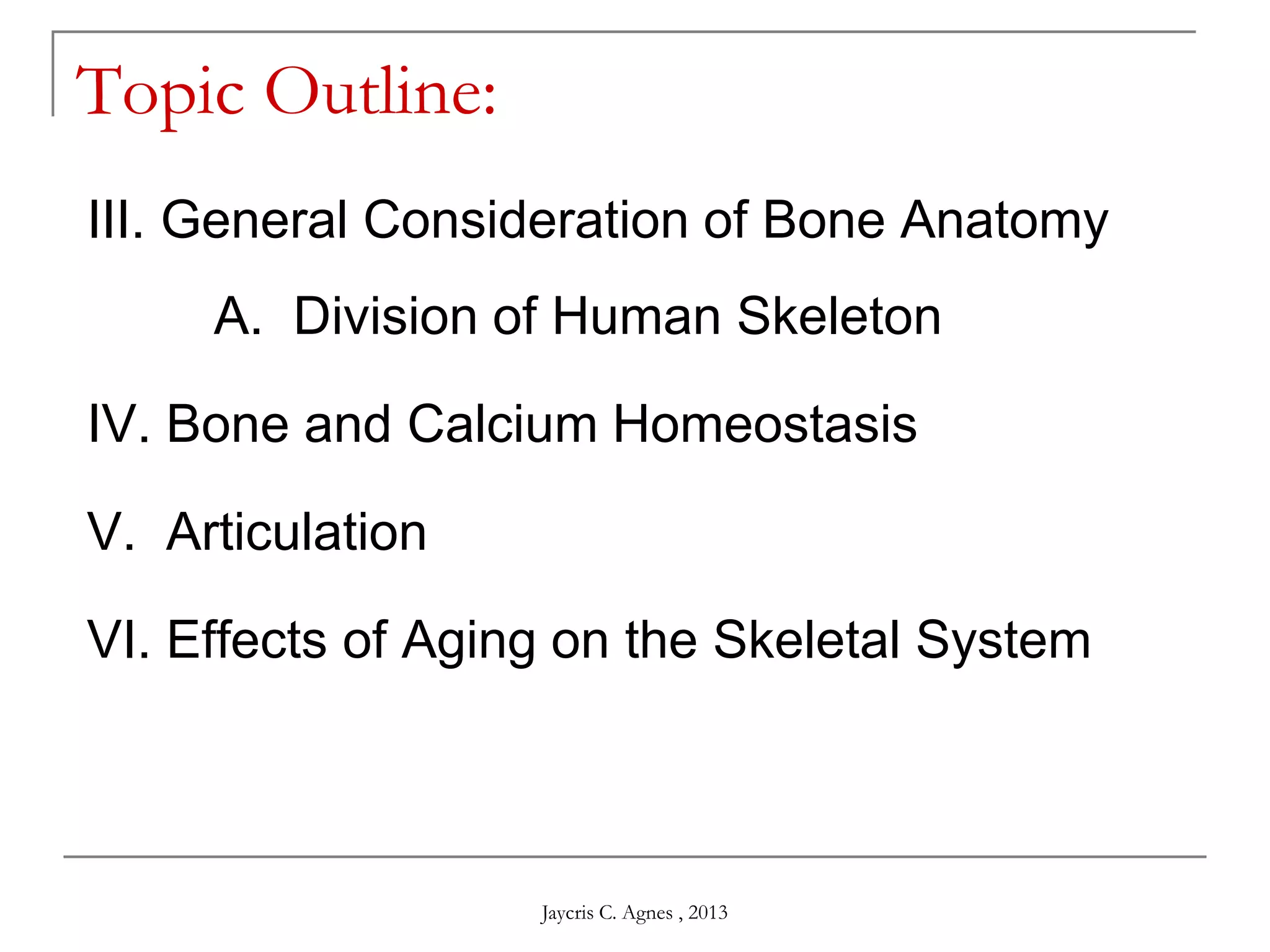
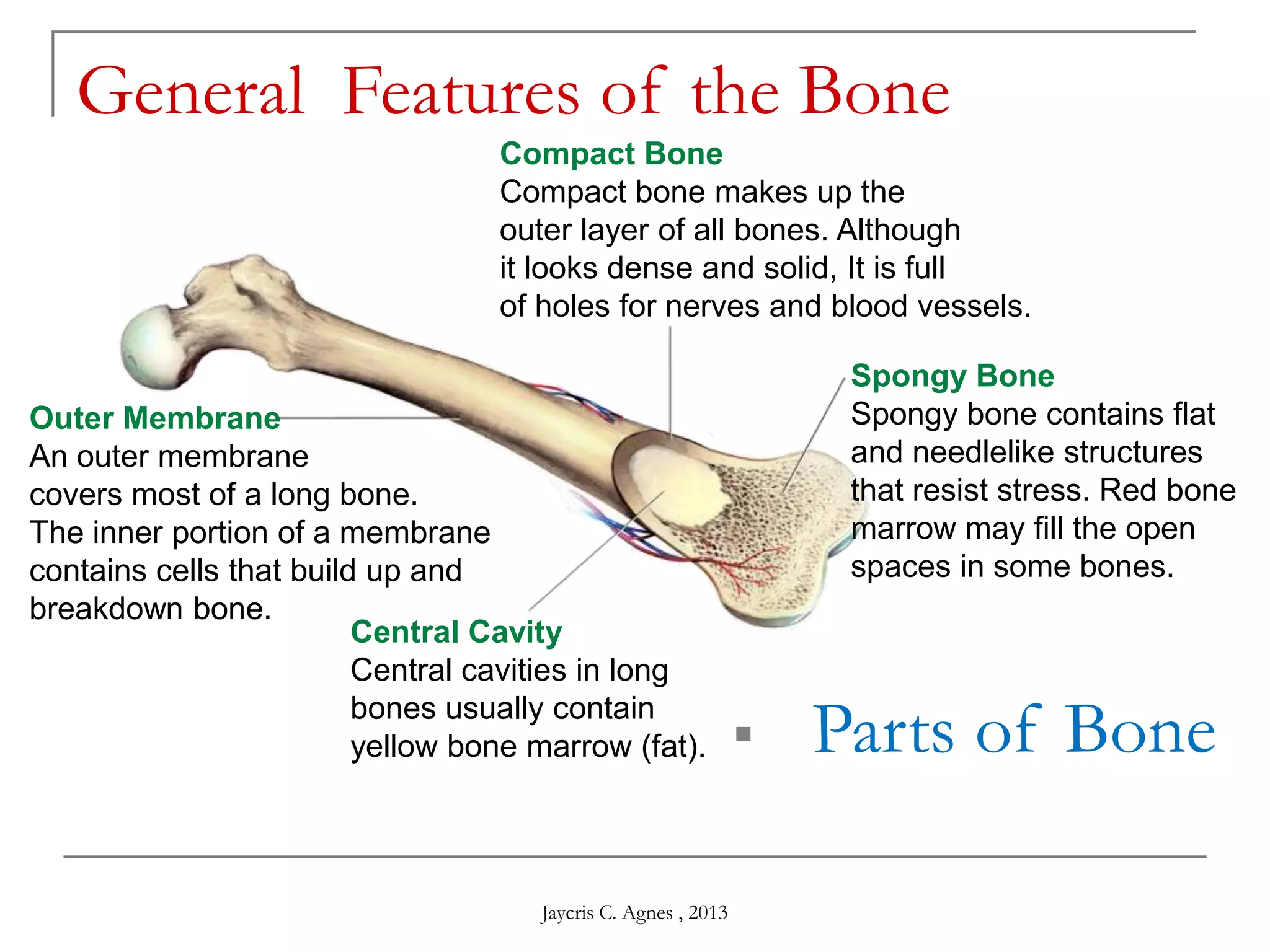
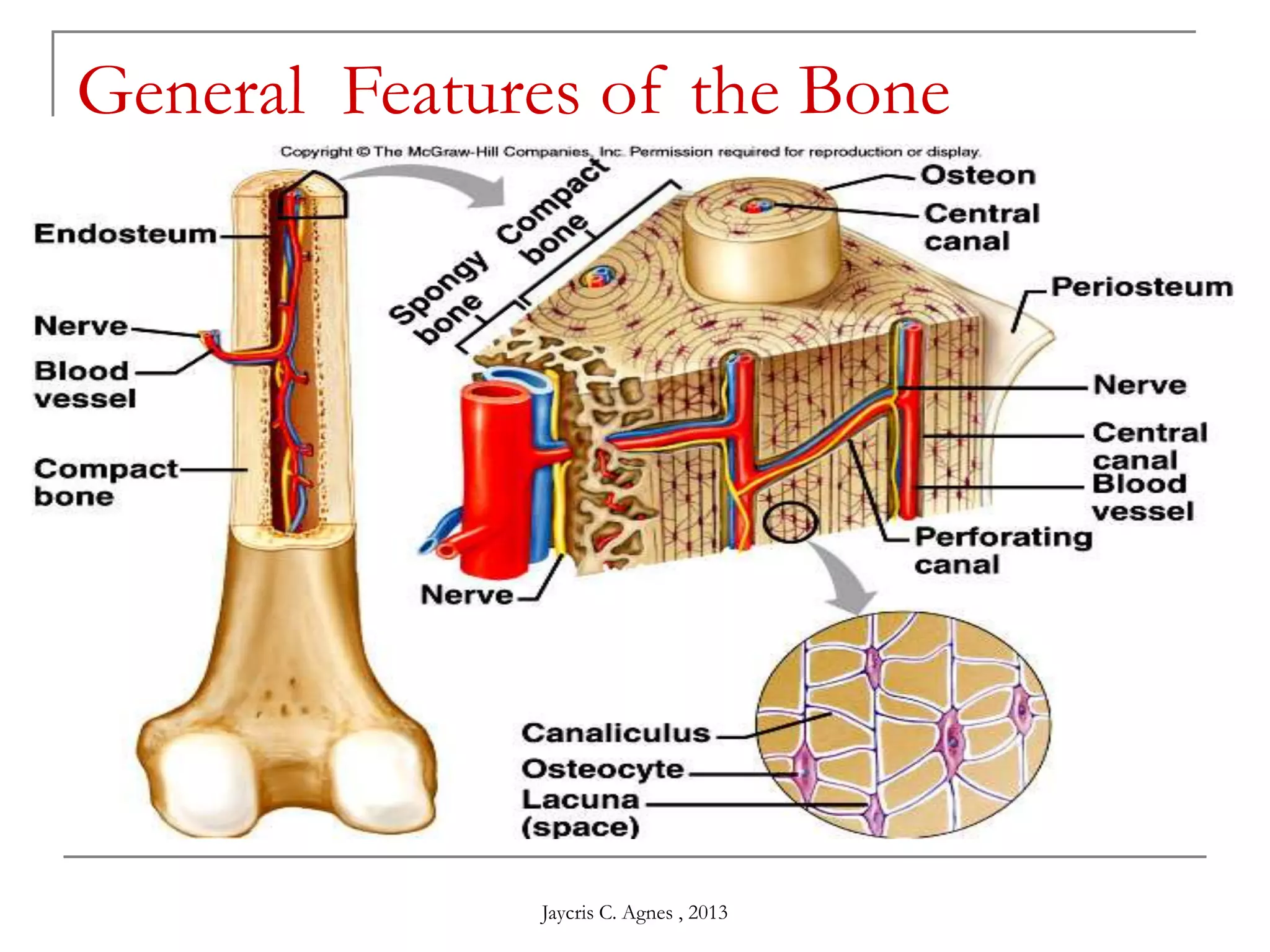
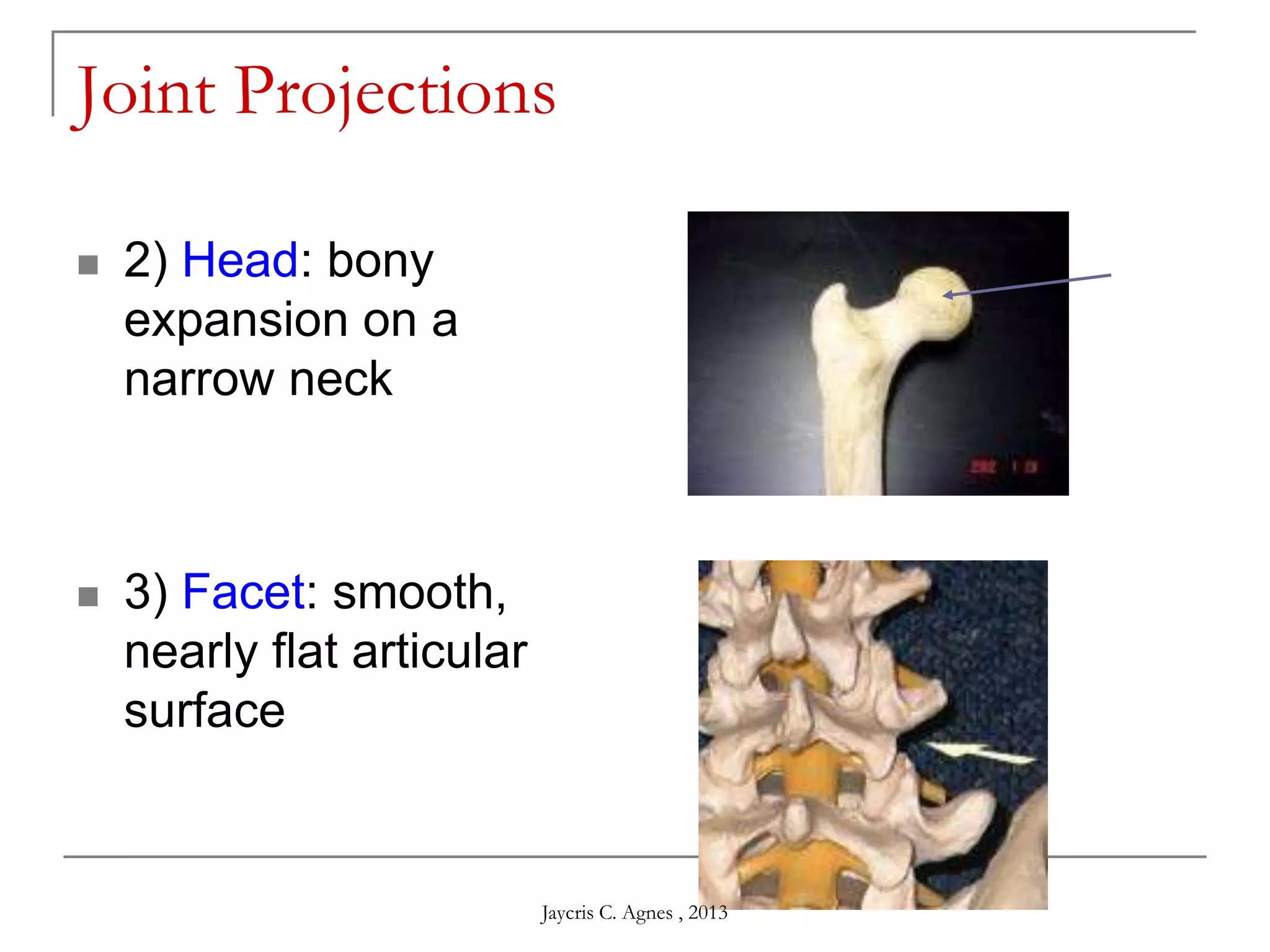
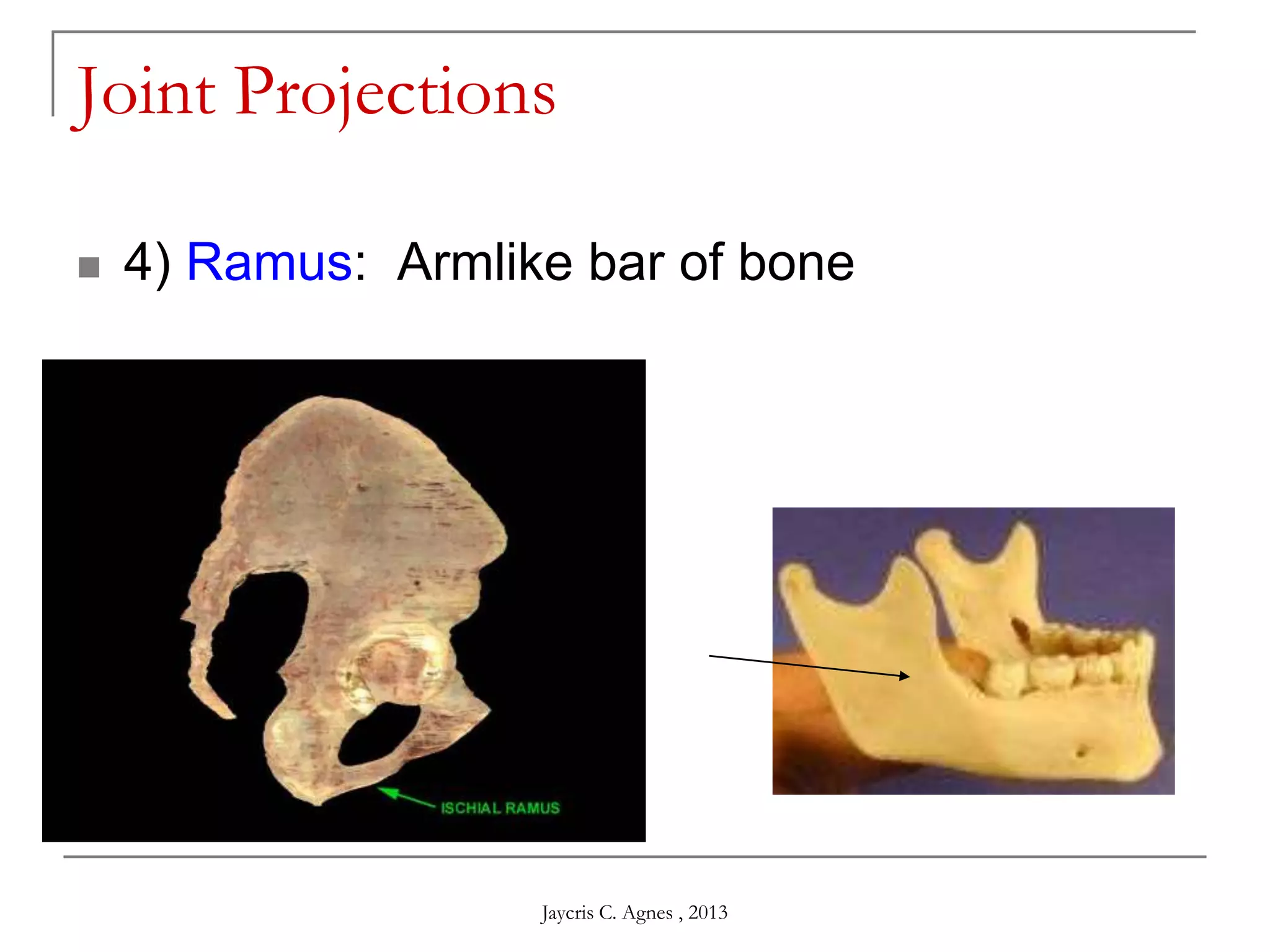
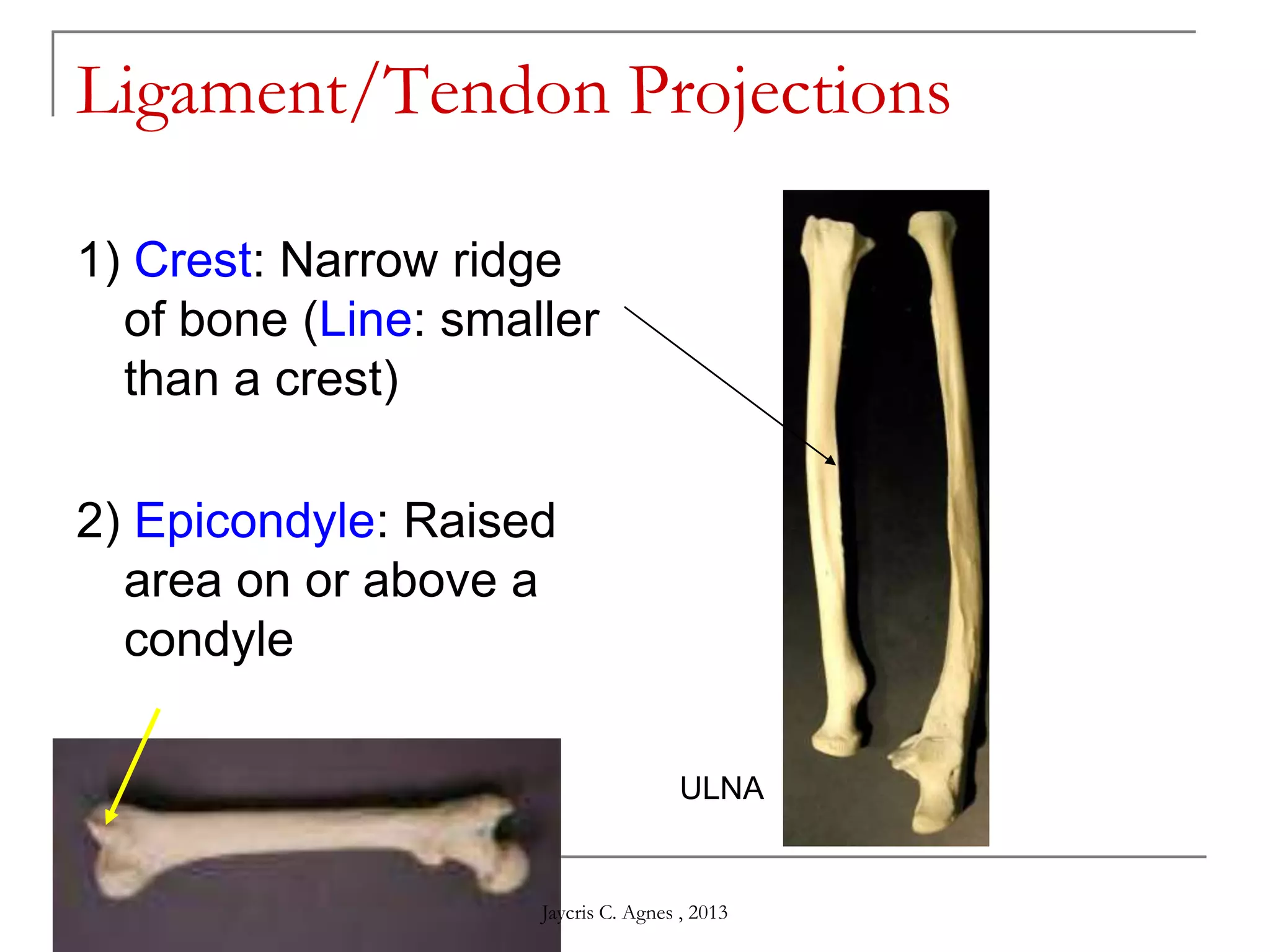
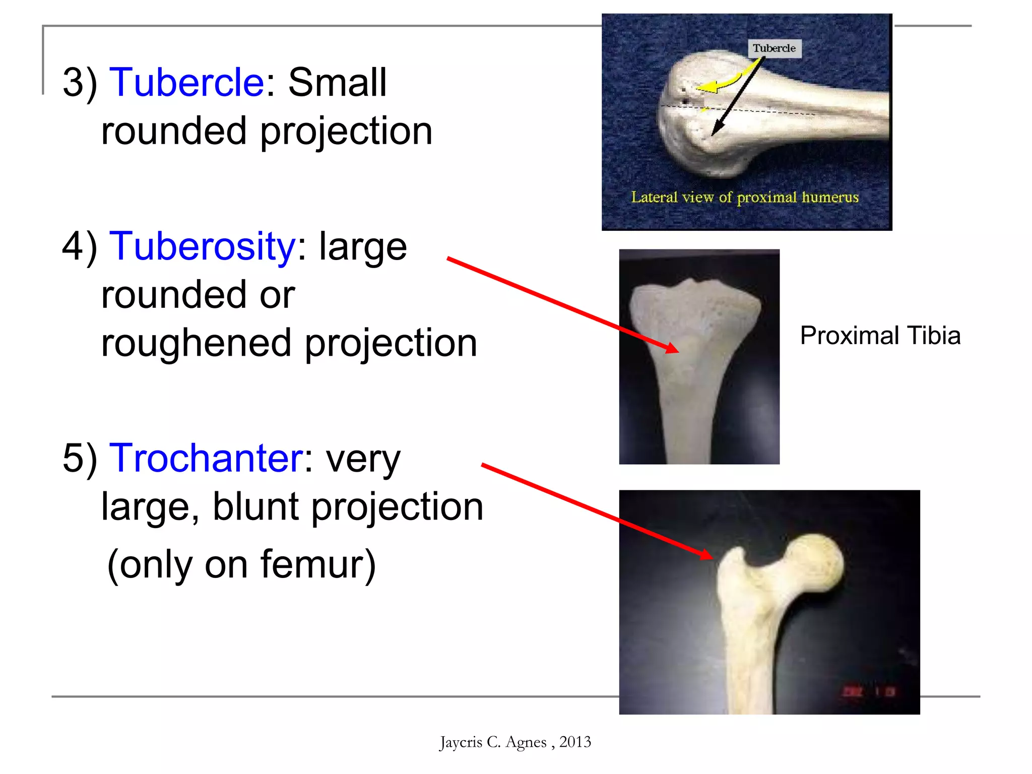
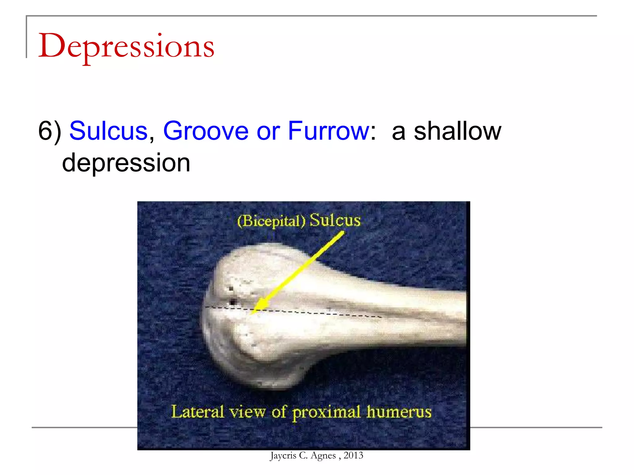
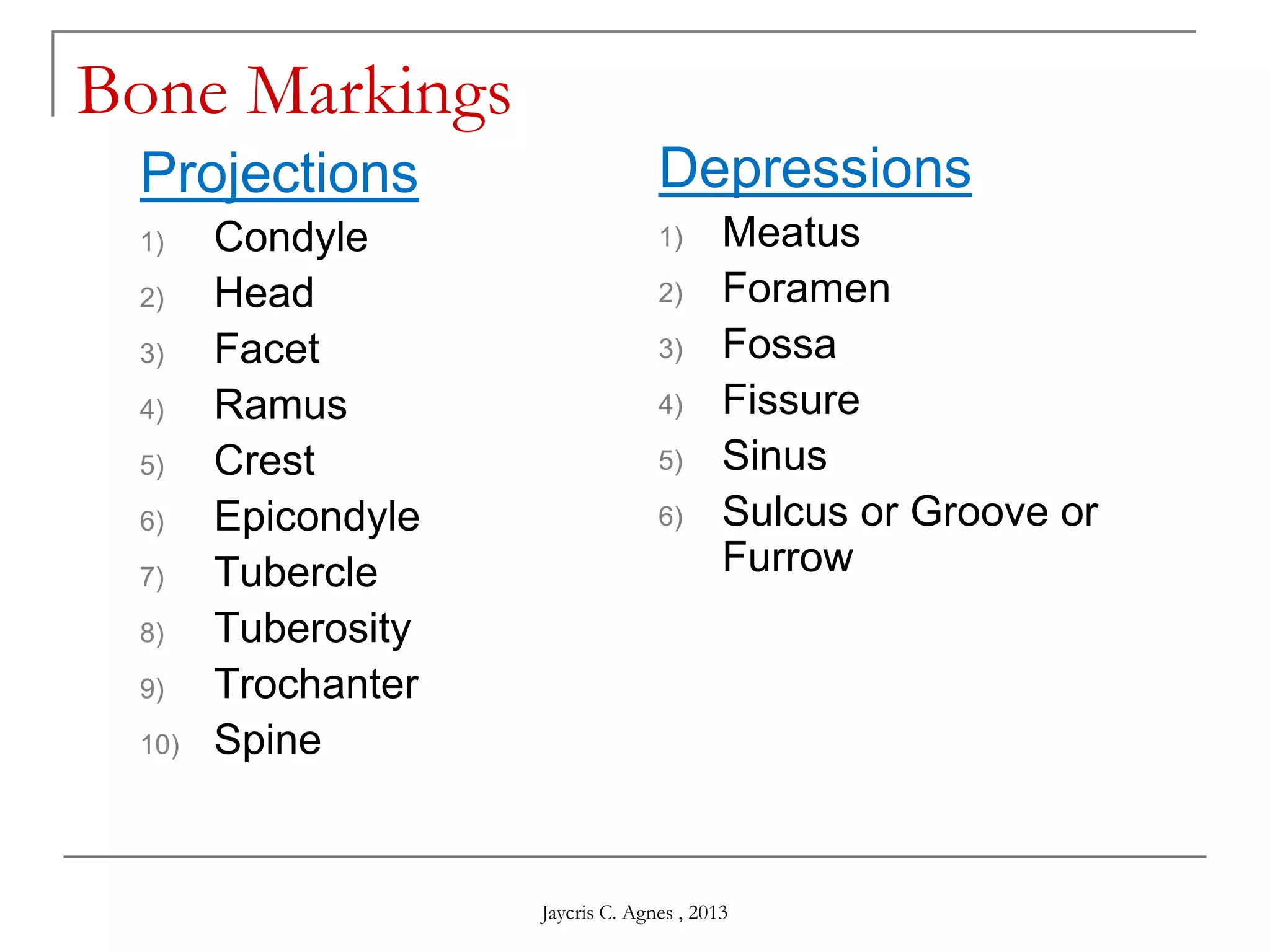
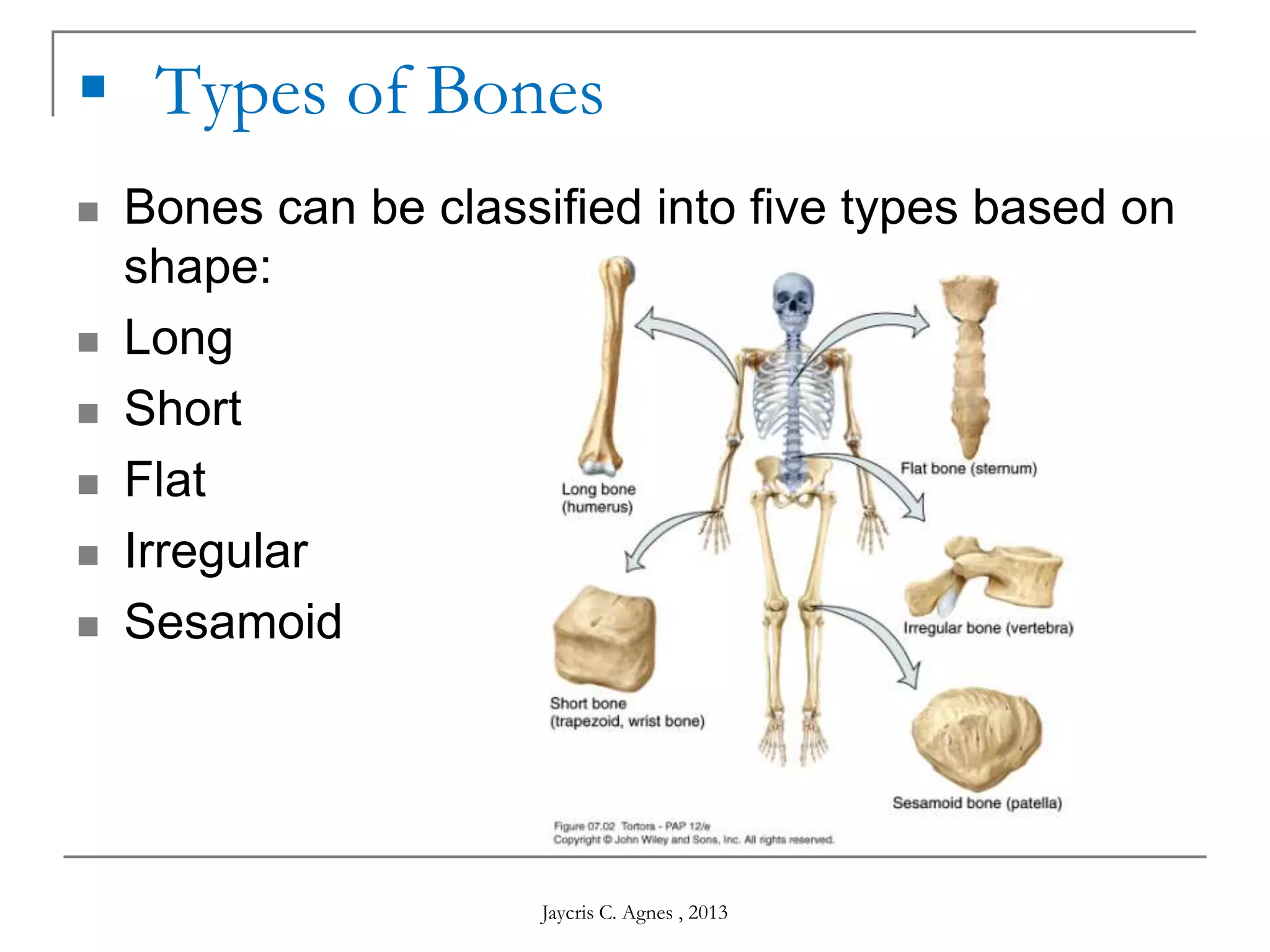
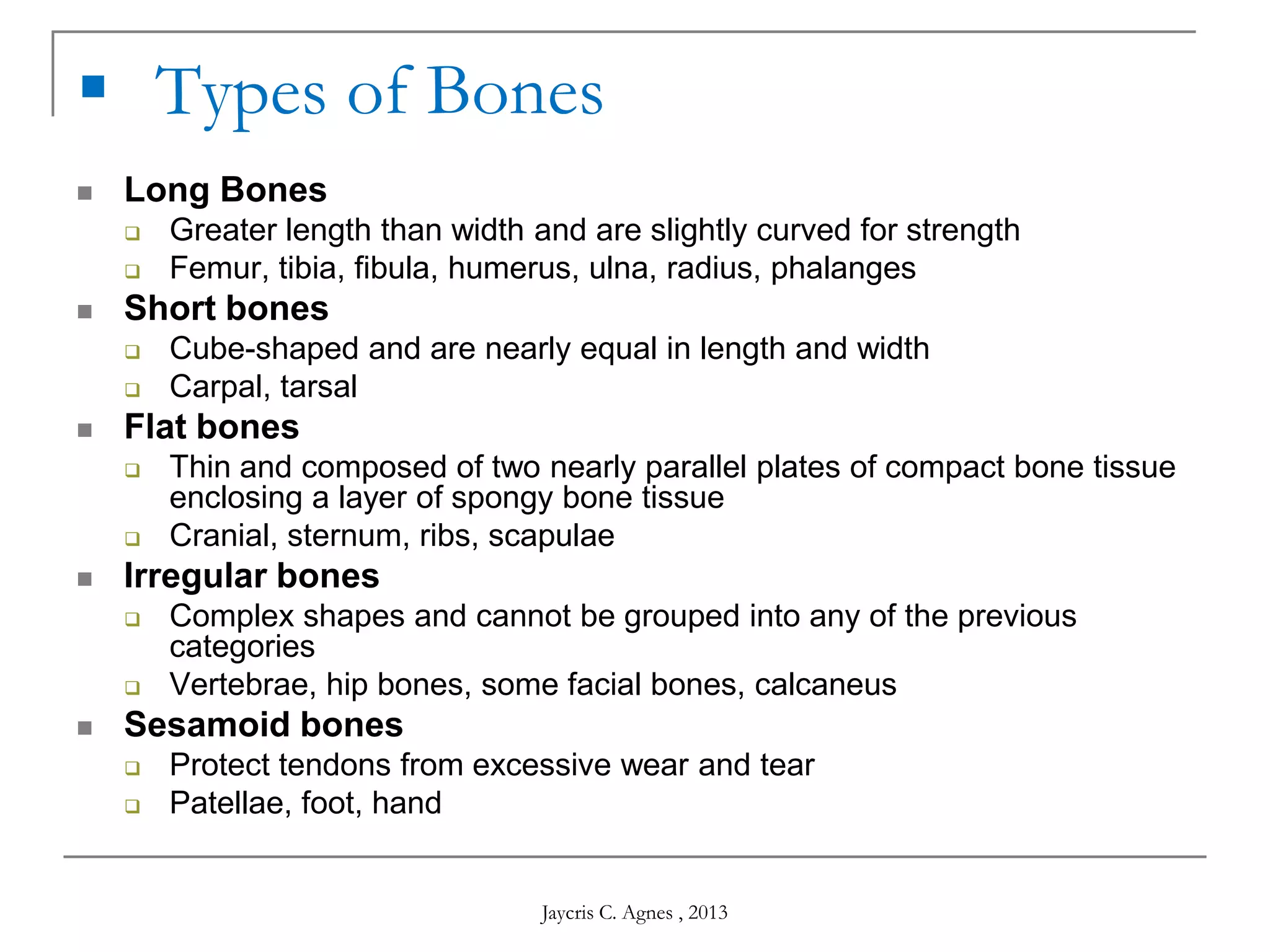
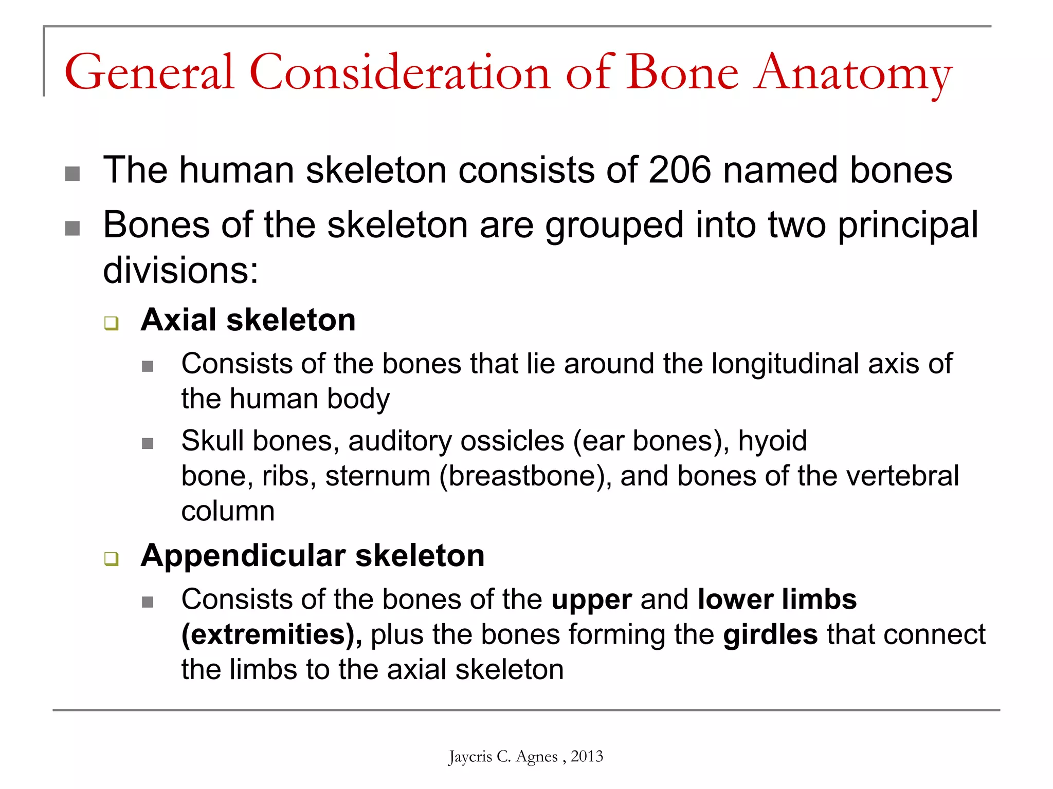
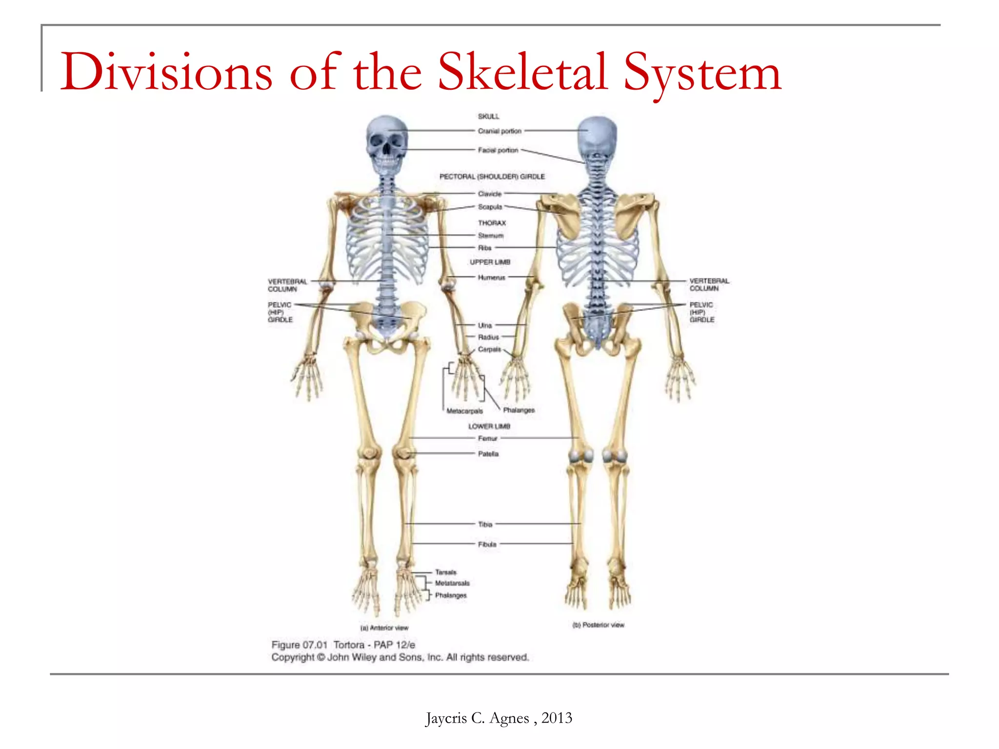
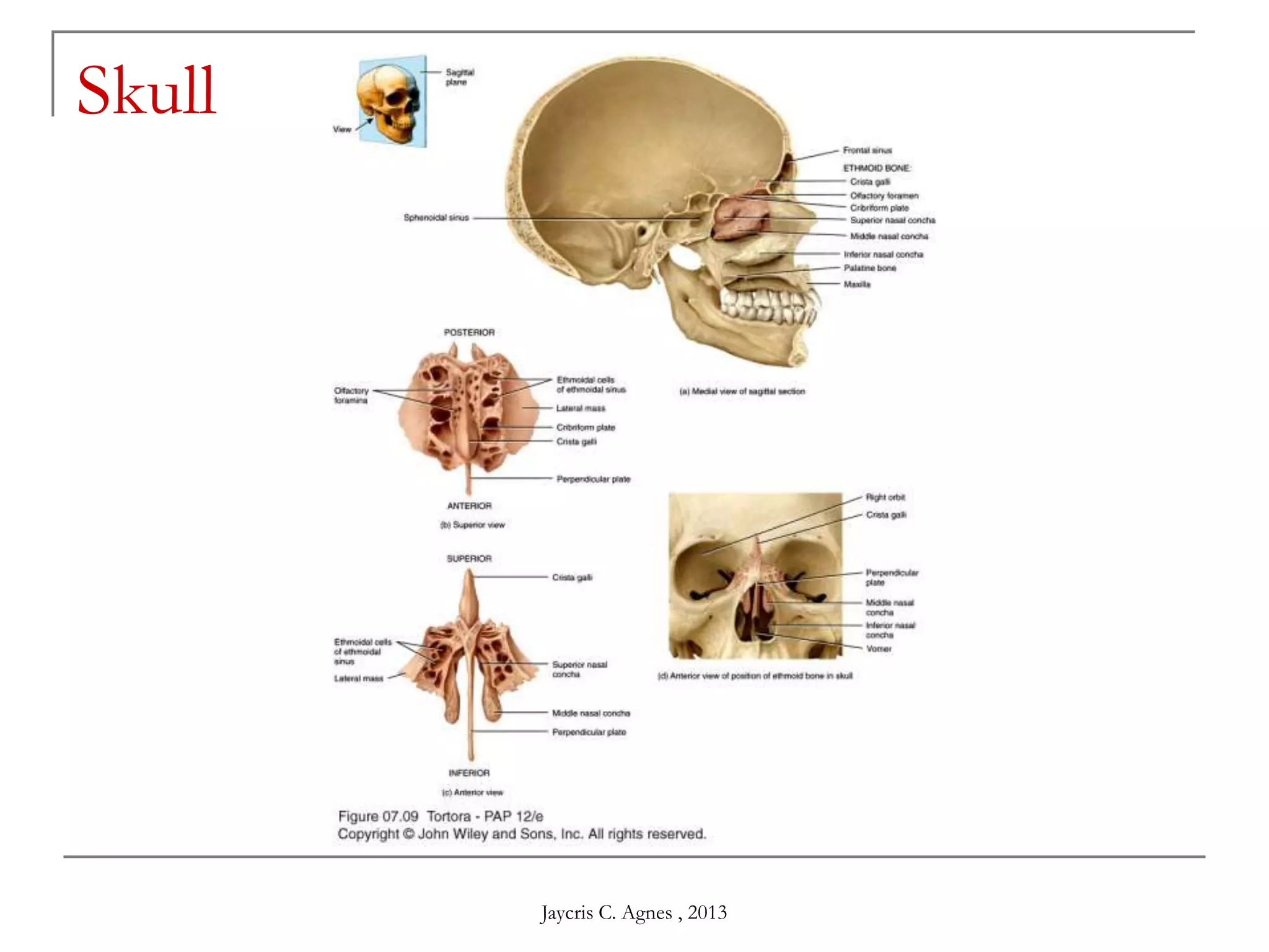
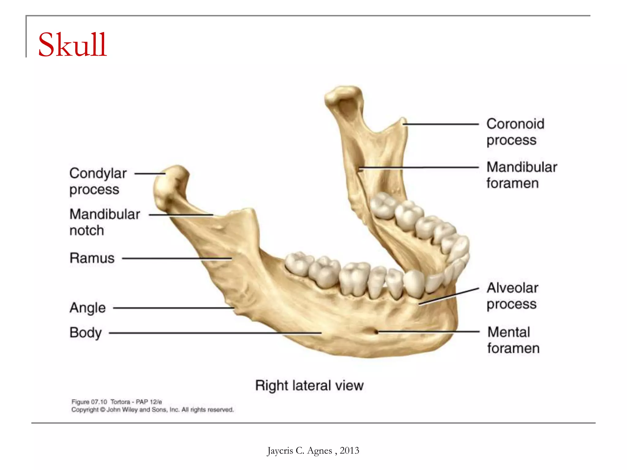
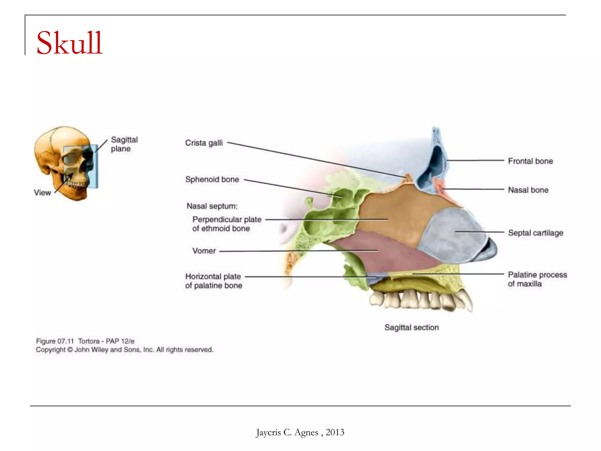
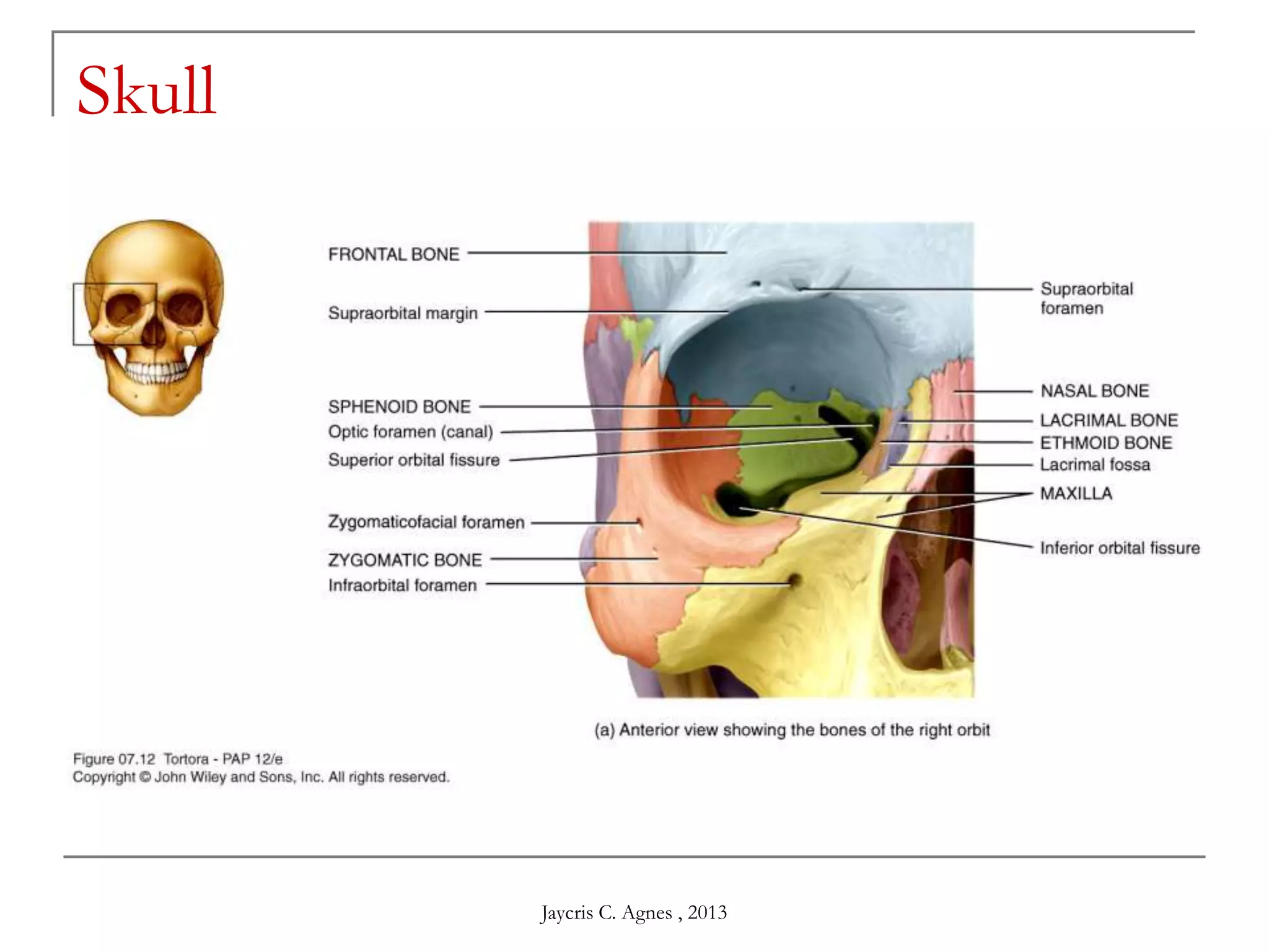
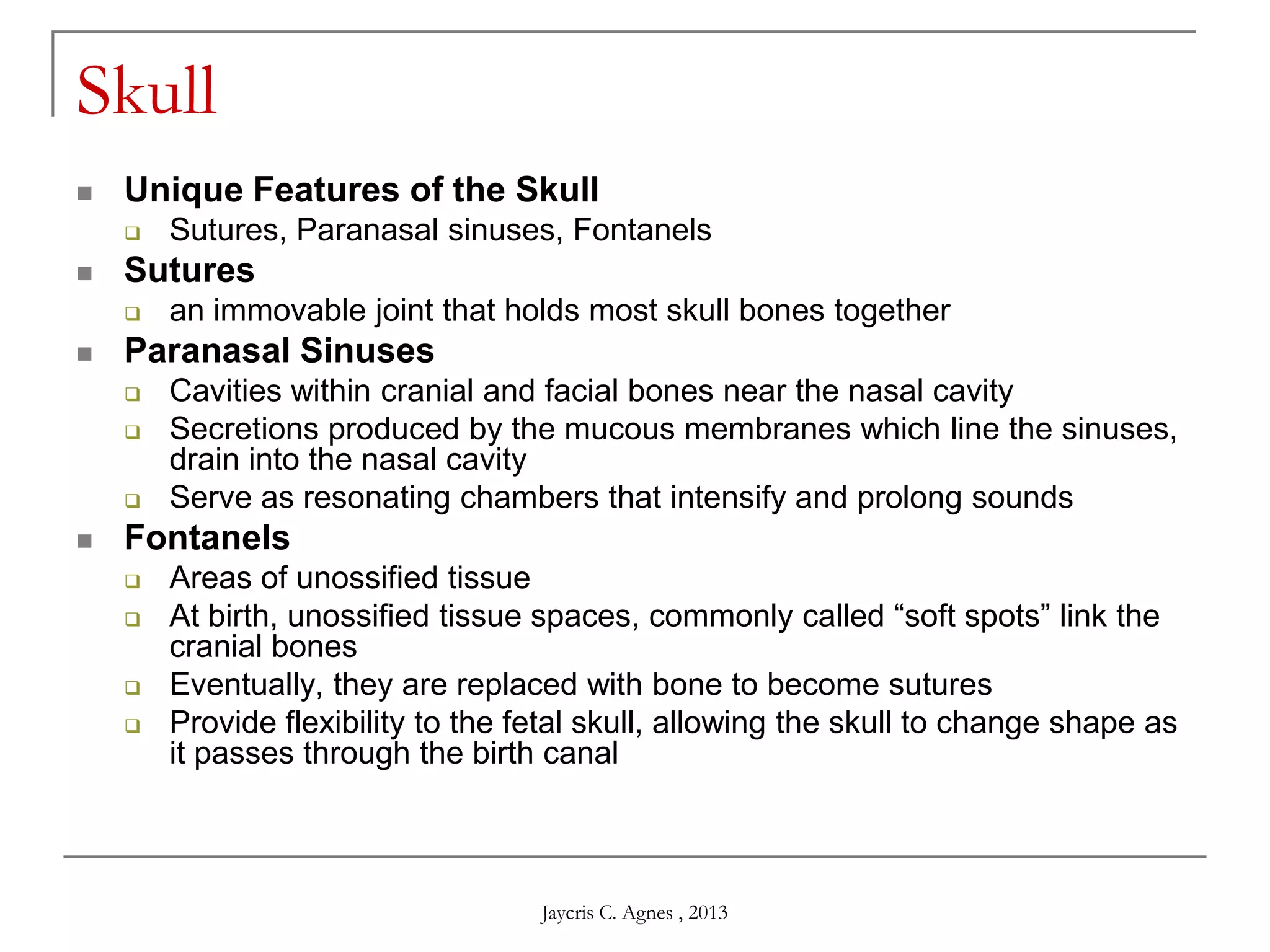
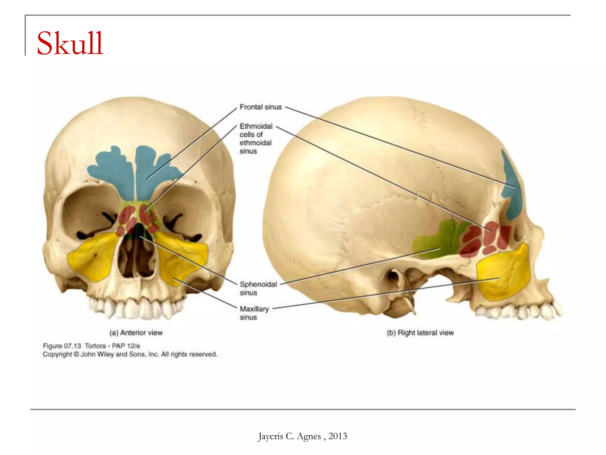
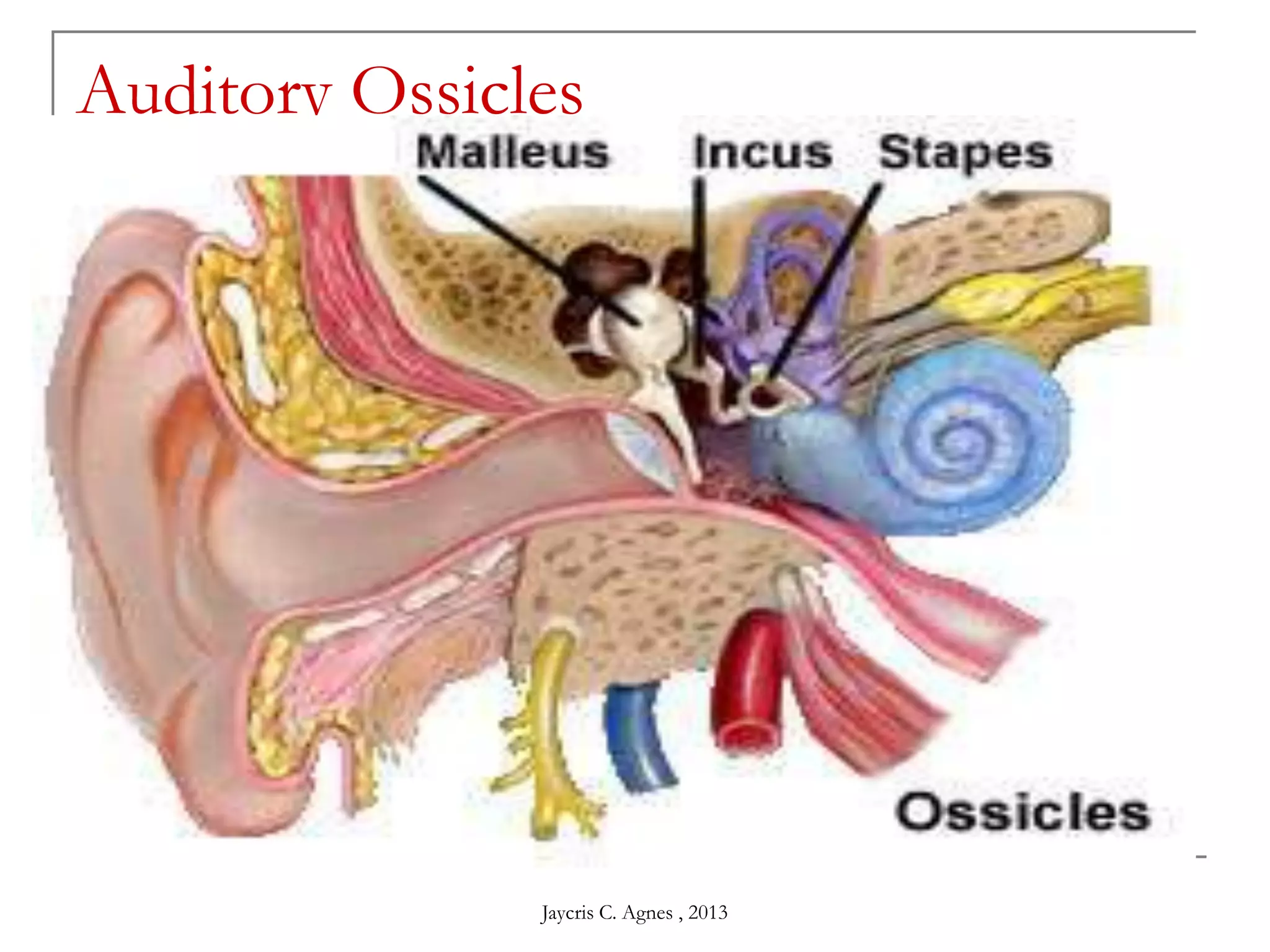
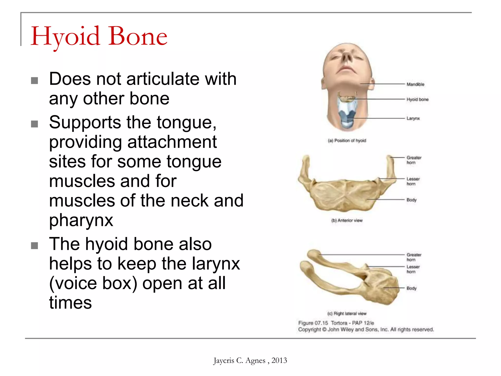
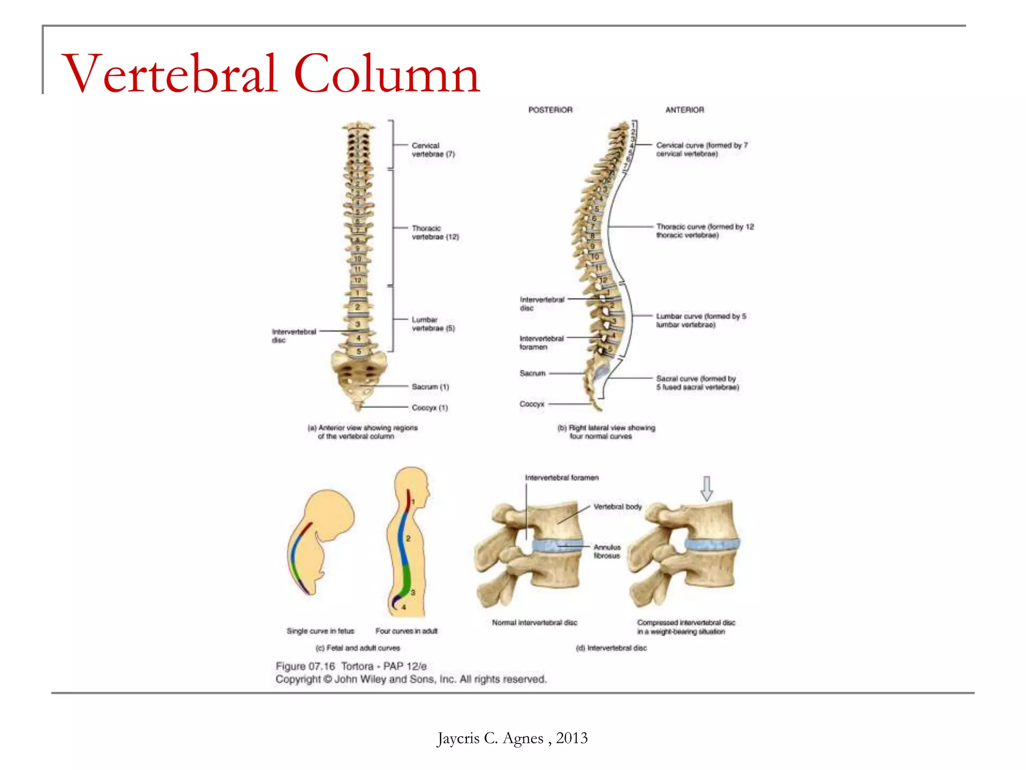
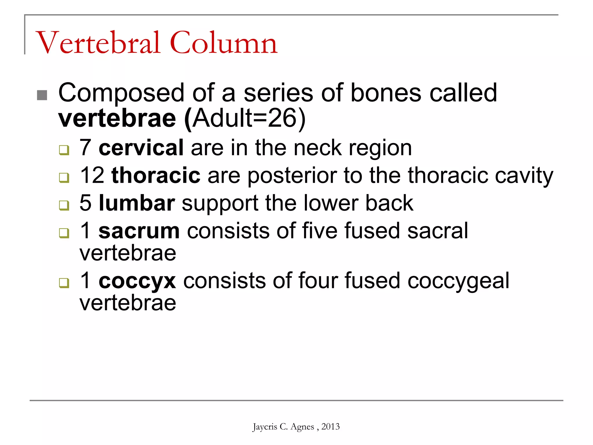
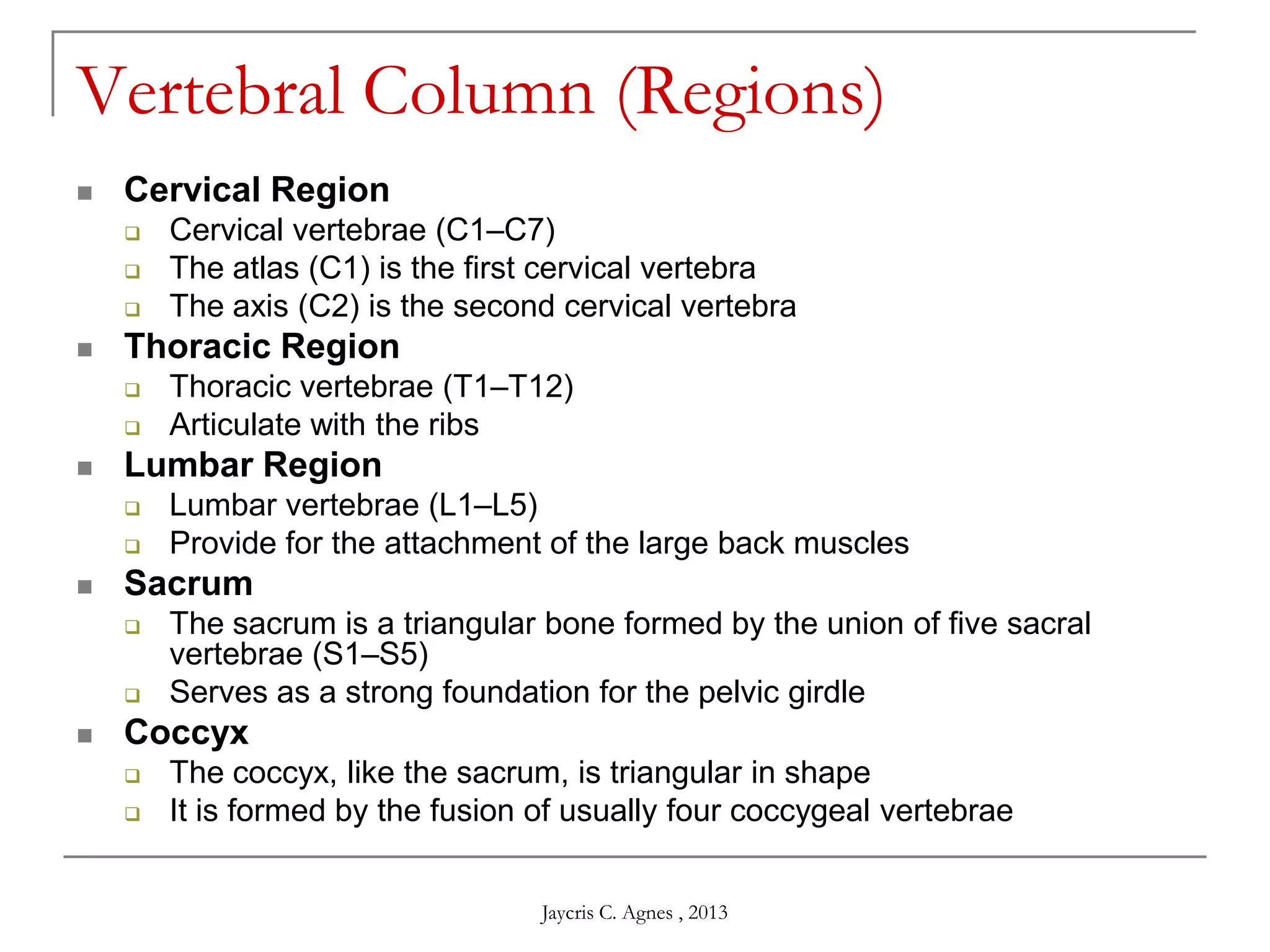
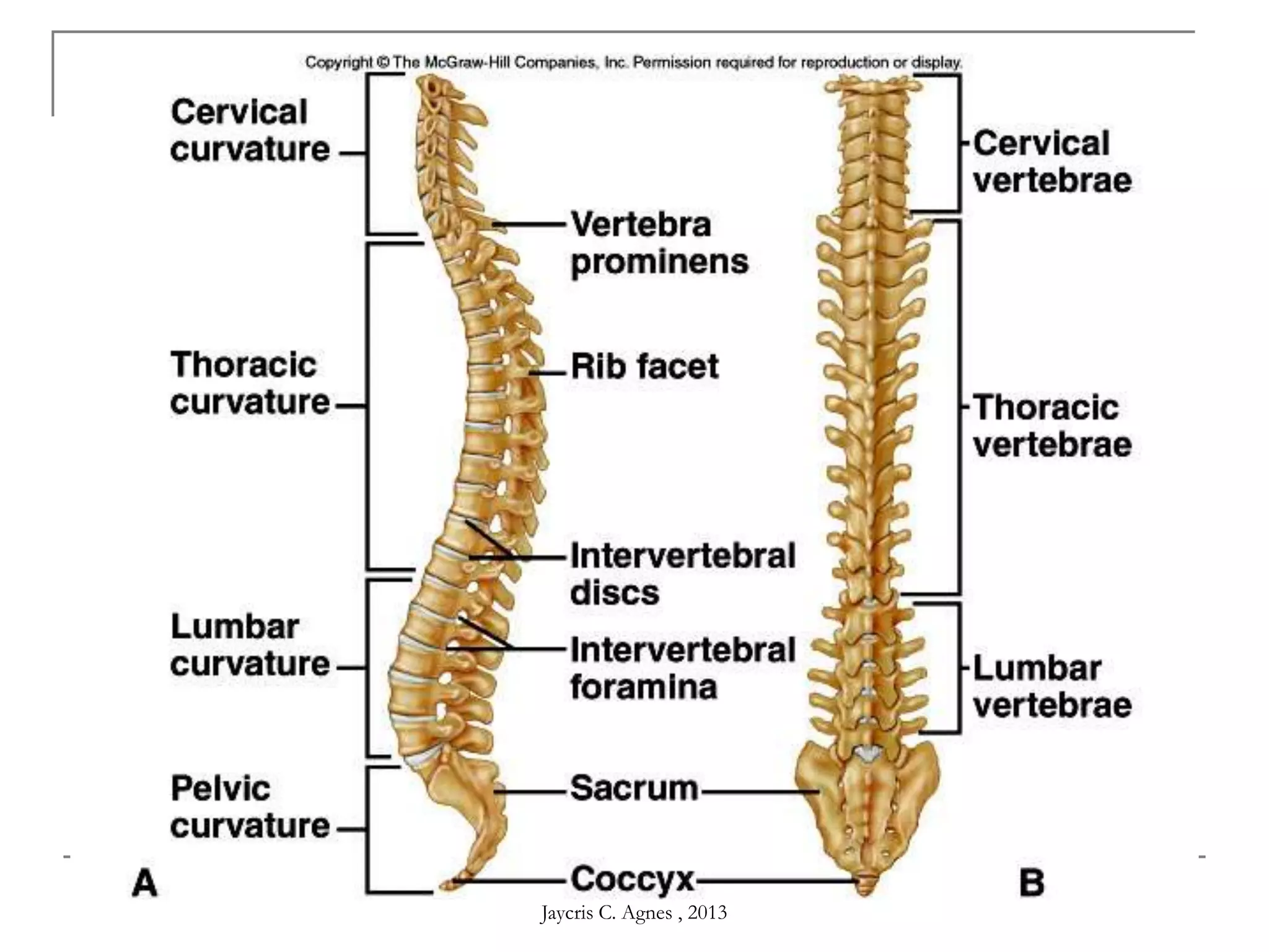
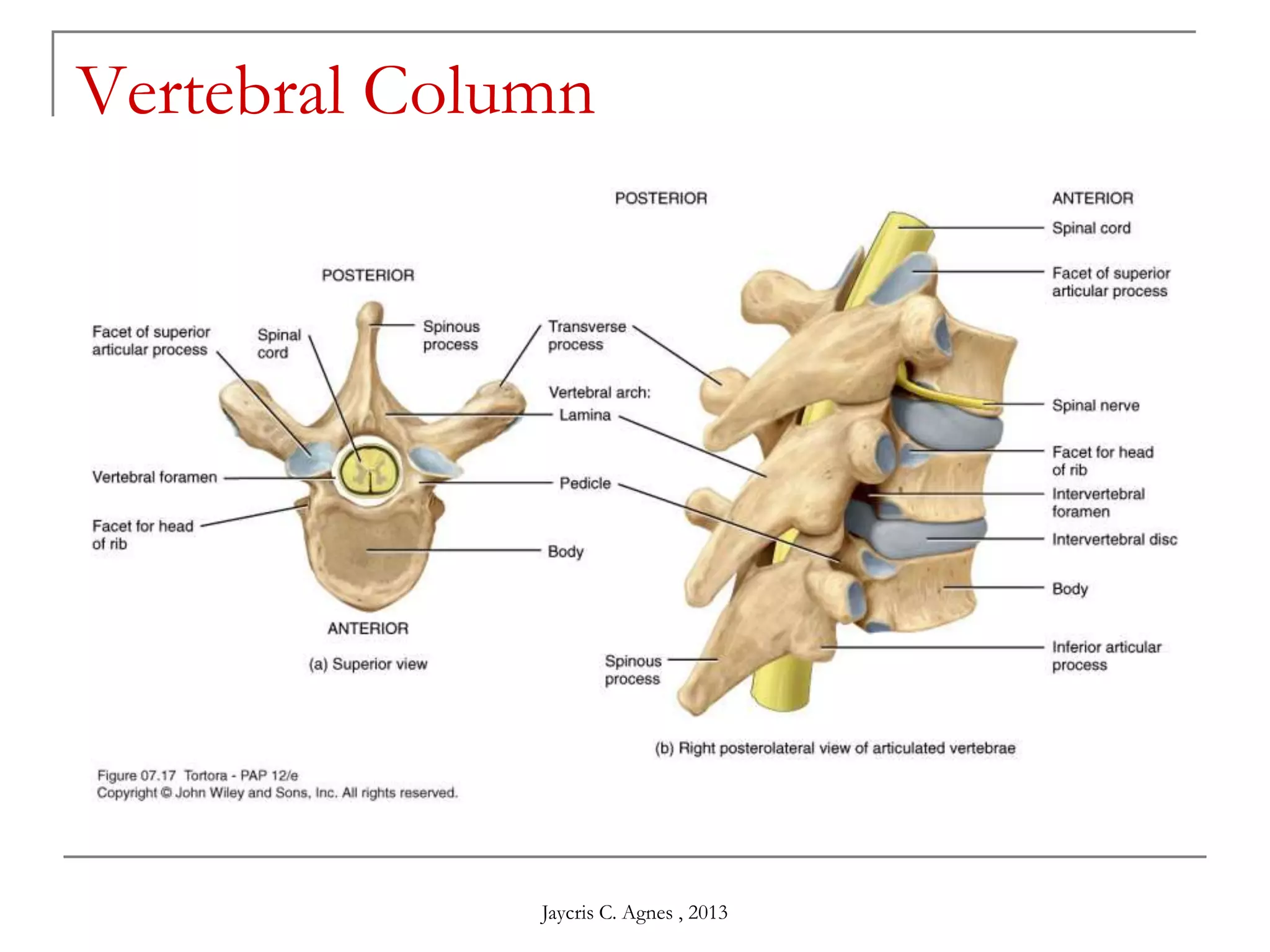
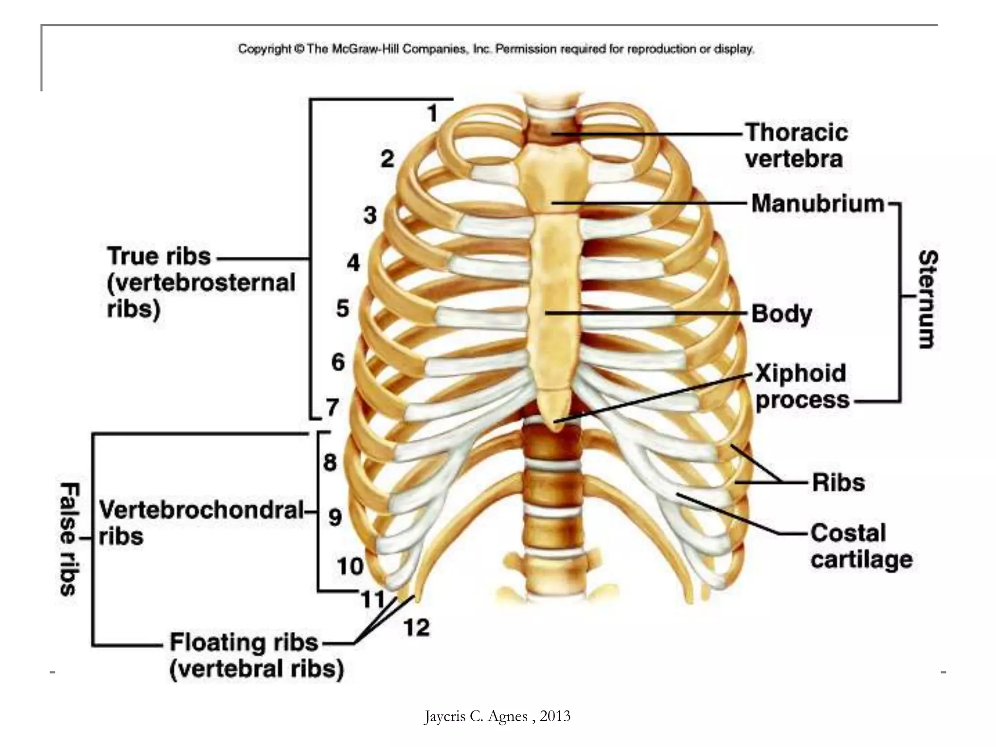
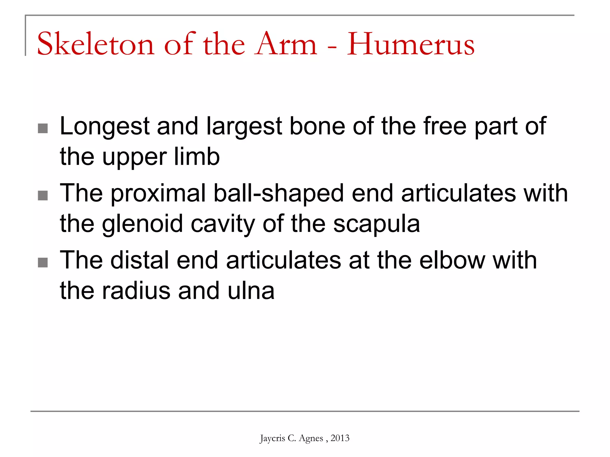
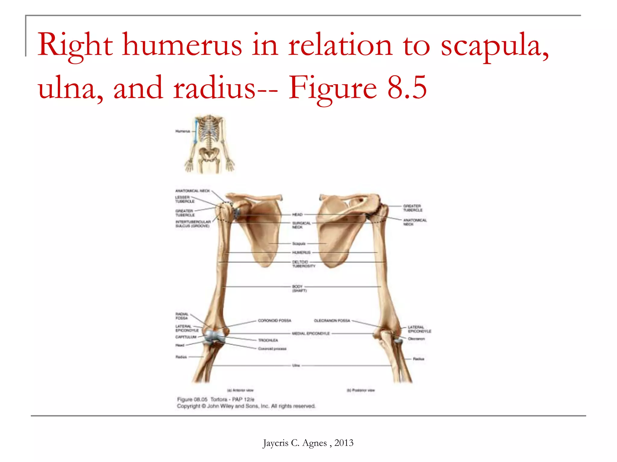
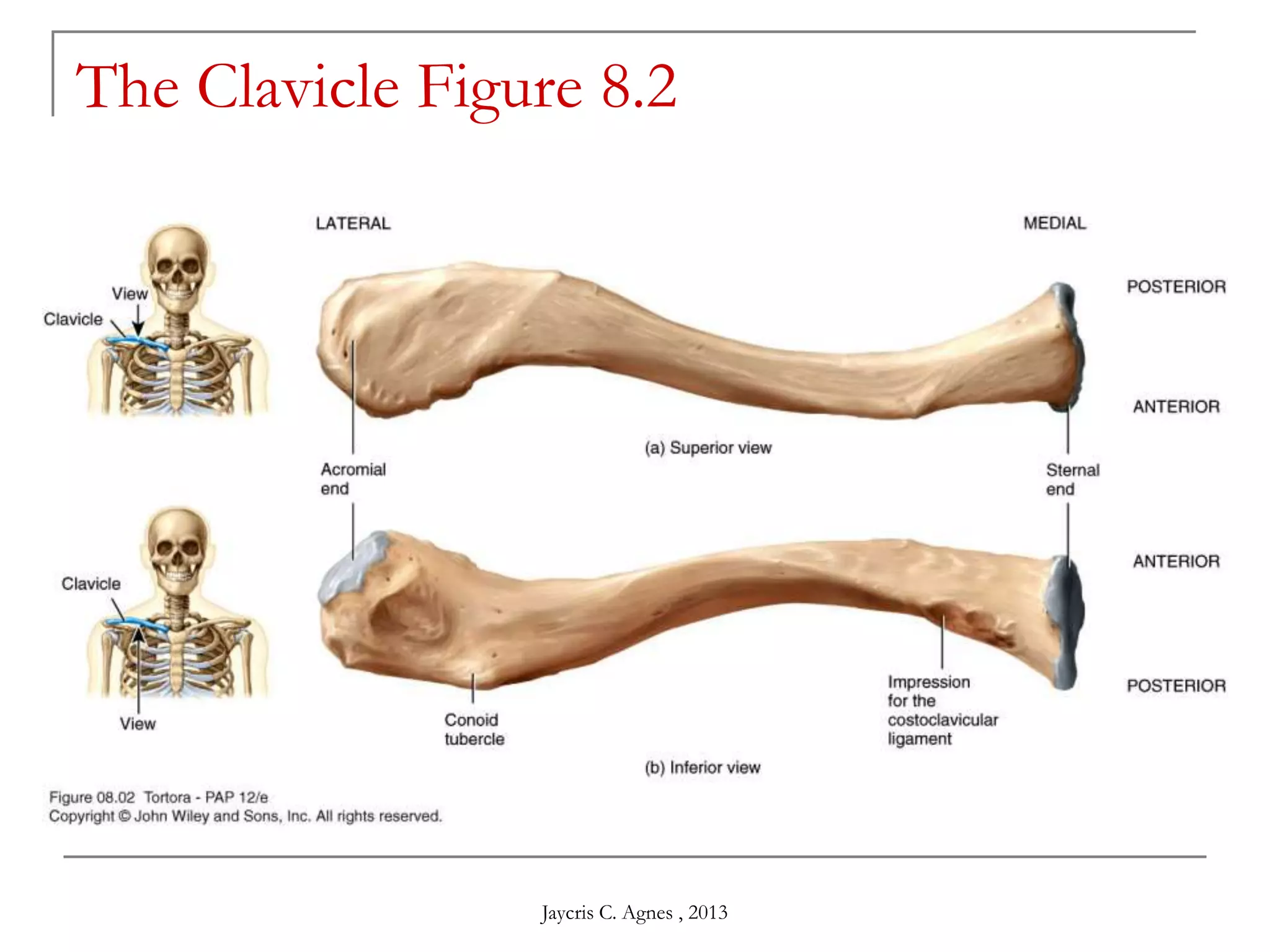
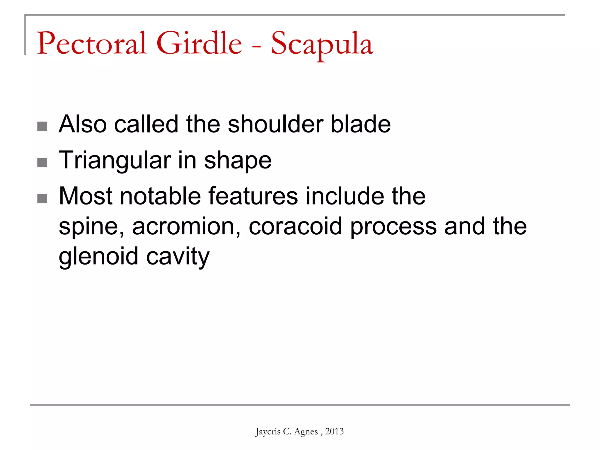
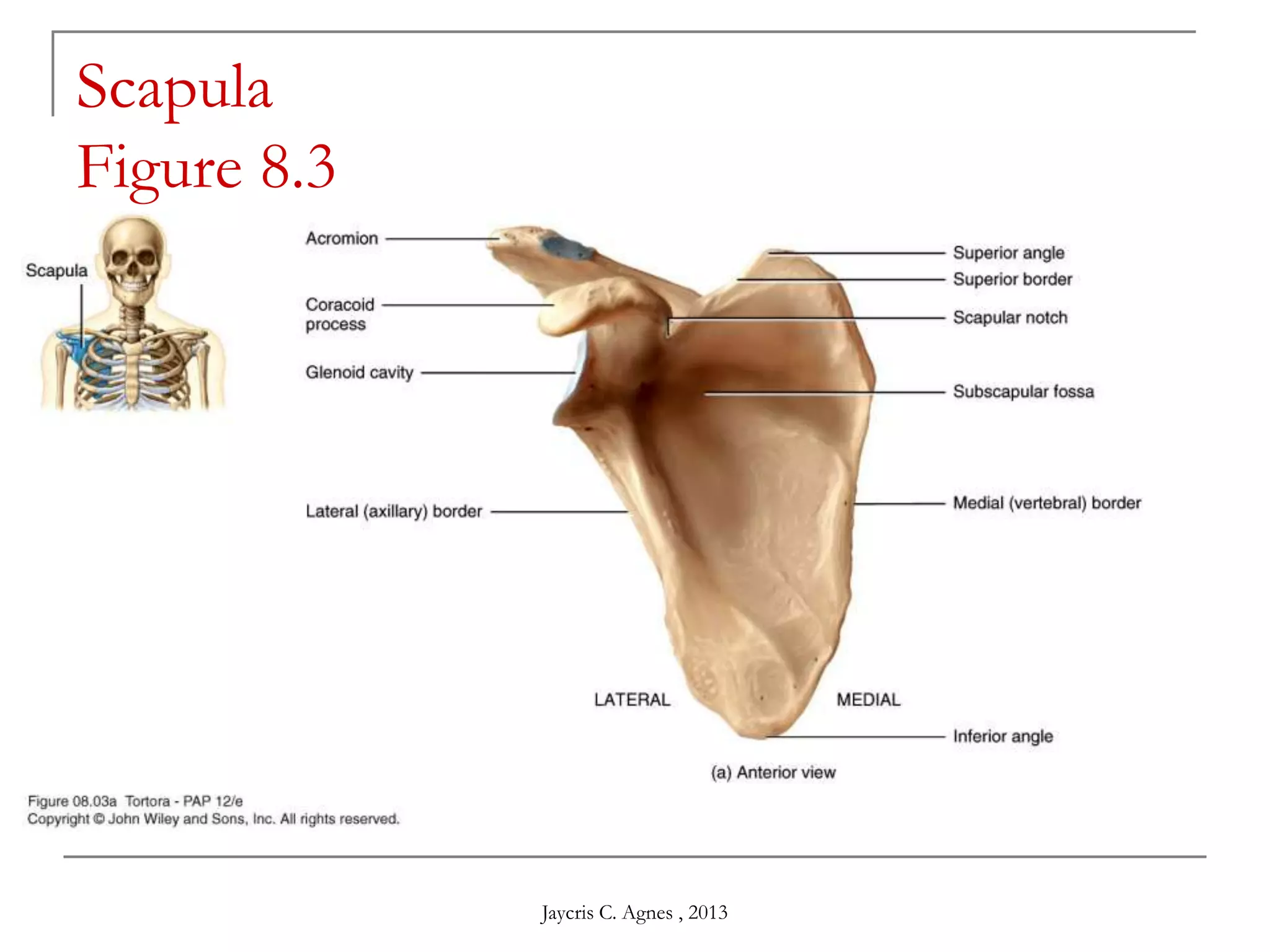
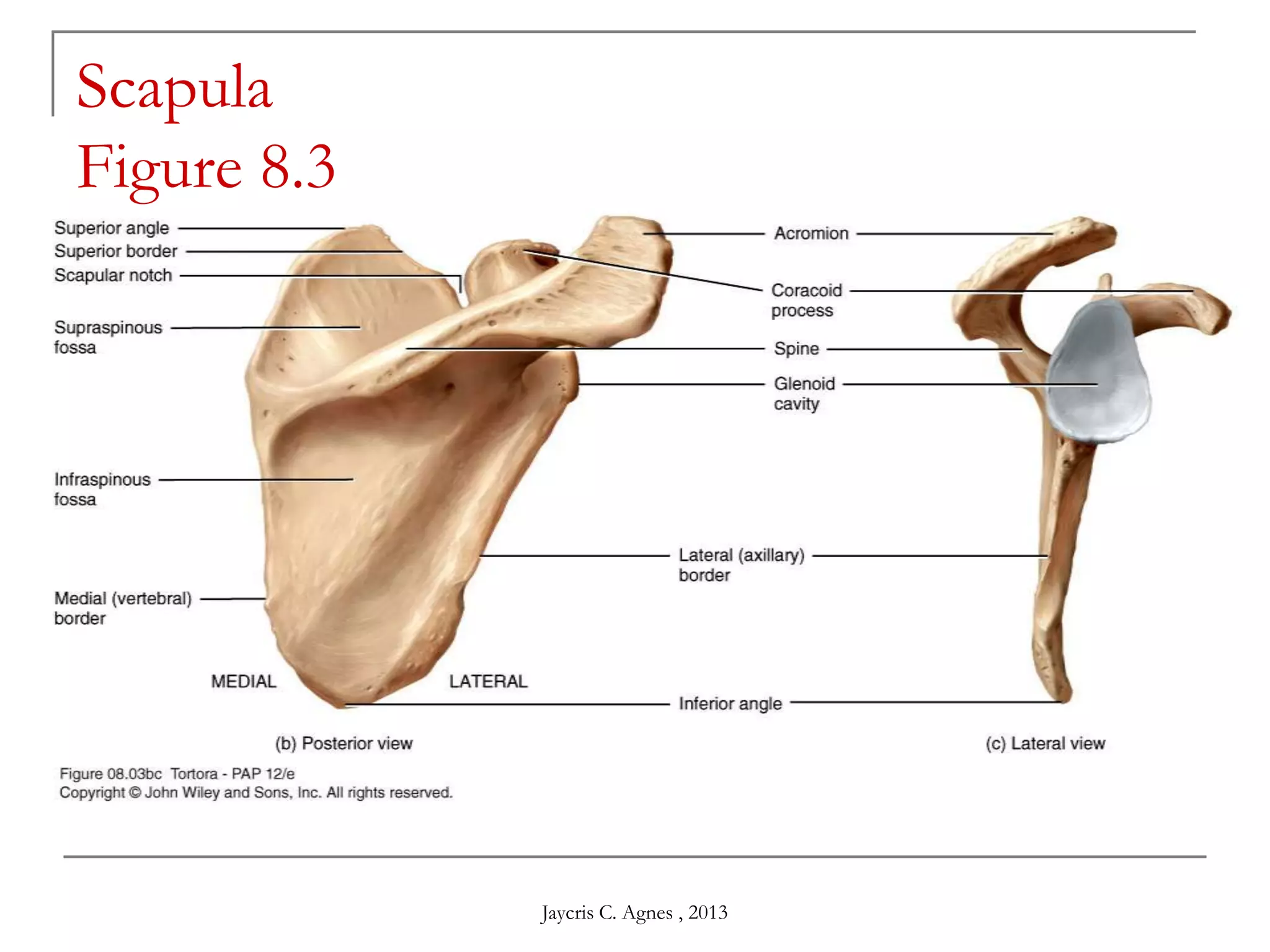
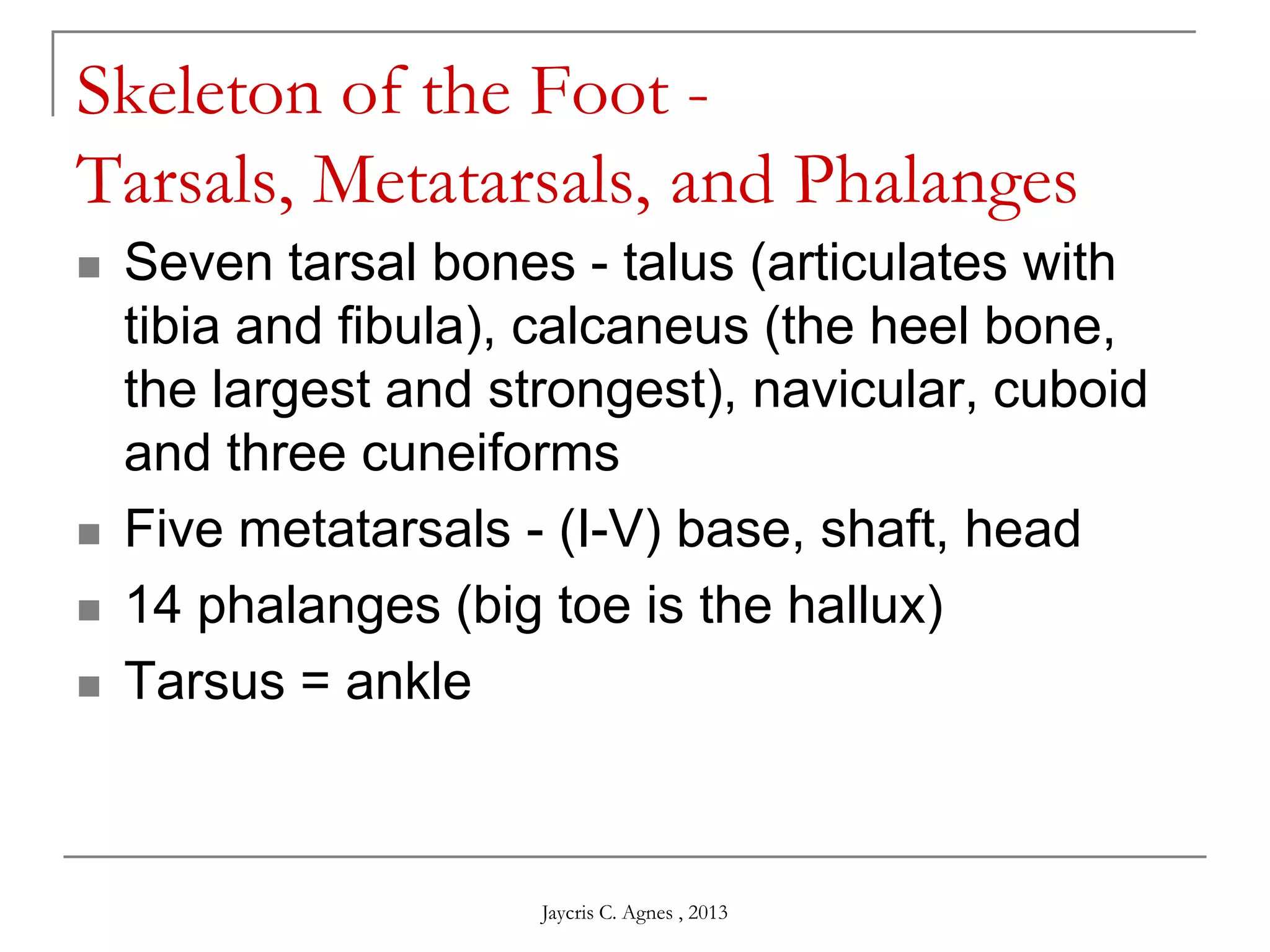
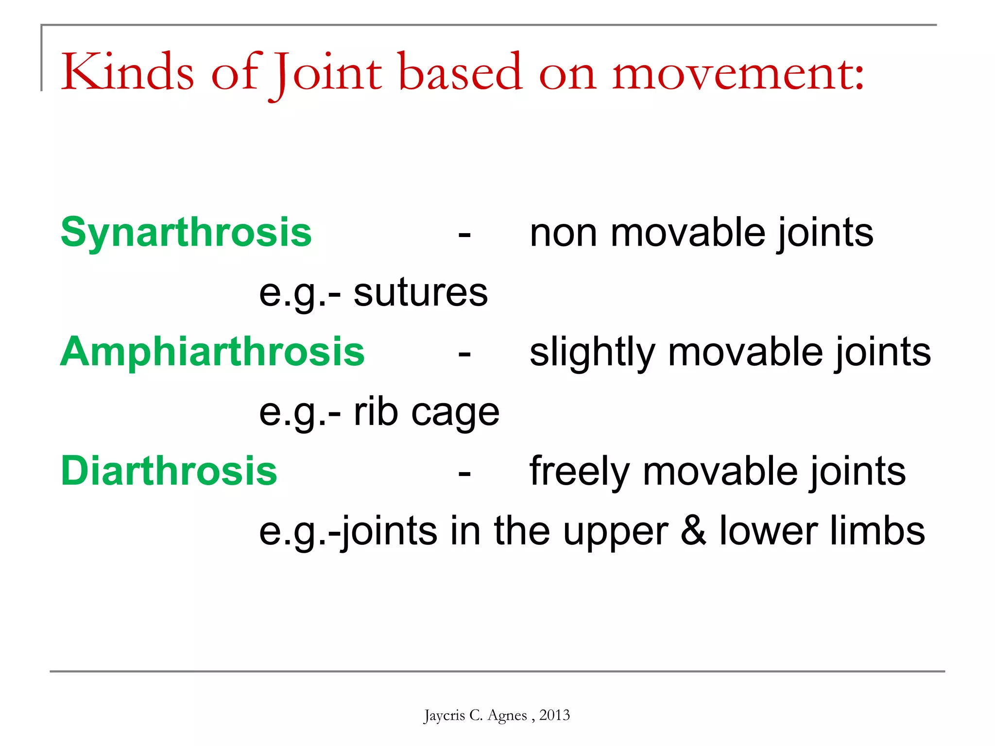
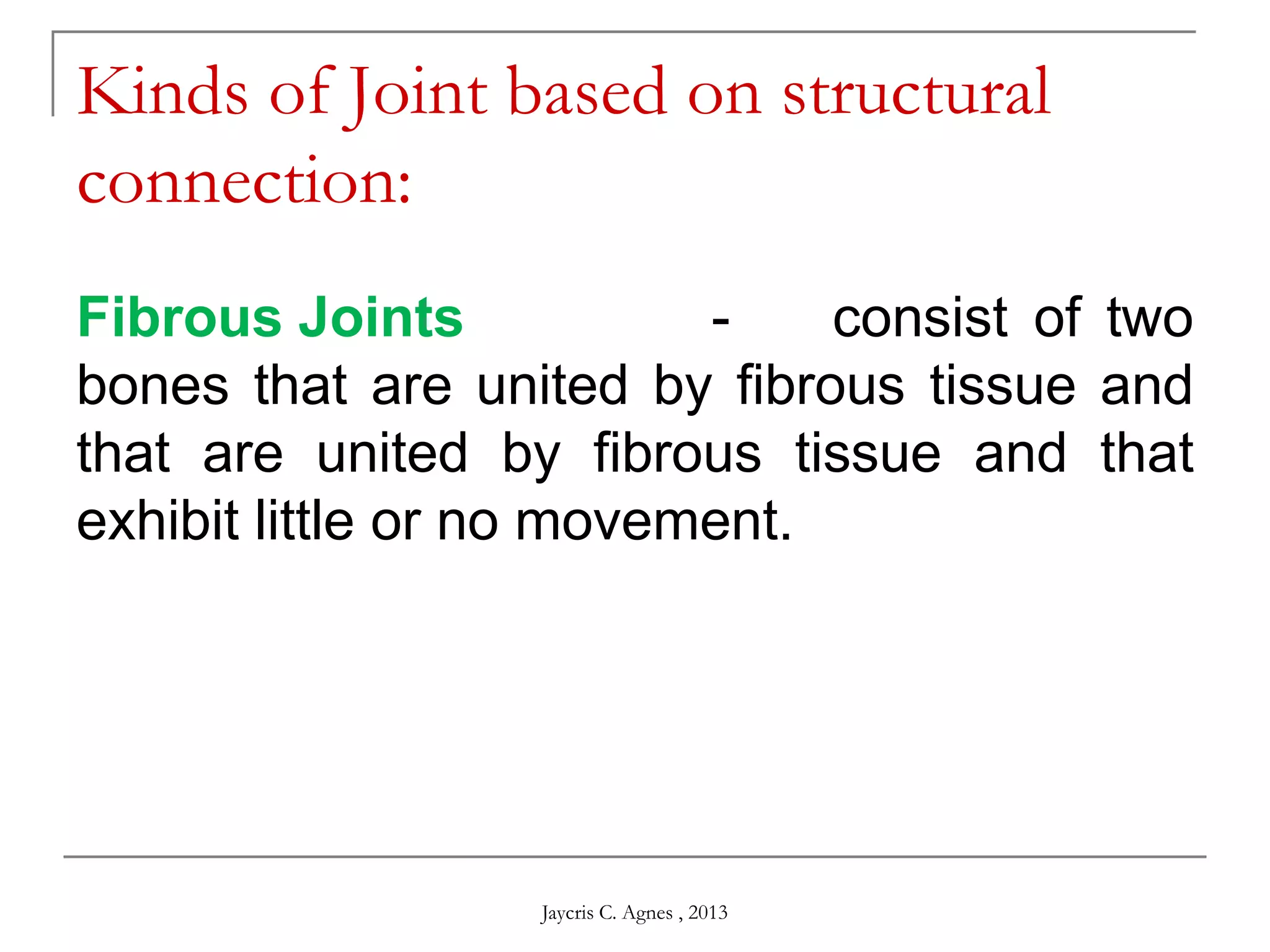
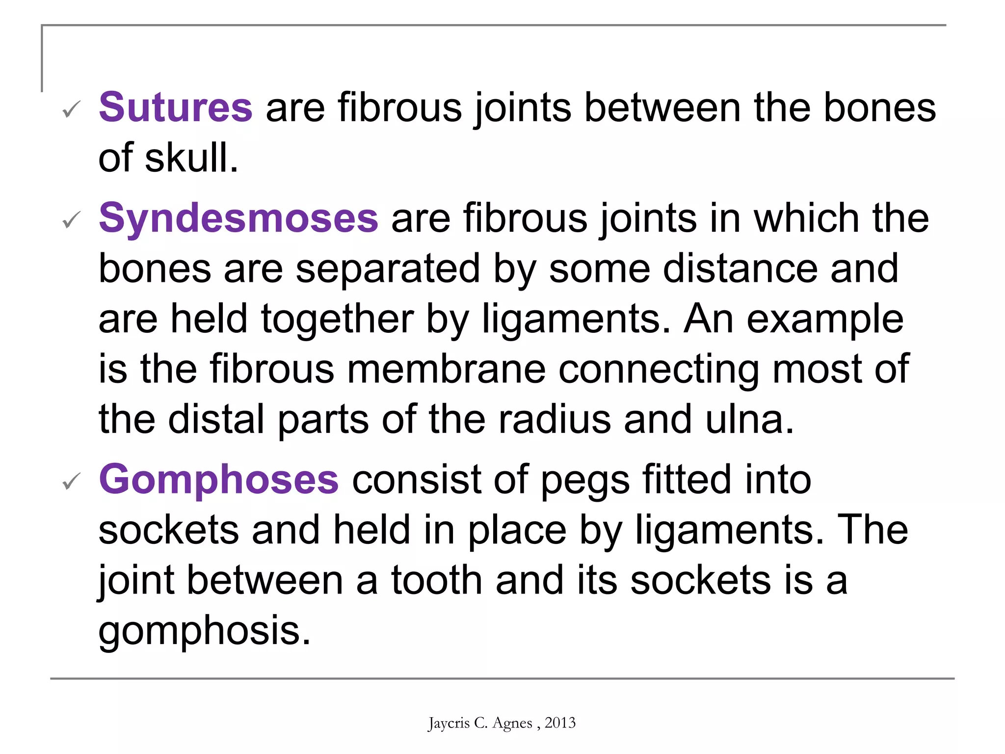
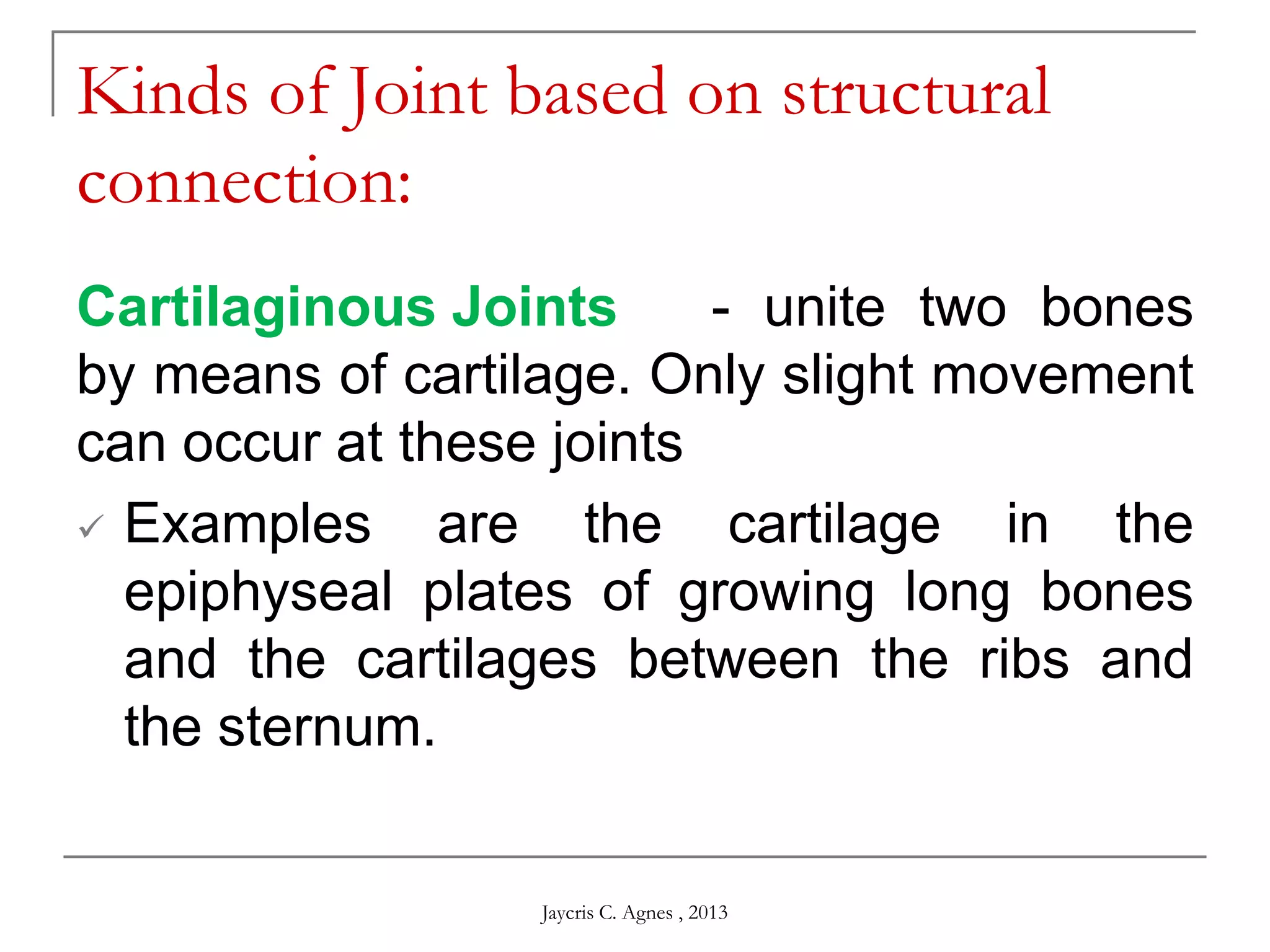
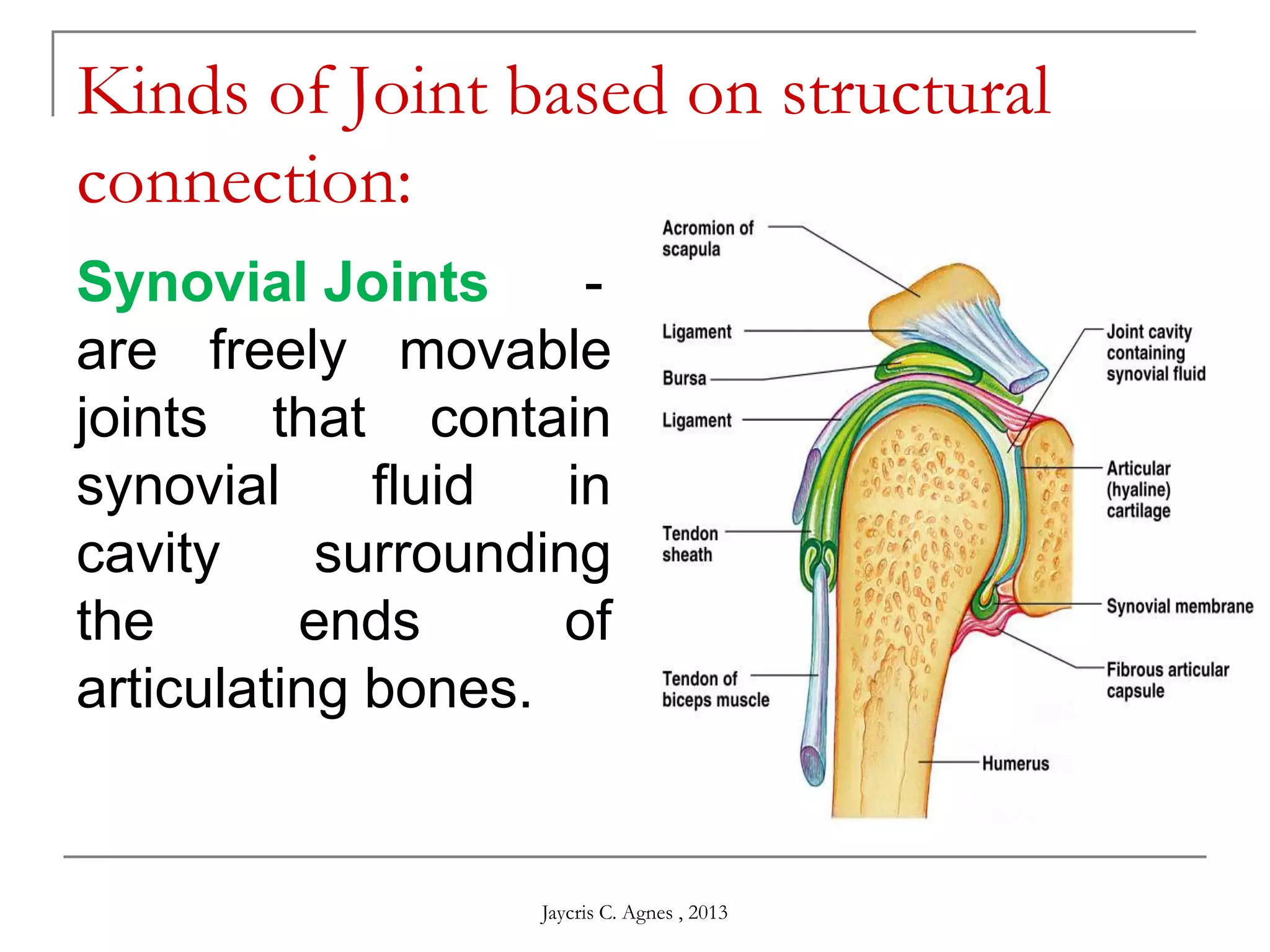
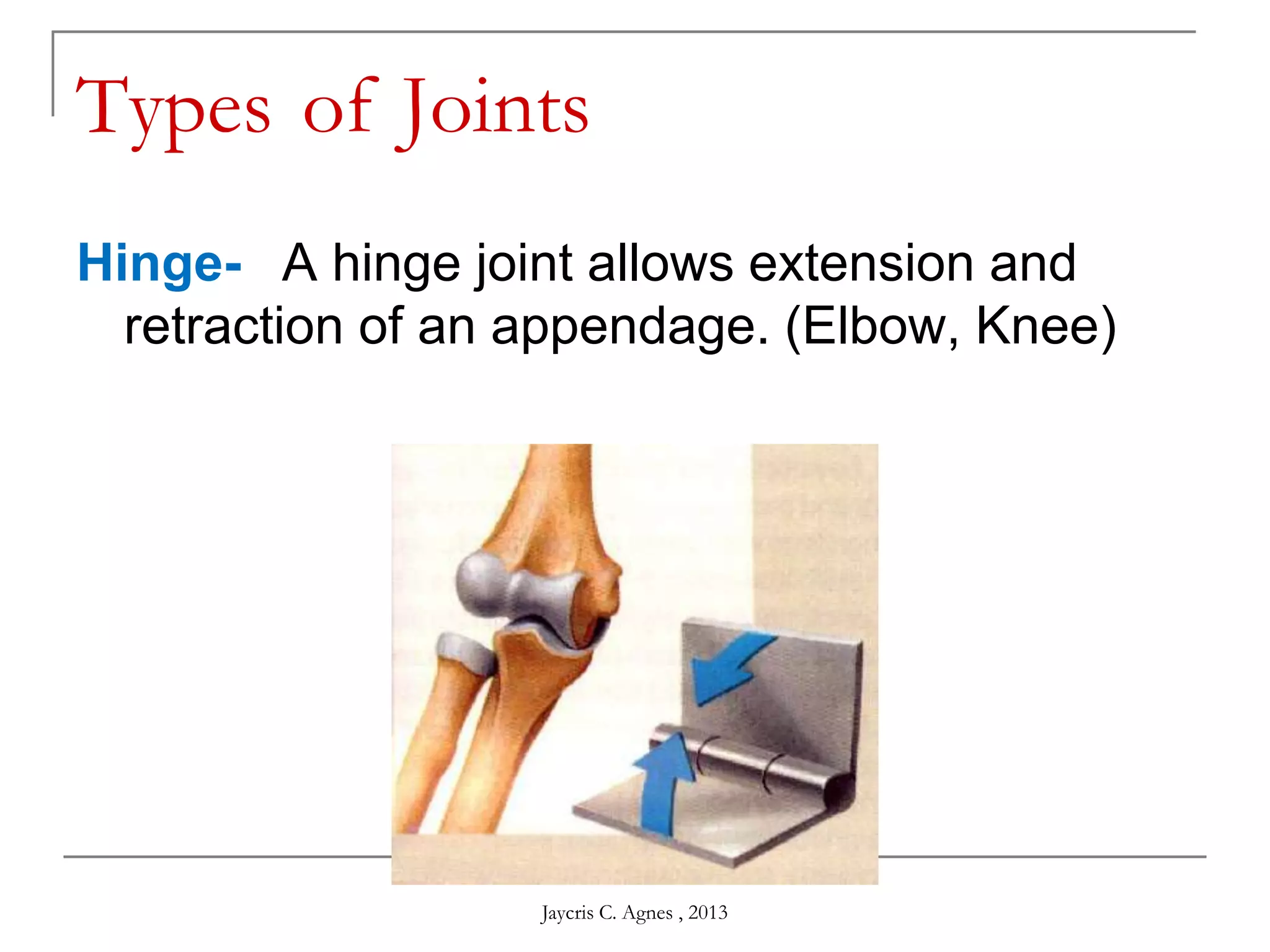
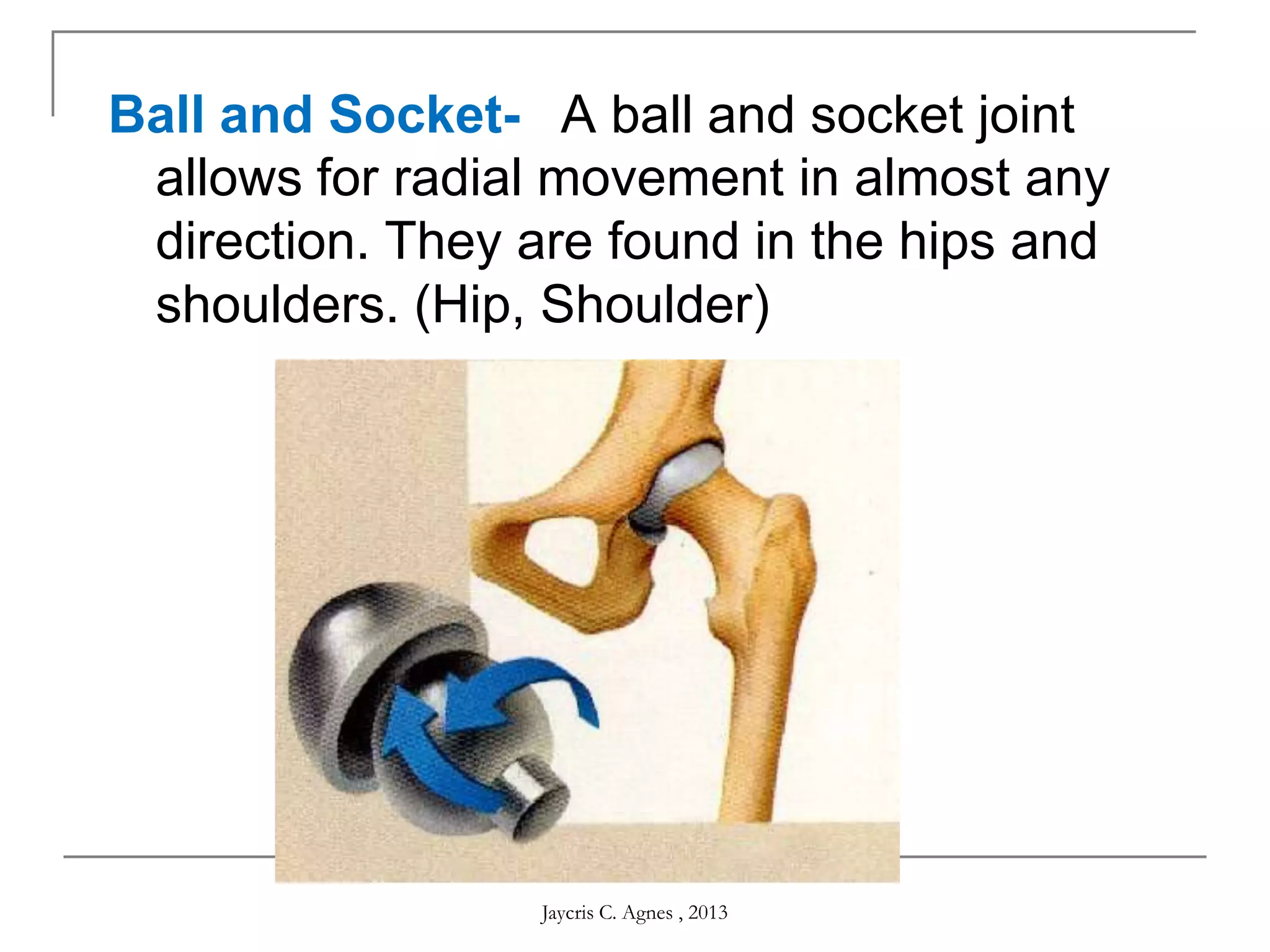
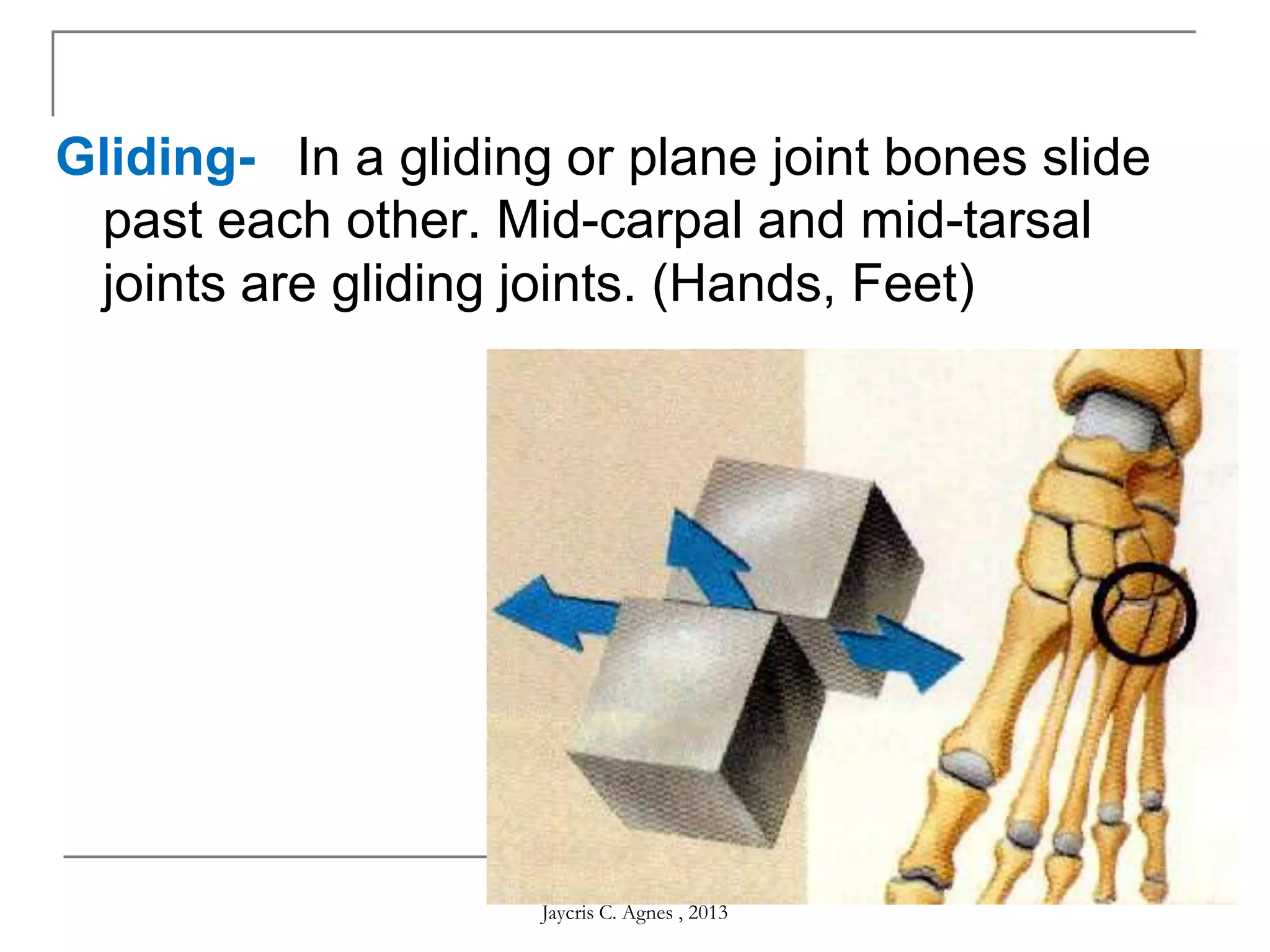
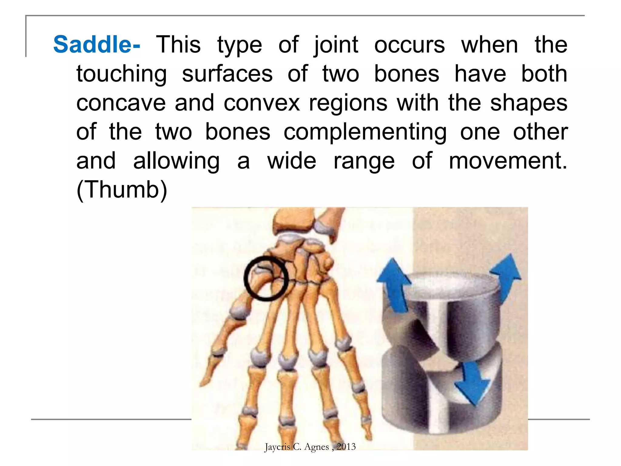
Jaycris C. Agnes
BSED- Biological Science
Skeletal System
Jaycris C. Agnes , 2013
Learning Objectives:
At the end of the discussion the students must be able to:
 Enumerate the major roles of skeletal system
and its parts,
 Explain the process of bone formation,
remodelling, repair and aging; and
 Acclaim the roles of skeletal system to
human survival.
Jaycris C. Agnes , 2013
Topic Outline:
I. Function of Skeletal System
A. Bones
B. Cartilages
C. Tendons and Ligaments
D. Joints
II. General Features of Bone
A. Parts of Bone
B. Bone Cells
D. Bone Surface Markings
E. Bone Ossification, Remodelling and Repair
F. Types of Bones
Jaycris C. Agnes , 2013
Topic Outline:
III. General Consideration of Bone Anatomy
A. Division of Human Skeleton
IV. Bone and Calcium Homeostasis
V. Articulation
VI. Effects of Aging on the Skeletal System
Jaycris C. Agnes , 2013
Functions of the Skeletal System
 Bone
 Mechanical
 Protection — bones can serve to protect
internal organs, such as the skull protecting
the brain or the ribs protecting
the heart and lungs.
 Structure — bones provide a frame to keep
the body supported.
Jaycris C. Agnes , 2013
 Movement — bones, skeletal
muscles, tendons, ligaments and joints functi
on together to generate and transfer forces
so that individual body parts or the whole
body can be manipulated in three-
dimensional space. The interaction between
bone and muscle is studied in biomechanics.
 Sound transduction — bones are important
in the mechanical aspect of
overshadowed hearing.
Jaycris C. Agnes , 2013
 Synthetic
 Blood production — the marrow, located
within the medullary cavity of long bones and
interstices of cancellous bone, produces
blood cells in a process called
hematopoiesis.
Jaycris C. Agnes , 2013
 Metabolic
 Mineral storage — bones act as reserves of
minerals important for the body, most
notably calcium and phosphorus.
 Growth factor storage — mineralized bone
matrix stores important growth factors such
as insulin-like growth factors, transforming
growth factor, bone morphogenetic
proteins and others.
Jaycris C. Agnes , 2013
 Fat storage — the yellow bone marrow acts
as a storage reserve of fatty acids.
 Acid-base balance — bone buffers the
blood against excessive pH changes by
absorbing or releasing alkaline salts.
 Detoxification — bone tissues can also
store heavy metals and other foreign
elements, removing them from the blood and
reducing their effects on other tissues. These
can later be gradually released for excretion.
Jaycris C. Agnes , 2013
 Endocrine organ — bone
controls phosphate metabolism by
releasing fibroblast growth factor – 23, which
acts on kidneys to reduce phosphate
reabsorption. Bone cells also release a
hormone called osteocalcin, which
contributes to the regulation of blood
sugar (glucose) and fat deposition.
Osteocalcin increases both
the insulin secretion and sensitivity, in
addition to boosting the number of insulin-
producing cells and reducing stores of fat.
Jaycris C. Agnes , 2013
Functions of the Skeletal System
 Cartilage
 Cartilage provides a model for bone growth
and formation, provides a smooth cushion
between adjacent bones, and provides firm
and flexible support.
Jaycris C. Agnes , 2013
Functions of the Skeletal System
 Tendons and Ligaments
 Tendons attach muscles to bones, and
ligaments attach bones to bones.
 Joints
 Joints allow movement between bones.
Jaycris C. Agnes , 2013
Compact Bone
Compact bone makes up the
outer layer of all bones. Although
it looks dense and solid, It is full
of holes for nerves and blood vessels.
Spongy Bone
Spongy bone contains flat
and needlelike structures
that resist stress. Red bone
marrow may fill the open
spaces in some bones.
Central Cavity
Central cavities in long
bones usually contain
yellow bone marrow (fat).
Outer Membrane
An outer membrane
covers most of a long bone.
The inner portion of a membrane
contains cells that build up and
breakdown bone.
General Features of the Bone
 Parts of Bone
Jaycris C. Agnes , 2013
General Features of the Bone
Jaycris C. Agnes , 2013
General Features of the Bone
 Bone Cells
There are several types of cells constituting the
typical bone:
 Osteocytes
 Mature bone cells
 Osteoblasts
 Bone-forming cells
 Osteoclasts
 Bone-destroying cells
 Break down bone matrix for remodeling and release
of calcium
Jaycris C. Agnes , 2013
 Bone Surface Markings
 Bones have characteristic surface markings
 Structural features adapted for specific functions
 There are two major types of surface markings:
 1) Depressions and openings
 Allow the passage of blood vessels and nerves or form
joints
 2) Processes/ Projections
 Projections or outgrowths that form joints or serve as
attachment points for ligaments and tendons
Jaycris C. Agnes , 2013
Joint Projections
 1) Condyle: Rounded articular projection
Condyle
Jaycris C. Agnes , 2013
Joint Projections
 2) Head: bony
expansion on a
narrow neck
 3) Facet: smooth,
nearly flat articular
surface
Jaycris C. Agnes , 2013
Joint Projections
 4) Ramus: Armlike bar of bone
Jaycris C. Agnes , 2013
Ligament/Tendon Projections
1) Crest: Narrow ridge
of bone (Line: smaller
than a crest)
2) Epicondyle: Raised
area on or above a
condyle
ULNA
Jaycris C. Agnes , 2013
3) Tubercle: Small
rounded projection
4) Tuberosity: large
rounded or
roughened projection
5) Trochanter: very
large, blunt projection
(only on femur)
Proximal Tibia
Jaycris C. Agnes , 2013
6) Spine: Sharp, pointed
projection
Thoracic Vertebrae
Jaycris C. Agnes , 2013
DEPRESSIONS
 Allow blood vessels or nerves to pass
through.
1) Meatus: (me - A- tus) Canal or tube
Jaycris C. Agnes , 2013
Depressions
2) Fossa: shallow basin
3) Fissure: narrow, slit-
like opening
Jaycris C. Agnes , 2013
Depressions
4) Sinus: Cavity within a
bone; filled with air
and lined with
mucous membranes
5) Foramen: Round or
oval opening
Foramen Magnum
Jaycris C. Agnes , 2013
Depressions
6) Sulcus, Groove or Furrow: a shallow
depression
Jaycris C. Agnes , 2013
Bone Markings
Projections
1) Condyle
2) Head
3) Facet
4) Ramus
5) Crest
6) Epicondyle
7) Tubercle
8) Tuberosity
9) Trochanter
10) Spine
Depressions
1) Meatus
2) Foramen
3) Fossa
4) Fissure
5) Sinus
6) Sulcus or Groove or
Furrow
Jaycris C. Agnes , 2013
Bone Ossification
 The formation of bone during the fetal stage
of development occurs by two
processes: Intramembranous
Ossification and Endochondral
Ossification.
Jaycris C. Agnes , 2013
Intramembranous Ossification
Intramembranous ossification mainly occurs
during formation of the flat bones of
the skull but also the mandible, maxilla, and
clavicles; the bone is formed from
connective tissue such
as mesenchyme tissue rather than from
cartilage.
Jaycris C. Agnes , 2013
Intramembranous Ossification
 The steps in intramembranous ossification
are:
1. Development of ossification center.
2. Calcification.
3. Formation of trabeculae.
4. Development of periosteum.
Jaycris C. Agnes , 2013
Endochondral Ossification
Endochondral ossification, on the other
hand, occurs in long bones and most of the
rest of the bones in the body; it involves an
initial hyaline cartilage that continues to
grow.
Jaycris C. Agnes , 2013
Endochondral Ossification
 The steps in endochondral ossification
are:
1. Development of cartilage model
2. Growth of cartilage model
3. Development of the primary ossification center
4. Development of the secondary ossification
center
5. Formation of articular cartilage and epiphyseal
plate
Jaycris C. Agnes , 2013
Bone Growth
 Does drinking milk can make you
taller?
Jaycris C. Agnes , 2013
Bone Remodelling
Bone remodelling consists of removal of
existing bone by osteoclasts and deposition
of new bone by osteoblasts.
Jaycris C. Agnes , 2013
Bone Repair
Jaycris C. Agnes , 2013
 Types of Bones
 Bones can be classified into five types based on
shape:
 Long
 Short
 Flat
 Irregular
 Sesamoid
Jaycris C. Agnes , 2013
 Types of Bones
 Long Bones
 Greater length than width and are slightly curved for strength
 Femur, tibia, fibula, humerus, ulna, radius, phalanges
 Short bones
 Cube-shaped and are nearly equal in length and width
 Carpal, tarsal
 Flat bones
 Thin and composed of two nearly parallel plates of compact bone tissue
enclosing a layer of spongy bone tissue
 Cranial, sternum, ribs, scapulae
 Irregular bones
 Complex shapes and cannot be grouped into any of the previous
categories
 Vertebrae, hip bones, some facial bones, calcaneus
 Sesamoid bones
 Protect tendons from excessive wear and tear
 Patellae, foot, hand
Jaycris C. Agnes , 2013
General Consideration of Bone Anatomy
 The human skeleton consists of 206 named bones
 Bones of the skeleton are grouped into two principal
divisions:
 Axial skeleton
 Consists of the bones that lie around the longitudinal axis of
the human body
 Skull bones, auditory ossicles (ear bones), hyoid
bone, ribs, sternum (breastbone), and bones of the vertebral
column
 Appendicular skeleton
 Consists of the bones of the upper and lower limbs
(extremities), plus the bones forming the girdles that connect
the limbs to the axial skeleton
Jaycris C. Agnes , 2013
Divisions of the Skeletal System
Jaycris C. Agnes , 2013
Divisions of the Skeletal System
Jaycris C. Agnes , 2013
The Axial Skeleton
Jaycris C. Agnes , 2013
Skull
 Skull (cranium)
 Consists of 22 bones
 Bones of the skull are grouped into two
categories:
 Cranial bones
 Eight cranial bones form the cranial cavity
 Frontal bone, two parietal bones, two temporal bones, the
occipital bone, the sphenoid bone, ethmoid bone
 Facial bones
 Fourteen facial bones form the face
 Two nasal bones, two maxillae, two zygomatic bones, the
mandible, two lacrimal bones, two palatine bones, two inferior
nasal conchae, vomer
Jaycris C. Agnes , 2013
Skull
Jaycris C. Agnes , 2013
Skull
 The cranial and facial bones protect and support
special sense organs and the brain
 Besides forming the large cranial cavity, the skull
also forms several smaller cavities
 Nasal cavity
 Orbits (eye sockets)
 Paranasal sinuses
 Small cavities which house organs involved in hearing
and equilibrium
Jaycris C. Agnes , 2013
Skull
 Immovable joints called sutures fuse most of the
skull bones together
 The skull provides large areas of attachment for
muscles that move various parts of the head
 Skull and facial bones provide attachment for
muscles that produce facial expressions
 The facial bones form the framework of the face
and provide support for the entrances to the
digestive and respiratory systems
Jaycris C. Agnes , 2013
Skull (Cranial Bones)
 Frontal Bone
 Forms the forehead
 Parietal Bones
 Form the sides and roof of the cranial cavity
 Temporal Bones
 Form the lateral aspects and floor of the cranium
 Occipital Bone
 Forms the posterior part and most of the base of the cranium
 Sphenoid Bone
 Lies at the middle part of the base of the skull
 Ethmoid Bone
 Located on the midline in the anterior part of the cranial floor medial to
the orbits
 A major superior supporting structure of the nasal cavity
 Contain thin projections called conchae which are lined by mucous
membranes
 Increased surface area in the nasal cavity helps to humidify inhaled air
trapping inhaled particles
Jaycris C. Agnes , 2013
Skull
Jaycris C. Agnes , 2013
Skull
Jaycris C. Agnes , 2013
Skull
Jaycris C. Agnes , 2013
Skull (Facial Bones)
 Nasal Bones
 Form the bridge of the nose
 Maxillae
 Form the upper jawbone
 Form most of the hard palate
 Separates the nasal cavity from the oral cavity
 Zygomatic Bones
 commonly called cheekbones, form the prominences of the
cheeks
 Lacrimal Bones
 Form a part of the medial wall of each orbit
 Palatine Bones
 Form the posterior portion of the hard palate
 Inferior Nasal Conchae
 Form a part of the inferior lateral wall of the nasal cavity
Jaycris C. Agnes , 2013
Skull (Facial Bones)
 Vomer
 Forms the inferior portion of the nasal septum
 Mandible
 Lower jawbone
 The largest, strongest facial bone
 The only movable skull bone
 Nasal Septum
 Divides the interior of the nasal cavity into right and left sides
 ―Broken nose,‖ in most cases, refers to septal damage rather
than the nasal bones themselves
 Orbits
 Eye socket
 Foramina
 Openings for blood vessels , nerves , or ligaments of the skull
Jaycris C. Agnes , 2013
Skull
Jaycris C. Agnes , 2013
Skull
Jaycris C. Agnes , 2013
Skull
Jaycris C. Agnes , 2013
Skull
Jaycris C. Agnes , 2013
Skull
Jaycris C. Agnes , 2013
Skull
Jaycris C. Agnes , 2013
Skull
 Unique Features of the Skull
 Sutures, Paranasal sinuses, Fontanels
 Sutures
 an immovable joint that holds most skull bones together
 Paranasal Sinuses
 Cavities within cranial and facial bones near the nasal cavity
 Secretions produced by the mucous membranes which line the sinuses,
drain into the nasal cavity
 Serve as resonating chambers that intensify and prolong sounds
 Fontanels
 Areas of unossified tissue
 At birth, unossified tissue spaces, commonly called ―soft spots‖ link the
cranial bones
 Eventually, they are replaced with bone to become sutures
 Provide flexibility to the fetal skull, allowing the skull to change shape as
it passes through the birth canal
Jaycris C. Agnes , 2013
Skull
Jaycris C. Agnes , 2013
Skull
Jaycris C. Agnes , 2013
Auditory Ossicles
 The middle ear has 3 tiny bones: the
Malleus, Incus and Stapes.
 Stapes is the smallest bone in the body
 Helps in hearing.
Jaycris C. Agnes , 2013
Auditory Ossicles
Jaycris C. Agnes , 2013
Auditory Ossicles
Jaycris C. Agnes , 2013
Hyoid Bone
 Does not articulate with
any other bone
 Supports the tongue,
providing attachment
sites for some tongue
muscles and for
muscles of the neck and
pharynx
 The hyoid bone also
helps to keep the larynx
(voice box) open at all
times
Jaycris C. Agnes , 2013
Vertebral Column
 Also called the spine, backbone, or spinal
column
 Functions to:
 Protect the spinal cord
 Support the head
 Serve as a point of attachment for the ribs, pelvic
girdle, and muscles
 The vertebral column is curved to varying
degrees in different locations
 Curves increase the column strength
 Help maintain balance in the upright position
 Absorb shocks during walking, and help protect the
vertebrae from fracture
Jaycris C. Agnes , 2013
Vertebral Column
Jaycris C. Agnes , 2013
Vertebral Column
 Composed of a series of bones called
vertebrae (Adult=26)
 7 cervical are in the neck region
 12 thoracic are posterior to the thoracic cavity
 5 lumbar support the lower back
 1 sacrum consists of five fused sacral
vertebrae
 1 coccyx consists of four fused coccygeal
vertebrae
Jaycris C. Agnes , 2013
Vertebral Column (Regions)
 Cervical Region
 Cervical vertebrae (C1–C7)
 The atlas (C1) is the first cervical vertebra
 The axis (C2) is the second cervical vertebra
 Thoracic Region
 Thoracic vertebrae (T1–T12)
 Articulate with the ribs
 Lumbar Region
 Lumbar vertebrae (L1–L5)
 Provide for the attachment of the large back muscles
 Sacrum
 The sacrum is a triangular bone formed by the union of five sacral
vertebrae (S1–S5)
 Serves as a strong foundation for the pelvic girdle
 Coccyx
 The coccyx, like the sacrum, is triangular in shape
 It is formed by the fusion of usually four coccygeal vertebrae
Jaycris C. Agnes , 2013
Vertebral Column
Jaycris C. Agnes , 2013
Vertebral Column
Jaycris C. Agnes , 2013
Vertebral Column
Jaycris C. Agnes , 2013
Vertebral Column
Jaycris C. Agnes , 2013
Vertebral Column
Jaycris C. Agnes , 2013
Vertebral Column
Jaycris C. Agnes , 2013
Thorax
 Thoracic cage is formed by the:
 Sternum
 Ribs
 Costal cartilages
 Thoracic vertebrae
 Functions to:
 Enclose and protect the organs in the thoracic
and abdominal cavities
 Provide support for the bones of the upper
limbs
 Play a role in breathing
Jaycris C. Agnes , 2013
Thorax
 Sternum
 ―Breastbone‖ located in the center of the
thoracic wall
 Consists of the manubrium, body, xiphoid
process
 Ribs
 Twelve pairs of ribs give structural support to
the sides of the thoracic cavity
 Costal cartilages
 Costal cartilages contribute to the elasticity of
the thoracic cage
Jaycris C. Agnes , 2013
Thorax
Jaycris C. Agnes , 2013
The Appendicular Skeleton
Jaycris C. Agnes , 2013
Appendicular Skeleton
 The primary function is movement
 It includes bones of the upper and lower
limbs
 Girdles attach the limbs to the axial skeleton
Jaycris C. Agnes , 2013
Skeleton of the Upper Limb
 Each upper limb has 30 bones
 The upper limb consists of the bones of the
arm, forearm, wrist, and hand.
Jaycris C. Agnes , 2013
Upper Limb
 1 humerus (arm)
 1 ulna (forearm)
 1 radius (forearm)
 8 carpals (wrist)
 5 metacarpal and 14 phalanges (hand)
Jaycris C. Agnes , 2013
Skeleton of the Arm - Humerus
 Longest and largest bone of the free part of
the upper limb
 The proximal ball-shaped end articulates with
the glenoid cavity of the scapula
 The distal end articulates at the elbow with
the radius and ulna
Jaycris C. Agnes , 2013
Humerus and Glenohumeral Joint
Figure 8.4
Jaycris C. Agnes , 2013
Right humerus in relation to scapula,
ulna, and radius-- Figure 8.5
Jaycris C. Agnes , 2013
Right ulna and radius in relation to the
humerus and carpals -- Figure 8.6
Skeleton of the Hand
 The carpus (wrist) consists of 8 small bones
(carpals)
 Two rows of carpal bones (So Long Top Part, Here
Comes The Thumb)
 Proximal row (lateral-medial) - scaphoid, lunate,
triquetrum, pisiform
 Distal row (medial-lateral) - hamate ,
capitate,trapezoid, trapezium
 Scaphoid - most commonly fractured
 Carpal tunnel - space between carpal bones and
flexor retinaculum
Jaycris C. Agnes , 2013
Metacarpals and Phalanges
 Five metacarpals - numbered I-V, lateral to
medial
 14 phalanges - two in the thumb (pollex) and
three in each of the other fingers
 Each phalanx has a base, shaft, and head
 Joints - carpometacarpal,
metacarpophalangeal, interphalangeal
Jaycris C. Agnes , 2013
Right wrist and hand in relation to ulna
and radius -- Figure 8.8
Jaycris C. Agnes , 2013
Pectoral Girdle - Clavicle
 The clavicle is ―S‖ shaped
 The medial end articulates with the
manubrium of the sternum forming the
sternoclavicular joint
 The lateral end articulates with the acromion
forming the acromioclavicular joint
Jaycris C. Agnes , 2013
The Pectoral (or Shoulder) Girdle
Figure 8.1
Jaycris C. Agnes , 2013
The Clavicle Figure 8.2
Jaycris C. Agnes , 2013
Pectoral Girdle - Scapula
 Also called the shoulder blade
 Triangular in shape
 Most notable features include the
spine, acromion, coracoid process and the
glenoid cavity
Jaycris C. Agnes , 2013
Scapula
Figure 8.3
Jaycris C. Agnes , 2013
Scapula
Figure 8.3
Jaycris C. Agnes , 2013
Skeleton of the Lower Limb
 Skeleton of the Lower Limb
 Consist of bones of thigh, leg, ankle and foot.
 Each lower limb has 30 bones.
Jaycris C. Agnes , 2013
Pelvic (Hip) Girdle
 Each coxal (hip) bone consists of three bones
that fuse together: ilium, pubis, and ischium
 The two coxal bones are joined anteriorly by
the pubic symphysis (fibrocartilage)
 Joined posteriorly by the sacrum forming the
sacroiliac joints (Fig 8.9)
Jaycris C. Agnes , 2013
Bony Pelvis Figure 8.9
Jaycris C. Agnes , 2013
Right Hip Bone Figure 8.10
Comparing Male and Female Pelves
 Males - bone are larger and heavier
 Pelvic inlet is smaller and heart shaped
 Pubic arch is less the 90°
 Female - wider and shallower
 Pubic arch is greater than 90°
 More space in the true pelvis (Table 8.1)
Jaycris C. Agnes , 2013
Comparing Male and Female Pelves
Table 8.1
Jaycris C. Agnes , 2013
Comparing Male and Female Pelves
Table 8.1
Right Lower Limb
Figure 8.12
Jaycris C. Agnes , 2013
Skeleton of the Thigh - Femur and Patella
 Femur - longest, heaviest, and strongest bone in the
body
 Proximally, the head articulates with the acetabulum
of the hip bone forming the hip (coxal) joint
 Neck - distal to head, common site of fracture
 Distally, the medial and lateral condyles articulate
with the condyles of the tibia forming the knee joint
 Also articulates with patella
Jaycris C. Agnes , 2013
Femur
 Greater and lesser trochanters are projections
where large muscles attach
 Gluteal tuberosity and linea aspera - attachment
sites for the large hip muscles
 Intercondylar fossa - depression between the
condyles
 Medial and lateral epicondyles - muscle site
attachments for the knee muscles
Jaycris C. Agnes , 2013
Right Femur Figure 8.13
Patella
 Largest sesamoid bone in the body
 Forms the patellofemoral joint
 Superior surface is the base
 Inferior, narrower surface is the apex
 Thick articular cartilage lines the posterior
surface
 Increases the leverage of the quadriceps
femoris muscle
 Patellofemoral stress syndrome - ―runner’s
knee‖
Jaycris C. Agnes , 2013
Patella Figure 8.14
Jaycris C. Agnes , 2013
Tibia and Fibula Figure 8.15
Jaycris C. Agnes , 2013
Tibia and Fibula Figure 8.15
Jaycris C. Agnes , 2013
Skeleton of the Foot -
Tarsals, Metatarsals, and Phalanges
 Seven tarsal bones - talus (articulates with
tibia and fibula), calcaneus (the heel bone,
the largest and strongest), navicular, cuboid
and three cuneiforms
 Five metatarsals - (I-V) base, shaft, head
 14 phalanges (big toe is the hallux)
 Tarsus = ankle
Jaycris C. Agnes , 2013
Right Foot Figure 8.16
Jaycris C. Agnes , 2013
Bone and Calcium Homeostasis
 When blood calcium levels are too low,
osteoclast activity increases.
 When blood calcium levels are too high
osteoblast activity decreases.
Jaycris C. Agnes , 2013
Bone and Calcium Homeostasis
 Parathyroid hormones (PTH) – stimulates
increased bone breakdown and increased
blood calcium levels by indirectly stimulating
osteoclast activities.
 Calcitonin – decreases osteoclast activity and
thus decreases blood calcium level.
Jaycris C. Agnes , 2013
Articulation
An articulation, or
joint, is a place where
two bones come
together.
Jaycris C. Agnes , 2013
Kinds of Joint based on movement:
Synarthrosis - non movable joints
e.g.- sutures
Amphiarthrosis - slightly movable joints
e.g.- rib cage
Diarthrosis - freely movable joints
e.g.-joints in the upper & lower limbs
Jaycris C. Agnes , 2013
Kinds of Joint based on structural
connection:
Fibrous Joints - consist of two
bones that are united by fibrous tissue and
that are united by fibrous tissue and that
exhibit little or no movement.
Jaycris C. Agnes , 2013
 Sutures are fibrous joints between the bones
of skull.
 Syndesmoses are fibrous joints in which the
bones are separated by some distance and
are held together by ligaments. An example
is the fibrous membrane connecting most of
the distal parts of the radius and ulna.
 Gomphoses consist of pegs fitted into
sockets and held in place by ligaments. The
joint between a tooth and its sockets is a
gomphosis.
Jaycris C. Agnes , 2013
Cartilaginous Joints - unite two bones
by means of cartilage. Only slight movement
can occur at these joints
 Examples are the cartilage in the
epiphyseal plates of growing long bones
and the cartilages between the ribs and
the sternum.
Kinds of Joint based on structural
connection:
Jaycris C. Agnes , 2013
Kinds of Joint based on structural
connection:
Synovial Joints -
are freely movable
joints that contain
synovial fluid in
cavity surrounding
the ends of
articulating bones.
Jaycris C. Agnes , 2013
Types of Joints
Hinge- 
A hinge joint allows extension and
retraction of an appendage. (Elbow, Knee)
Jaycris C. Agnes , 2013
Ball and Socket- 
A ball and socket joint
allows for radial movement in almost any
direction. They are found in the hips and
shoulders. (Hip, Shoulder)
Jaycris C. Agnes , 2013
Gliding- 
In a gliding or plane joint bones slide
past each other. Mid-carpal and mid-tarsal
joints are gliding joints. (Hands, Feet)
Jaycris C. Agnes , 2013
Saddle- This type of joint occurs when the
touching surfaces of two bones have both
concave and convex regions with the shapes
of the two bones complementing one other
and allowing a wide range of movement.
(Thumb)
Jaycris C. Agnes , 2013
Types of Movement
Jaycris C. Agnes , 2013
Effects of aging on the Skeletal System
 Bone matrix becomes more brittle and
decreases in total amount during aging.
 Joints lose articular cartilage and become
less flexible with age.
Jaycris C. Agnes , 2013
End….Jaycris C. Agnes , 2013

Skeletal System

  • 1.
    Jaycris C. Agnes BSED-Biological Science Skeletal System Jaycris C. Agnes , 2013
  • 2.
    Learning Objectives: At theend of the discussion the students must be able to:  Enumerate the major roles of skeletal system and its parts,  Explain the process of bone formation, remodelling, repair and aging; and  Acclaim the roles of skeletal system to human survival. Jaycris C. Agnes , 2013
  • 3.
    Topic Outline: I. Functionof Skeletal System A. Bones B. Cartilages C. Tendons and Ligaments D. Joints II. General Features of Bone A. Parts of Bone B. Bone Cells D. Bone Surface Markings E. Bone Ossification, Remodelling and Repair F. Types of Bones Jaycris C. Agnes , 2013
  • 4.
    Topic Outline: III. GeneralConsideration of Bone Anatomy A. Division of Human Skeleton IV. Bone and Calcium Homeostasis V. Articulation VI. Effects of Aging on the Skeletal System Jaycris C. Agnes , 2013
  • 5.
    Functions of theSkeletal System  Bone  Mechanical  Protection — bones can serve to protect internal organs, such as the skull protecting the brain or the ribs protecting the heart and lungs.  Structure — bones provide a frame to keep the body supported. Jaycris C. Agnes , 2013
  • 6.
     Movement —bones, skeletal muscles, tendons, ligaments and joints functi on together to generate and transfer forces so that individual body parts or the whole body can be manipulated in three- dimensional space. The interaction between bone and muscle is studied in biomechanics.  Sound transduction — bones are important in the mechanical aspect of overshadowed hearing. Jaycris C. Agnes , 2013
  • 7.
     Synthetic  Bloodproduction — the marrow, located within the medullary cavity of long bones and interstices of cancellous bone, produces blood cells in a process called hematopoiesis. Jaycris C. Agnes , 2013
  • 8.
     Metabolic  Mineralstorage — bones act as reserves of minerals important for the body, most notably calcium and phosphorus.  Growth factor storage — mineralized bone matrix stores important growth factors such as insulin-like growth factors, transforming growth factor, bone morphogenetic proteins and others. Jaycris C. Agnes , 2013
  • 9.
     Fat storage— the yellow bone marrow acts as a storage reserve of fatty acids.  Acid-base balance — bone buffers the blood against excessive pH changes by absorbing or releasing alkaline salts.  Detoxification — bone tissues can also store heavy metals and other foreign elements, removing them from the blood and reducing their effects on other tissues. These can later be gradually released for excretion. Jaycris C. Agnes , 2013
  • 10.
     Endocrine organ— bone controls phosphate metabolism by releasing fibroblast growth factor – 23, which acts on kidneys to reduce phosphate reabsorption. Bone cells also release a hormone called osteocalcin, which contributes to the regulation of blood sugar (glucose) and fat deposition. Osteocalcin increases both the insulin secretion and sensitivity, in addition to boosting the number of insulin- producing cells and reducing stores of fat. Jaycris C. Agnes , 2013
  • 11.
    Functions of theSkeletal System  Cartilage  Cartilage provides a model for bone growth and formation, provides a smooth cushion between adjacent bones, and provides firm and flexible support. Jaycris C. Agnes , 2013
  • 12.
    Functions of theSkeletal System  Tendons and Ligaments  Tendons attach muscles to bones, and ligaments attach bones to bones.  Joints  Joints allow movement between bones. Jaycris C. Agnes , 2013
  • 13.
    Compact Bone Compact bonemakes up the outer layer of all bones. Although it looks dense and solid, It is full of holes for nerves and blood vessels. Spongy Bone Spongy bone contains flat and needlelike structures that resist stress. Red bone marrow may fill the open spaces in some bones. Central Cavity Central cavities in long bones usually contain yellow bone marrow (fat). Outer Membrane An outer membrane covers most of a long bone. The inner portion of a membrane contains cells that build up and breakdown bone. General Features of the Bone  Parts of Bone Jaycris C. Agnes , 2013
  • 14.
    General Features ofthe Bone Jaycris C. Agnes , 2013
  • 15.
    General Features ofthe Bone  Bone Cells There are several types of cells constituting the typical bone:  Osteocytes  Mature bone cells  Osteoblasts  Bone-forming cells  Osteoclasts  Bone-destroying cells  Break down bone matrix for remodeling and release of calcium Jaycris C. Agnes , 2013
  • 16.
     Bone SurfaceMarkings  Bones have characteristic surface markings  Structural features adapted for specific functions  There are two major types of surface markings:  1) Depressions and openings  Allow the passage of blood vessels and nerves or form joints  2) Processes/ Projections  Projections or outgrowths that form joints or serve as attachment points for ligaments and tendons Jaycris C. Agnes , 2013
  • 17.
    Joint Projections  1)Condyle: Rounded articular projection Condyle Jaycris C. Agnes , 2013
  • 18.
    Joint Projections  2)Head: bony expansion on a narrow neck  3) Facet: smooth, nearly flat articular surface Jaycris C. Agnes , 2013
  • 19.
    Joint Projections  4)Ramus: Armlike bar of bone Jaycris C. Agnes , 2013
  • 20.
    Ligament/Tendon Projections 1) Crest:Narrow ridge of bone (Line: smaller than a crest) 2) Epicondyle: Raised area on or above a condyle ULNA Jaycris C. Agnes , 2013
  • 21.
    3) Tubercle: Small roundedprojection 4) Tuberosity: large rounded or roughened projection 5) Trochanter: very large, blunt projection (only on femur) Proximal Tibia Jaycris C. Agnes , 2013
  • 22.
    6) Spine: Sharp,pointed projection Thoracic Vertebrae Jaycris C. Agnes , 2013
  • 23.
    DEPRESSIONS  Allow bloodvessels or nerves to pass through. 1) Meatus: (me - A- tus) Canal or tube Jaycris C. Agnes , 2013
  • 24.
    Depressions 2) Fossa: shallowbasin 3) Fissure: narrow, slit- like opening Jaycris C. Agnes , 2013
  • 25.
    Depressions 4) Sinus: Cavitywithin a bone; filled with air and lined with mucous membranes 5) Foramen: Round or oval opening Foramen Magnum Jaycris C. Agnes , 2013
  • 26.
    Depressions 6) Sulcus, Grooveor Furrow: a shallow depression Jaycris C. Agnes , 2013
  • 27.
    Bone Markings Projections 1) Condyle 2)Head 3) Facet 4) Ramus 5) Crest 6) Epicondyle 7) Tubercle 8) Tuberosity 9) Trochanter 10) Spine Depressions 1) Meatus 2) Foramen 3) Fossa 4) Fissure 5) Sinus 6) Sulcus or Groove or Furrow Jaycris C. Agnes , 2013
  • 28.
    Bone Ossification  Theformation of bone during the fetal stage of development occurs by two processes: Intramembranous Ossification and Endochondral Ossification. Jaycris C. Agnes , 2013
  • 29.
    Intramembranous Ossification Intramembranous ossificationmainly occurs during formation of the flat bones of the skull but also the mandible, maxilla, and clavicles; the bone is formed from connective tissue such as mesenchyme tissue rather than from cartilage. Jaycris C. Agnes , 2013
  • 30.
    Intramembranous Ossification  Thesteps in intramembranous ossification are: 1. Development of ossification center. 2. Calcification. 3. Formation of trabeculae. 4. Development of periosteum. Jaycris C. Agnes , 2013
  • 31.
    Endochondral Ossification Endochondral ossification,on the other hand, occurs in long bones and most of the rest of the bones in the body; it involves an initial hyaline cartilage that continues to grow. Jaycris C. Agnes , 2013
  • 32.
    Endochondral Ossification  Thesteps in endochondral ossification are: 1. Development of cartilage model 2. Growth of cartilage model 3. Development of the primary ossification center 4. Development of the secondary ossification center 5. Formation of articular cartilage and epiphyseal plate Jaycris C. Agnes , 2013
  • 33.
  • 34.
    Bone Growth  Doesdrinking milk can make you taller? Jaycris C. Agnes , 2013
  • 35.
    Bone Remodelling Bone remodellingconsists of removal of existing bone by osteoclasts and deposition of new bone by osteoblasts. Jaycris C. Agnes , 2013
  • 36.
  • 37.
     Types ofBones  Bones can be classified into five types based on shape:  Long  Short  Flat  Irregular  Sesamoid Jaycris C. Agnes , 2013
  • 38.
     Types ofBones  Long Bones  Greater length than width and are slightly curved for strength  Femur, tibia, fibula, humerus, ulna, radius, phalanges  Short bones  Cube-shaped and are nearly equal in length and width  Carpal, tarsal  Flat bones  Thin and composed of two nearly parallel plates of compact bone tissue enclosing a layer of spongy bone tissue  Cranial, sternum, ribs, scapulae  Irregular bones  Complex shapes and cannot be grouped into any of the previous categories  Vertebrae, hip bones, some facial bones, calcaneus  Sesamoid bones  Protect tendons from excessive wear and tear  Patellae, foot, hand Jaycris C. Agnes , 2013
  • 39.
    General Consideration ofBone Anatomy  The human skeleton consists of 206 named bones  Bones of the skeleton are grouped into two principal divisions:  Axial skeleton  Consists of the bones that lie around the longitudinal axis of the human body  Skull bones, auditory ossicles (ear bones), hyoid bone, ribs, sternum (breastbone), and bones of the vertebral column  Appendicular skeleton  Consists of the bones of the upper and lower limbs (extremities), plus the bones forming the girdles that connect the limbs to the axial skeleton Jaycris C. Agnes , 2013
  • 40.
    Divisions of theSkeletal System Jaycris C. Agnes , 2013
  • 41.
    Divisions of theSkeletal System Jaycris C. Agnes , 2013
  • 42.
  • 43.
    Skull  Skull (cranium) Consists of 22 bones  Bones of the skull are grouped into two categories:  Cranial bones  Eight cranial bones form the cranial cavity  Frontal bone, two parietal bones, two temporal bones, the occipital bone, the sphenoid bone, ethmoid bone  Facial bones  Fourteen facial bones form the face  Two nasal bones, two maxillae, two zygomatic bones, the mandible, two lacrimal bones, two palatine bones, two inferior nasal conchae, vomer Jaycris C. Agnes , 2013
  • 44.
  • 45.
    Skull  The cranialand facial bones protect and support special sense organs and the brain  Besides forming the large cranial cavity, the skull also forms several smaller cavities  Nasal cavity  Orbits (eye sockets)  Paranasal sinuses  Small cavities which house organs involved in hearing and equilibrium Jaycris C. Agnes , 2013
  • 46.
    Skull  Immovable jointscalled sutures fuse most of the skull bones together  The skull provides large areas of attachment for muscles that move various parts of the head  Skull and facial bones provide attachment for muscles that produce facial expressions  The facial bones form the framework of the face and provide support for the entrances to the digestive and respiratory systems Jaycris C. Agnes , 2013
  • 47.
    Skull (Cranial Bones) Frontal Bone  Forms the forehead  Parietal Bones  Form the sides and roof of the cranial cavity  Temporal Bones  Form the lateral aspects and floor of the cranium  Occipital Bone  Forms the posterior part and most of the base of the cranium  Sphenoid Bone  Lies at the middle part of the base of the skull  Ethmoid Bone  Located on the midline in the anterior part of the cranial floor medial to the orbits  A major superior supporting structure of the nasal cavity  Contain thin projections called conchae which are lined by mucous membranes  Increased surface area in the nasal cavity helps to humidify inhaled air trapping inhaled particles Jaycris C. Agnes , 2013
  • 48.
  • 49.
  • 50.
  • 51.
    Skull (Facial Bones) Nasal Bones  Form the bridge of the nose  Maxillae  Form the upper jawbone  Form most of the hard palate  Separates the nasal cavity from the oral cavity  Zygomatic Bones  commonly called cheekbones, form the prominences of the cheeks  Lacrimal Bones  Form a part of the medial wall of each orbit  Palatine Bones  Form the posterior portion of the hard palate  Inferior Nasal Conchae  Form a part of the inferior lateral wall of the nasal cavity Jaycris C. Agnes , 2013
  • 52.
    Skull (Facial Bones) Vomer  Forms the inferior portion of the nasal septum  Mandible  Lower jawbone  The largest, strongest facial bone  The only movable skull bone  Nasal Septum  Divides the interior of the nasal cavity into right and left sides  ―Broken nose,‖ in most cases, refers to septal damage rather than the nasal bones themselves  Orbits  Eye socket  Foramina  Openings for blood vessels , nerves , or ligaments of the skull Jaycris C. Agnes , 2013
  • 53.
  • 54.
  • 55.
  • 56.
  • 57.
  • 58.
  • 59.
    Skull  Unique Featuresof the Skull  Sutures, Paranasal sinuses, Fontanels  Sutures  an immovable joint that holds most skull bones together  Paranasal Sinuses  Cavities within cranial and facial bones near the nasal cavity  Secretions produced by the mucous membranes which line the sinuses, drain into the nasal cavity  Serve as resonating chambers that intensify and prolong sounds  Fontanels  Areas of unossified tissue  At birth, unossified tissue spaces, commonly called ―soft spots‖ link the cranial bones  Eventually, they are replaced with bone to become sutures  Provide flexibility to the fetal skull, allowing the skull to change shape as it passes through the birth canal Jaycris C. Agnes , 2013
  • 60.
  • 61.
  • 62.
    Auditory Ossicles  Themiddle ear has 3 tiny bones: the Malleus, Incus and Stapes.  Stapes is the smallest bone in the body  Helps in hearing. Jaycris C. Agnes , 2013
  • 63.
  • 64.
  • 65.
    Hyoid Bone  Doesnot articulate with any other bone  Supports the tongue, providing attachment sites for some tongue muscles and for muscles of the neck and pharynx  The hyoid bone also helps to keep the larynx (voice box) open at all times Jaycris C. Agnes , 2013
  • 66.
    Vertebral Column  Alsocalled the spine, backbone, or spinal column  Functions to:  Protect the spinal cord  Support the head  Serve as a point of attachment for the ribs, pelvic girdle, and muscles  The vertebral column is curved to varying degrees in different locations  Curves increase the column strength  Help maintain balance in the upright position  Absorb shocks during walking, and help protect the vertebrae from fracture Jaycris C. Agnes , 2013
  • 67.
  • 68.
    Vertebral Column  Composedof a series of bones called vertebrae (Adult=26)  7 cervical are in the neck region  12 thoracic are posterior to the thoracic cavity  5 lumbar support the lower back  1 sacrum consists of five fused sacral vertebrae  1 coccyx consists of four fused coccygeal vertebrae Jaycris C. Agnes , 2013
  • 69.
    Vertebral Column (Regions) Cervical Region  Cervical vertebrae (C1–C7)  The atlas (C1) is the first cervical vertebra  The axis (C2) is the second cervical vertebra  Thoracic Region  Thoracic vertebrae (T1–T12)  Articulate with the ribs  Lumbar Region  Lumbar vertebrae (L1–L5)  Provide for the attachment of the large back muscles  Sacrum  The sacrum is a triangular bone formed by the union of five sacral vertebrae (S1–S5)  Serves as a strong foundation for the pelvic girdle  Coccyx  The coccyx, like the sacrum, is triangular in shape  It is formed by the fusion of usually four coccygeal vertebrae Jaycris C. Agnes , 2013
  • 70.
  • 71.
  • 72.
  • 73.
  • 74.
  • 75.
  • 76.
  • 77.
    Thorax  Thoracic cageis formed by the:  Sternum  Ribs  Costal cartilages  Thoracic vertebrae  Functions to:  Enclose and protect the organs in the thoracic and abdominal cavities  Provide support for the bones of the upper limbs  Play a role in breathing Jaycris C. Agnes , 2013
  • 78.
    Thorax  Sternum  ―Breastbone‖located in the center of the thoracic wall  Consists of the manubrium, body, xiphoid process  Ribs  Twelve pairs of ribs give structural support to the sides of the thoracic cavity  Costal cartilages  Costal cartilages contribute to the elasticity of the thoracic cage Jaycris C. Agnes , 2013
  • 79.
  • 80.
  • 81.
  • 82.
    Appendicular Skeleton  Theprimary function is movement  It includes bones of the upper and lower limbs  Girdles attach the limbs to the axial skeleton Jaycris C. Agnes , 2013
  • 83.
    Skeleton of theUpper Limb  Each upper limb has 30 bones  The upper limb consists of the bones of the arm, forearm, wrist, and hand. Jaycris C. Agnes , 2013
  • 84.
    Upper Limb  1humerus (arm)  1 ulna (forearm)  1 radius (forearm)  8 carpals (wrist)  5 metacarpal and 14 phalanges (hand) Jaycris C. Agnes , 2013
  • 85.
    Skeleton of theArm - Humerus  Longest and largest bone of the free part of the upper limb  The proximal ball-shaped end articulates with the glenoid cavity of the scapula  The distal end articulates at the elbow with the radius and ulna Jaycris C. Agnes , 2013
  • 86.
    Humerus and GlenohumeralJoint Figure 8.4 Jaycris C. Agnes , 2013
  • 87.
    Right humerus inrelation to scapula, ulna, and radius-- Figure 8.5 Jaycris C. Agnes , 2013
  • 88.
    Jaycris C. Agnes, 2013 Right ulna and radius in relation to the humerus and carpals -- Figure 8.6
  • 89.
    Skeleton of theHand  The carpus (wrist) consists of 8 small bones (carpals)  Two rows of carpal bones (So Long Top Part, Here Comes The Thumb)  Proximal row (lateral-medial) - scaphoid, lunate, triquetrum, pisiform  Distal row (medial-lateral) - hamate , capitate,trapezoid, trapezium  Scaphoid - most commonly fractured  Carpal tunnel - space between carpal bones and flexor retinaculum Jaycris C. Agnes , 2013
  • 90.
    Metacarpals and Phalanges Five metacarpals - numbered I-V, lateral to medial  14 phalanges - two in the thumb (pollex) and three in each of the other fingers  Each phalanx has a base, shaft, and head  Joints - carpometacarpal, metacarpophalangeal, interphalangeal Jaycris C. Agnes , 2013
  • 91.
    Right wrist andhand in relation to ulna and radius -- Figure 8.8 Jaycris C. Agnes , 2013
  • 92.
    Pectoral Girdle -Clavicle  The clavicle is ―S‖ shaped  The medial end articulates with the manubrium of the sternum forming the sternoclavicular joint  The lateral end articulates with the acromion forming the acromioclavicular joint Jaycris C. Agnes , 2013
  • 93.
    The Pectoral (orShoulder) Girdle Figure 8.1 Jaycris C. Agnes , 2013
  • 94.
    The Clavicle Figure8.2 Jaycris C. Agnes , 2013
  • 95.
    Pectoral Girdle -Scapula  Also called the shoulder blade  Triangular in shape  Most notable features include the spine, acromion, coracoid process and the glenoid cavity Jaycris C. Agnes , 2013
  • 96.
  • 97.
  • 98.
    Skeleton of theLower Limb  Skeleton of the Lower Limb  Consist of bones of thigh, leg, ankle and foot.  Each lower limb has 30 bones. Jaycris C. Agnes , 2013
  • 99.
    Pelvic (Hip) Girdle Each coxal (hip) bone consists of three bones that fuse together: ilium, pubis, and ischium  The two coxal bones are joined anteriorly by the pubic symphysis (fibrocartilage)  Joined posteriorly by the sacrum forming the sacroiliac joints (Fig 8.9) Jaycris C. Agnes , 2013
  • 100.
    Bony Pelvis Figure8.9 Jaycris C. Agnes , 2013
  • 101.
    Jaycris C. Agnes, 2013 Right Hip Bone Figure 8.10
  • 102.
    Comparing Male andFemale Pelves  Males - bone are larger and heavier  Pelvic inlet is smaller and heart shaped  Pubic arch is less the 90°  Female - wider and shallower  Pubic arch is greater than 90°  More space in the true pelvis (Table 8.1) Jaycris C. Agnes , 2013
  • 103.
    Comparing Male andFemale Pelves Table 8.1 Jaycris C. Agnes , 2013
  • 104.
    Jaycris C. Agnes, 2013 Comparing Male and Female Pelves Table 8.1
  • 105.
    Right Lower Limb Figure8.12 Jaycris C. Agnes , 2013
  • 106.
    Skeleton of theThigh - Femur and Patella  Femur - longest, heaviest, and strongest bone in the body  Proximally, the head articulates with the acetabulum of the hip bone forming the hip (coxal) joint  Neck - distal to head, common site of fracture  Distally, the medial and lateral condyles articulate with the condyles of the tibia forming the knee joint  Also articulates with patella Jaycris C. Agnes , 2013
  • 107.
    Femur  Greater andlesser trochanters are projections where large muscles attach  Gluteal tuberosity and linea aspera - attachment sites for the large hip muscles  Intercondylar fossa - depression between the condyles  Medial and lateral epicondyles - muscle site attachments for the knee muscles Jaycris C. Agnes , 2013
  • 108.
    Jaycris C. Agnes, 2013 Right Femur Figure 8.13
  • 109.
    Patella  Largest sesamoidbone in the body  Forms the patellofemoral joint  Superior surface is the base  Inferior, narrower surface is the apex  Thick articular cartilage lines the posterior surface  Increases the leverage of the quadriceps femoris muscle  Patellofemoral stress syndrome - ―runner’s knee‖ Jaycris C. Agnes , 2013
  • 110.
  • 111.
    Tibia and FibulaFigure 8.15 Jaycris C. Agnes , 2013
  • 112.
    Tibia and FibulaFigure 8.15 Jaycris C. Agnes , 2013
  • 113.
    Skeleton of theFoot - Tarsals, Metatarsals, and Phalanges  Seven tarsal bones - talus (articulates with tibia and fibula), calcaneus (the heel bone, the largest and strongest), navicular, cuboid and three cuneiforms  Five metatarsals - (I-V) base, shaft, head  14 phalanges (big toe is the hallux)  Tarsus = ankle Jaycris C. Agnes , 2013
  • 114.
    Right Foot Figure8.16 Jaycris C. Agnes , 2013
  • 115.
    Bone and CalciumHomeostasis  When blood calcium levels are too low, osteoclast activity increases.  When blood calcium levels are too high osteoblast activity decreases. Jaycris C. Agnes , 2013
  • 116.
    Bone and CalciumHomeostasis  Parathyroid hormones (PTH) – stimulates increased bone breakdown and increased blood calcium levels by indirectly stimulating osteoclast activities.  Calcitonin – decreases osteoclast activity and thus decreases blood calcium level. Jaycris C. Agnes , 2013
  • 117.
    Articulation An articulation, or joint,is a place where two bones come together. Jaycris C. Agnes , 2013
  • 118.
    Kinds of Jointbased on movement: Synarthrosis - non movable joints e.g.- sutures Amphiarthrosis - slightly movable joints e.g.- rib cage Diarthrosis - freely movable joints e.g.-joints in the upper & lower limbs Jaycris C. Agnes , 2013
  • 119.
    Kinds of Jointbased on structural connection: Fibrous Joints - consist of two bones that are united by fibrous tissue and that are united by fibrous tissue and that exhibit little or no movement. Jaycris C. Agnes , 2013
  • 120.
     Sutures arefibrous joints between the bones of skull.  Syndesmoses are fibrous joints in which the bones are separated by some distance and are held together by ligaments. An example is the fibrous membrane connecting most of the distal parts of the radius and ulna.  Gomphoses consist of pegs fitted into sockets and held in place by ligaments. The joint between a tooth and its sockets is a gomphosis. Jaycris C. Agnes , 2013
  • 121.
    Cartilaginous Joints -unite two bones by means of cartilage. Only slight movement can occur at these joints  Examples are the cartilage in the epiphyseal plates of growing long bones and the cartilages between the ribs and the sternum. Kinds of Joint based on structural connection: Jaycris C. Agnes , 2013
  • 122.
    Kinds of Jointbased on structural connection: Synovial Joints - are freely movable joints that contain synovial fluid in cavity surrounding the ends of articulating bones. Jaycris C. Agnes , 2013
  • 123.
    Types of Joints Hinge-
A hinge joint allows extension and retraction of an appendage. (Elbow, Knee) Jaycris C. Agnes , 2013
  • 124.
    Ball and Socket-
A ball and socket joint allows for radial movement in almost any direction. They are found in the hips and shoulders. (Hip, Shoulder) Jaycris C. Agnes , 2013
  • 125.
    Gliding- 
In agliding or plane joint bones slide past each other. Mid-carpal and mid-tarsal joints are gliding joints. (Hands, Feet) Jaycris C. Agnes , 2013
  • 126.
    Saddle- This typeof joint occurs when the touching surfaces of two bones have both concave and convex regions with the shapes of the two bones complementing one other and allowing a wide range of movement. (Thumb) Jaycris C. Agnes , 2013
  • 127.
  • 128.
    Effects of agingon the Skeletal System  Bone matrix becomes more brittle and decreases in total amount during aging.  Joints lose articular cartilage and become less flexible with age. Jaycris C. Agnes , 2013
  • 129.