Downloaded 417 times
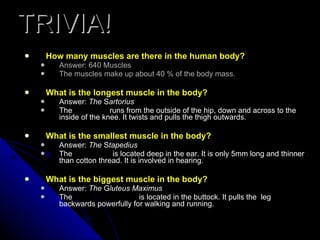

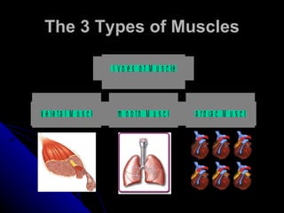


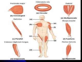
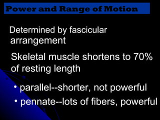
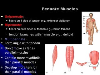


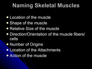


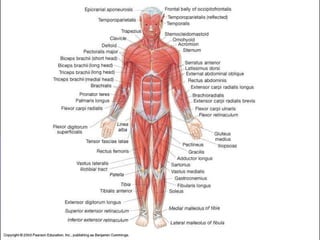
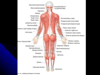
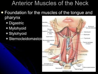
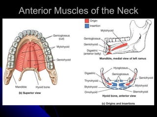
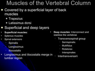
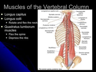


The document provides information about the muscular system, including: - There are approximately 640 muscles in the human body that make up 40% of body mass. - The longest muscle is the sartorius and the smallest is the stapedius. The largest is the gluteus maximus. - Muscles are classified by structure as striated, smooth or cardiac, and by function as voluntary or involuntary. - The main types of muscle are skeletal, smooth and cardiac muscle.


