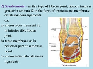
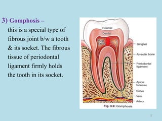
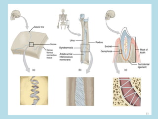
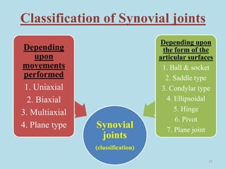
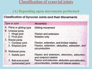
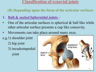
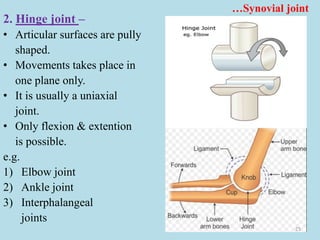
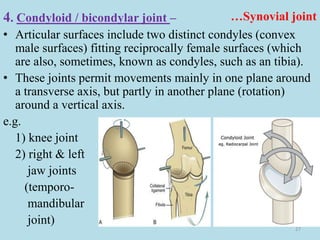
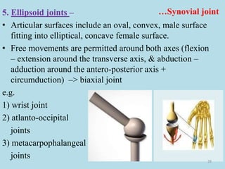
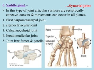
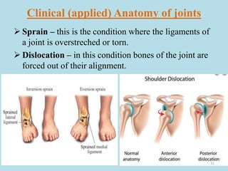
basic information about human body joints ( arthrology) .
topics cover under this ppt are- definition, classification and clinical anatomy of joints. this may be very helpful for the medical students for understand the basic concepts about joints.
2. Related terms –
Arthron (G) = a joint (arthrology – study of joints)
Articulatio (L) = a joint
Junctura (L) = a joint
Syndesmology (G - Syndesmosis = Ligament) = it is
the study of ligaments & related joints.
Joint /articulation – it is a junction b/w two or more
bones or cartilages. It is a device to permit movements.
2
4. Classification of joints
(A) Structural classification
1) Fibrous joints – the intervening
tissue b/w bones is fibrous.
a) Sutures
b) Syndesmosis
c) Gomphosis
2) Cartilaginous joints –
The intervening tissue b/w bones
is cartilaginous.
a) Primary catilaginous
joints (Synchondrosis)
b) Secondary catilaginous
joints /symphysis/
amphiarthrosis 4
5. `
3) Synovial Joints – where a cavity exists b/w the bones &
synovial membrane lines the cavity.
a) Ball & socket / spheroidal joints
b) Sellar or Saddle joints
c) Condylar or bicondylar joints
d) Ellipsoid joints
e) Hinge joints
f) Pivot or trochoid joints
g) Plane joints
(B) Regional classification
1) Skull type : immovable
2) Vertebral type : slightly movable
3) Limb type : freely movable
5
6. (C) Functional classification
(according to degree of mobility)
1) Synarthroses ( immovable) – these are fixed joints at
which there is no movements. e.g. sutures of the skull
2) Amphiarthroses (slightly movable) – a joint where
there is limited movements. e.g. joints b/w vertebral
bodies.
3) Diarthroses (freely movable) – a joint where there is a
wide range of movements. e.g. shoulder joint, hip joint.
6
7. (D) According to number of articulating
bones
1) Simple joint – a joint where
two bones articulate.
e.g. interphalangeal joints
2) Compound joint – a joints
where more than two bones
articulate within a single
capsule e.g. wrist &
elbow joint.
.
7
8. 3) Complex joint – A joint where the joint cavity is
completely or partially divided by an intra articular disc.
e.g.- temporomandibular joint, sternoclavicular joint
8
9. Fibrous joints (..Structural classi.)
• In fibrous joint the bones are joined by fibrous tissue.
• These joints are either immovable or permit a slight
degree of movement.
• These can be subdivided in to three subtypes.
1) Sutures : fibrous joint b/w skull bones.
sutures can be of many types depending upon the
shape & form of the opposing edges. These are-
i. Plane – internasal suture
ii. Serrate – interparietal suture
iii. Squamous – temporo-parietal suture
iv. Denticulate – lambdoid suture
v. Schindylesis type – joints b/w the upper border of
vomer & rostrum of sphenoid. 9
10. • Plane- opposing edges of the suture are plane.
• Serrate – projection of one bone fit in the gaps produced by the
projections of opposing bone & the projections are sloping.
• Dentate – projections are rectangular
• Squamous – the edge of one bone overlaps the edge of opposing bone.
• Schindylesis – the opposing edges are shaped like a wedge & its groove.
10
11. 2) Syndesmosis – in this type of fibrous joint, fibrous tissue is
greater in amount & in the form of interosseous membrane
or interosseous ligaments.
e.g.
a) interosseous ligament as
in inferior tibiofibular
joint.
b) tense membrane as in
posterior part of sarcoiliac
joint.
c) interosseous talocalcanean
ligaments.
11
12. 3) Gomphosis –
this is a special type of
fibrous joint b/w a tooth
& its socket. The fibrous
tissue of periodontal
ligament firmly holds
the tooth in its socket.
12
14. Cartilaginous joints (..Structural classi.)
• In this type of joints the bones are joined by cartilage. These are
two types-
1) Primary cartilaginous joint/ synchondrosis/ hyaline
cartilage joints –
• The bones are united by a plate of hyaline cartilage.
• The joint is immovable & strong.
• These are temporary in nature because after a certain age the
cartilaginous plate is replaced by bone.
e.g.
a) Joints b/w epiphysis & diaphysis
b) Spheno- occipital joint
c) First chondrosternal joint
d) Costochondral joint
14
16. 2) Secondary cartilaginous joint/ symphyses/ fibro-
cartilaginous joints –
• The articular surfaces are covered by a thin layer of
hyaline cartilage, and united by a disc of fibrocartilage.
• These joint are permanent & persist throughout life.
• Usually symphyses are found in the midline.
e.g.
1) Pubic symphysis
2) Manubriosternal joint
3) Intervertebral joints
16
18. Synovial joints (..Structural classi.)
• In this type of joint, the intervening tissue b/w the
articulating bones is the synovial membrane & synovial
fluid.
• The bones are separated by a cavity that permits a varying
degrees of movements.
• Synovial joints are most evolved & most mobile type.
Features of a typical synovial joints-
1. Joint cavity/ Articular cavity- between the articulating
bones there is a joint cavity filled with synovial fluid.
• The cavity may be partially or completely subdivided by
an articular disc or meniscus.
18
19. 2. Articular surface -
The surface is covered with hyaline (articular) cartilage.
3. Articular cartilage -
• articular cartilage is avascular, non-nervous & elastic.
• It is smooth & devoid of perichondrium.
• It is compressible & acts like a sponge, filling with the
synovial fluid when free & squeezing the fluid when
compressed by bones.
4. Articular capsule –
• The joint is surrounded by an articular capsule which is
made up of two layers, these are – (a) fiberous capsule &
(b) synovial membrane
19
21. a) fibrous capsule (outer layer) – made up of densely
packed collagen fibers.
b) synovial membrane (inner layer) – it is a thin but highly
vascular & lines the fibrous capsule on internal aspect.
• It is made up of areolar connective tissue with elastic
fibers & variable amount of adipose tissue & it secretes
synovial fluid
• The fibrous capsule is often reinforced by –
a) Capsular/ True ligament – representing thickenings of the
fiberous capsule.
b) The accessory ligaments – the articular bones are also
united by ligaments which stand apart from the capsule
which may be intracapsular or extracapsular.
21
22. Classification of Synovial joints
Synovial
joints
(classification)
Depending
upon
movements
performed
1. Uniaxial
2. Biaxial
3. Multiaxial
4. Plane type
Depending upon
the form of the
articular surfaces
1. Ball & socket
2. Saddle type
3. Condylar type
4. Ellipsoidal
5. Hinge
6. Pivot
7. Plane joint
22
24. Classification of synovial joints
(B) Depending upon the form of the articular surfaces
1. Ball & socket/Spheroidal joints –
• One of the articular surfaces is spherical & ball like while
other articular surface presents a cup like concavity.
• Movements can take place around many axes.
e.g.1) shoulder joint
2) hip joint
3) incudostapedial
joint
24
25. …Synovial joint
2. Hinge joint –
• Articular surfaces are pully
shaped.
• Movements takes place in
one plane only.
• It is usually a uniaxial
joint.
• Only flexion & extention
is possible.
e.g.
1) Elbow joint
2) Ankle joint
3) Interphalangeal
joints
25
26. …Synovial joint
3. Pivot joint –
• Articular surfaces comprise a central bony pivot (peg)
surrounded by an osteoligamentous ring.
• In these joints, a rounded process of bone rotates within a ring.
• Movements are permitted
in one plane around a vertical
axis. (rotation movement).
e.g.
1. atlanto-axial joint
2. Superior &
inferior
radio-ulnar
joints.
26
27. …Synovial joint
4. Condyloid / bicondylar joint –
• Articular surfaces include two distinct condyles (convex
male surfaces) fitting reciprocally female surfaces (which
are also, sometimes, known as condyles, such as an tibia).
• These joints permit movements mainly in one plane around
a transverse axis, but partly in another plane (rotation)
around a vertical axis.
e.g.
1) knee joint
2) right & left
jaw joints
(temporo-
mandibular
joint)
27
28. …Synovial joint
5. Ellipsoid joints –
• Articular surfaces include an oval, convex, male surface
fitting into elliptical, concave female surface.
• Free movements are permitted around both axes (flexion
– extension around the transverse axis, & abduction –
adduction around the antero-posterior axis +
circumduction) –> biaxial joint
e.g.
1) wrist joint
2) atlanto-occipital
joints
3) metacarpophalangeal
joints 28
29. …Synovial joint
6. Saddle joint –
• In this type of joint articular surfaces are reciprocally
concavo-convex & movements can occur in all planes.
1. First carpometacarpal joint.
2. sternoclavicular joint
3. Calcaneocuboid joint
4. Incudomalleolar joint
5. Joint b/w femer & patella
29
30. …Synovial joint
7. Plane synovial joints –
• Articular surfaces are more or less flat (plane).
• They permit gliding movements (translational) in various
directions.
e.g.
1. Intercarpal joints
2. Intertarsal jonts
3. Joints b/w articular
process of vertebrae
30
31. Clinical (applied) Anatomy of joints
Sprain – this is the condition where the ligaments of
a joint is overstreched or torn.
Dislocation – in this condition bones of the joint are
forced out of their alignment.
31
32. Arthritis – the term indicate all the inflammatory &
degenerative diseases of the joints.
(a) Osteoarthritis – is a degenerative condition related to
ageing process.
(b) Rheumatoid arthritis – is inflammatory in nature
32