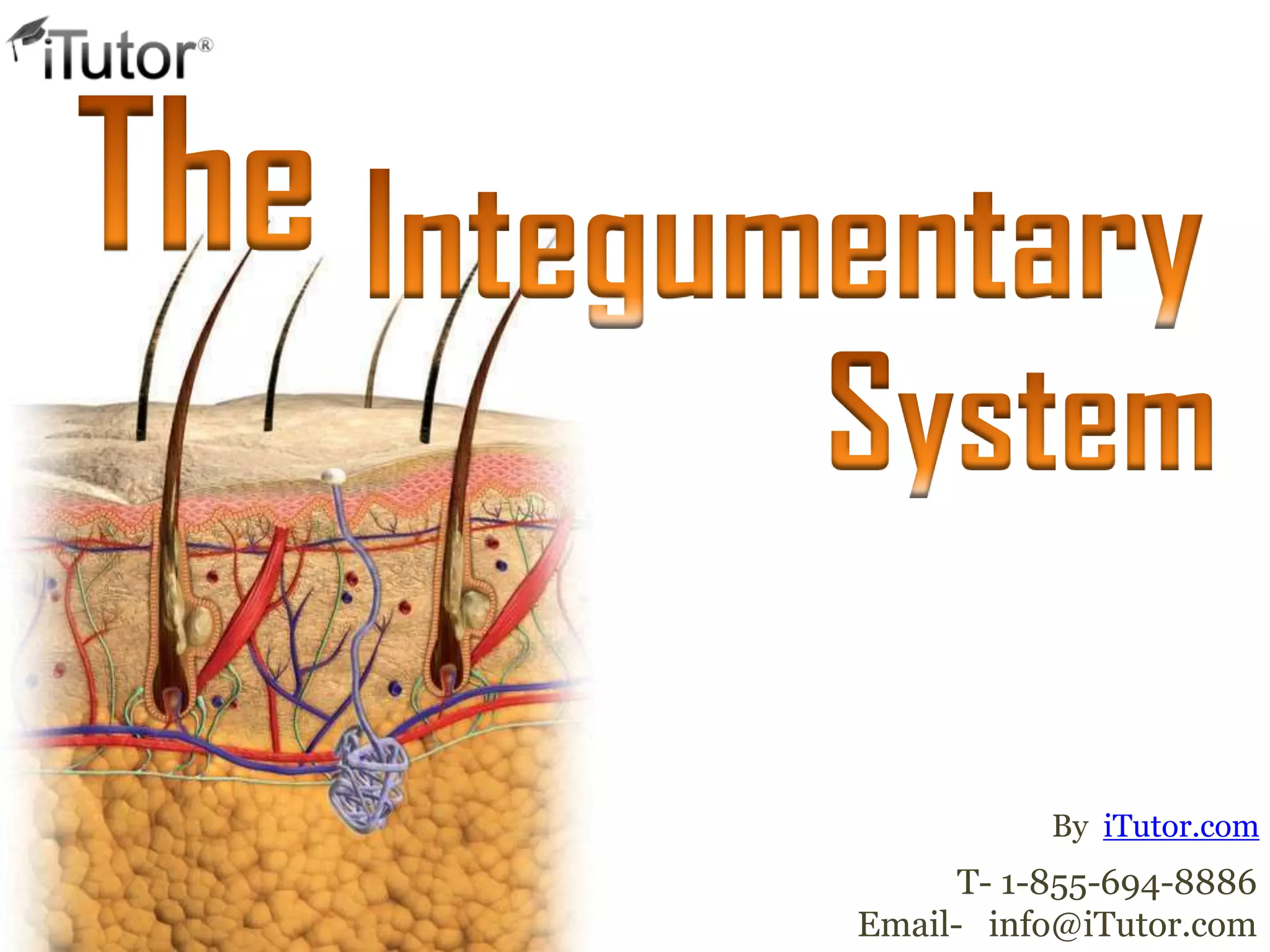
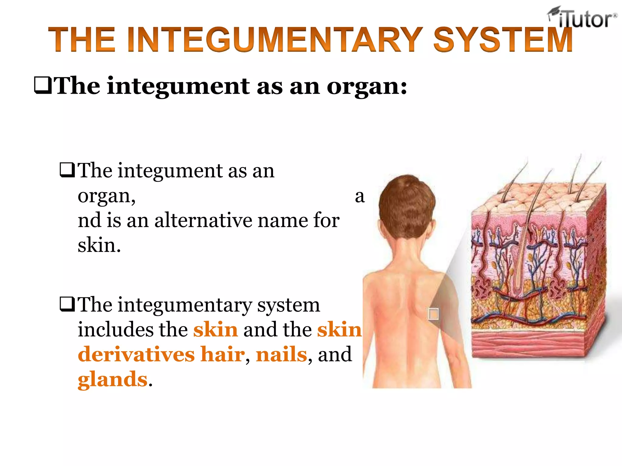
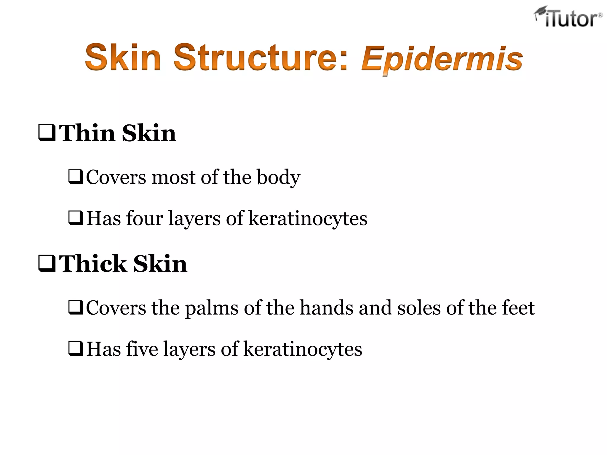
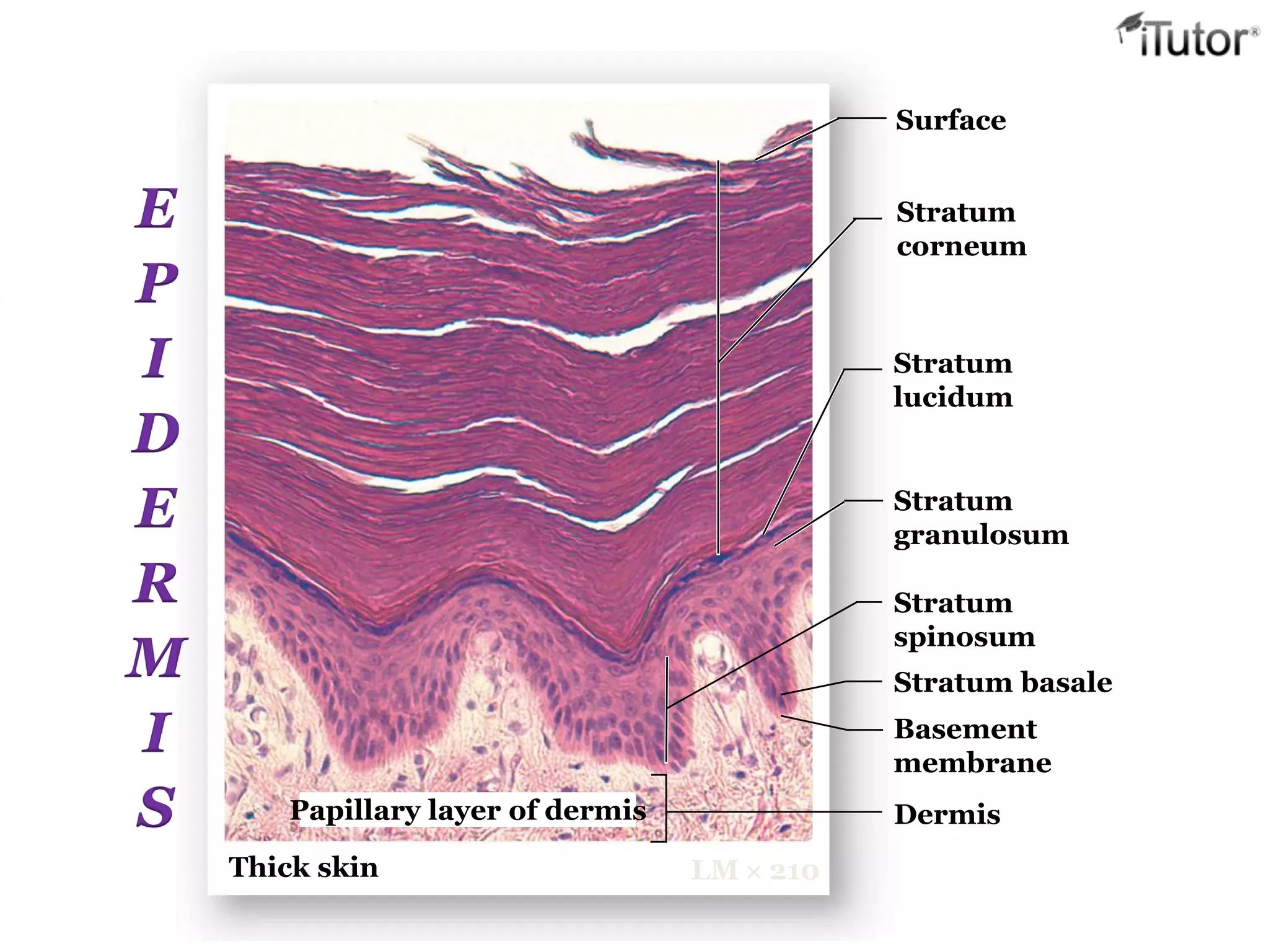
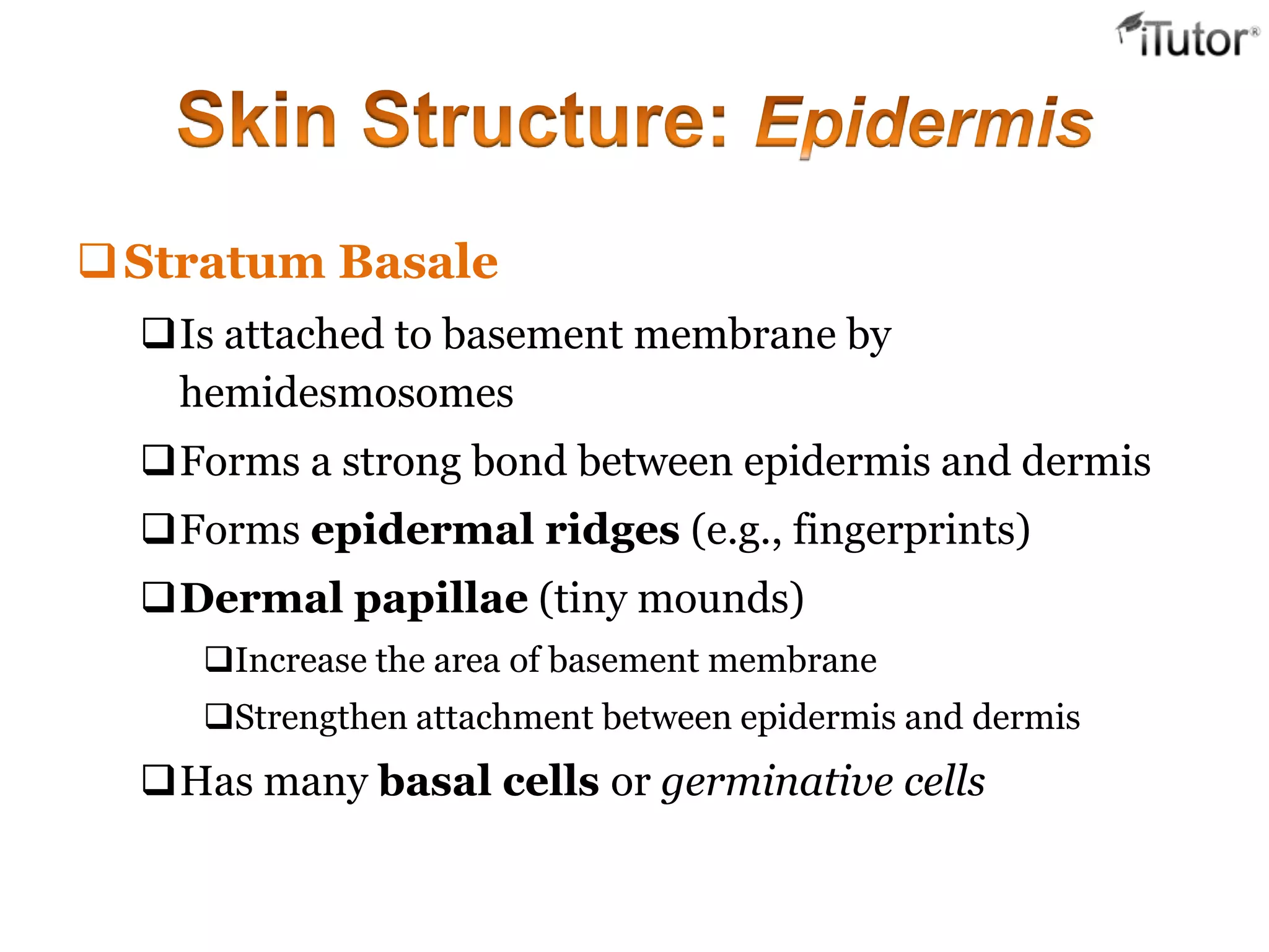
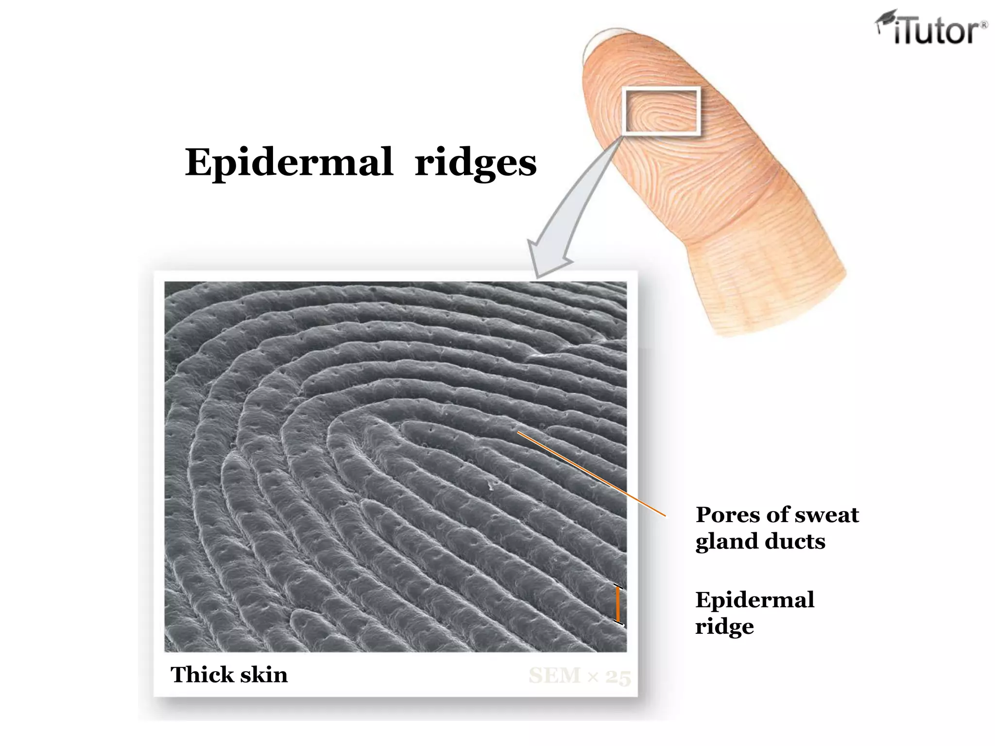
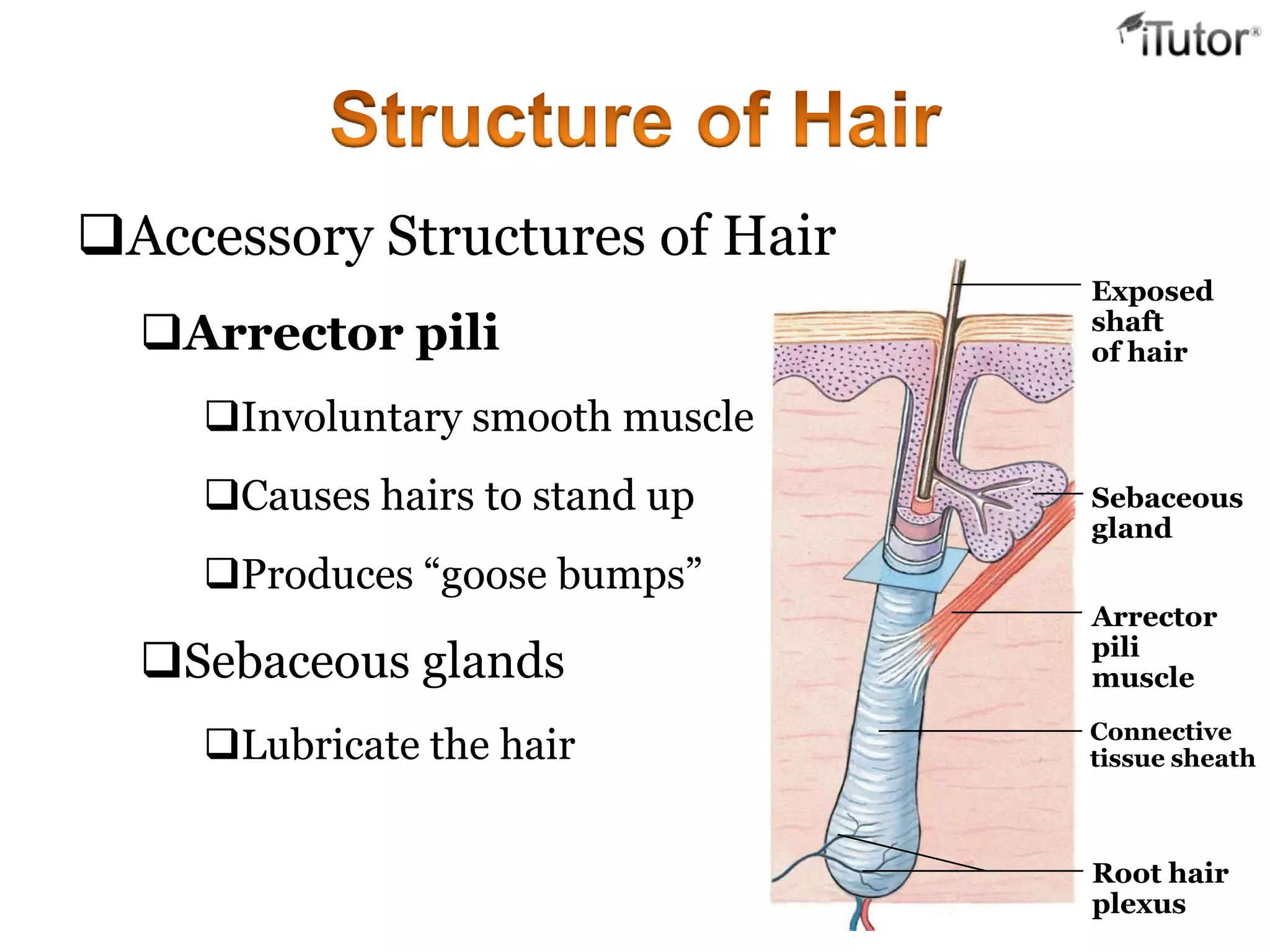
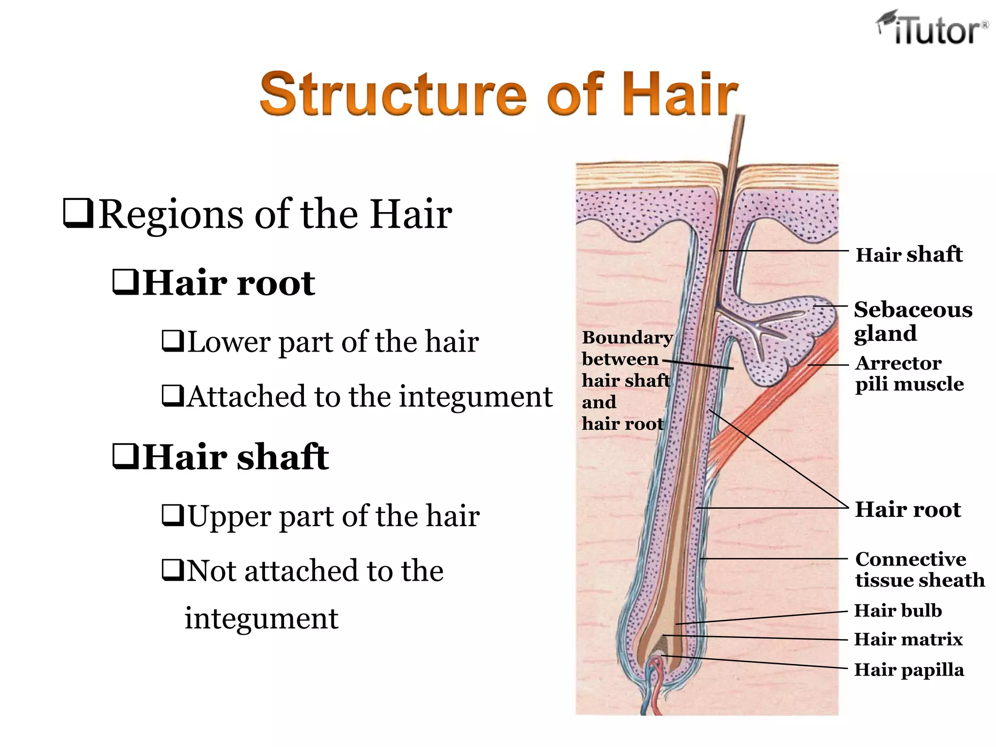
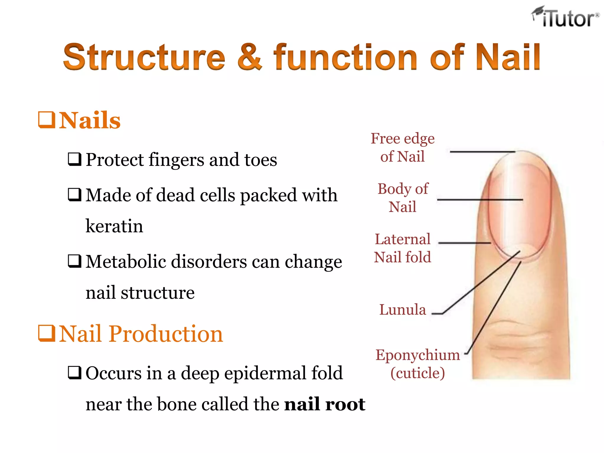
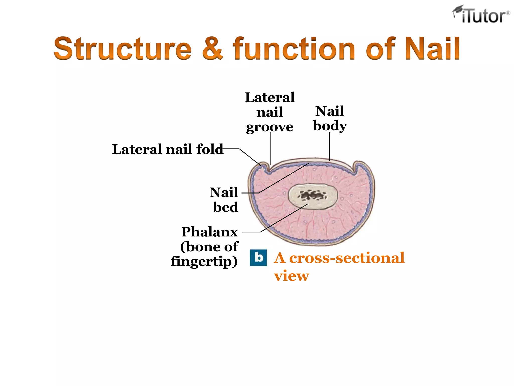
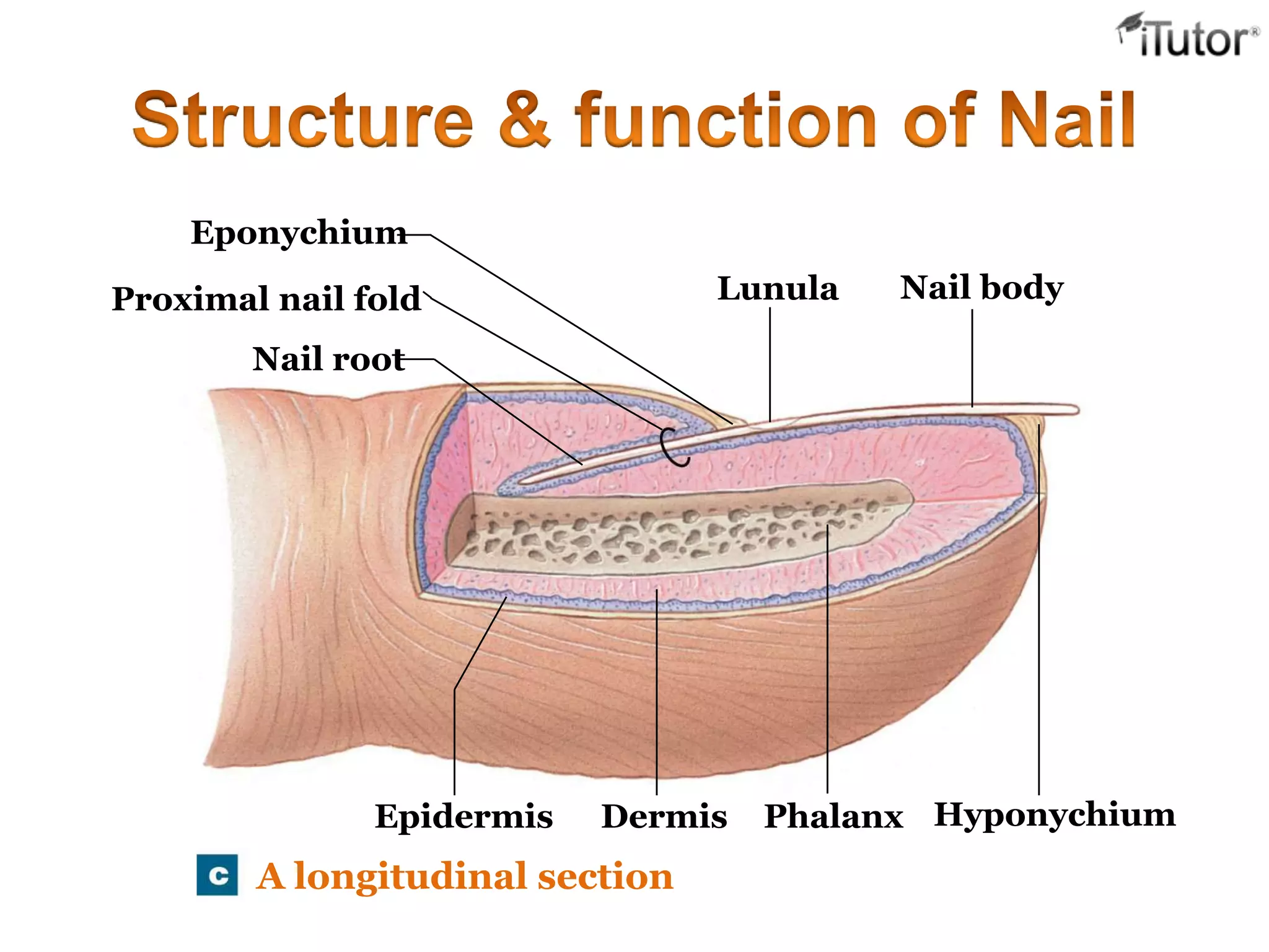
T- 1-855-694-8886
Email- info@iTutor.com
By iTutor.com
The integument as an
organ, a
nd is an alternative name for
skin.
The integumentary system
includes the skin and the skin
derivatives hair, nails, and
glands.
The integument as an organ:
The Integument
Is the largest system of the body
 16% of body weight,
 1.5 to 2m2
in area,
 The integument is made up of two parts:
1. Cutaneous membrane
a. Epidermis– Superficial epithelium
b. Dermis – underlying CT with blood supply
2. Accessory structures
a. Hair
b. Nails
c. Exocrine Glands
Protection
 First line of defense against
 Bacteria
 Viruses
 Protects underlying structures from
 Ultraviolet (UV) radiation
 Dehydration
Vitamin D production
 Needed for calcium absorption
Sensation
 Sensory receptors
 Body temperature regulation
If too hot
 Dermal blood vessels dilate
 Vessels carry more blood to surface so heat can escape
If too cold
 Dermal blood vessels constrict
 Prevents heat from escaping
 Excretion
Small amounts of waste products are lost through
perspiration
 Understanding how the skin can function in these many
ways starts with understanding the structure of the 3
layers of skin
The Epidermis
Epithelial tissue
Dermis
Dense connective tissue proper – irregular
Hypodermis
Subcutaneous tissue- loose connective tissue proper
and adipose tissue
Epidermis
Dermis
Hypodermis
or
subcutaneous layer
The Epidermis
Is a vascular stratified squamous epithelium
Nutrients and oxygen diffuse from capillaries in the dermis
Cells of the Epidermis
Keratinocytes
Contain large amounts of keratin
Are the most abundant cells in the epidermis
The structural relationship and
interface between the epidermis
and underlying dermis. The
proportions of the various layers
differ with the location sampled.
Dermis
Epidermis
Epidermal
ridge
Dermal
papilla
Thin Skin
Covers most of the body
Has four layers of keratinocytes
Thick Skin
Covers the palms of the hands and soles of the feet
Has five layers of keratinocytes
Structures of the Epidermis
 The five strata of keratinocytes in thick skin
 From basal lamina to free surface
1. Stratum basale
2. Stratum spinosum
3. Stratum granulosum
4. Stratum lucidum
5. Stratum corneum
Thick skin LM  210
Surface
Stratum
corneum
Stratum
lucidum
Stratum
granulosum
Stratum
spinosum
Stratum basale
Basement
membrane
DermisPapillary layer of dermis
E
P
I
D
E
R
M
I
S
Stratum Basale
Is attached to basement membrane by
hemidesmosomes
Forms a strong bond between epidermis and dermis
Forms epidermal ridges (e.g., fingerprints)
Dermal papillae (tiny mounds)
Increase the area of basement membrane
Strengthen attachment between epidermis and dermis
Has many basal cells or germinative cells
Thick skin SEM  25
Epidermal
ridge
Pores of sweat
gland ducts
Epidermal ridges
Stratum Spinosum — the ―spiny layer‖
Produced by division of stratum basale
Eight to ten layers of keratinocytes bound by
desmosomes
Cells shrink until cytoskeletons stick out (spiny)
Continue to divide, increasing thickness of epithelium
Contain dendritic (Langerhans) cells, active in
immune response
Stratum Granulosum — the ―grainy layer‖
Stops dividing, starts producing
Keratin
A tough, fibrous protein
Makes up hair and nails
Keratohyalin
Dense granules
Cross-link keratin fibers
Stratum Lucidum — the ―clear layer‖
Found only in thick skin
Covers stratum granulosum
Stratum Corneum — the ―horn layer‖
Exposed surface of skin
15 to 30 layers of keratinized cells
Water resistant
Shed and replaced every 2 weeks
The Dermis
 Located between epidermis
and subcutaneous layer
 Anchors epidermal accessory
structures (hair
follicles, sweat glands)
 Two components
1. Outer papillary layer
2. Deep reticular layer Dermis
 The Papillary Layer
Consists of areolar tissue
Contains smaller capillaries, lymphatics, and sensory
neurons
Has dermal papillae projecting between epidermal ridges
 The Reticular Layer
Consists of dense irregular connective tissue
Contains larger blood vessels, lymphatic vessels, and nerve
fibers
Contains collagen and elastic fibers
Contains connective tissue proper
An inflammation of the papillary layer
Caused by
 infection, radiation, mechanical irritation, or chemicals
(e.g., poison ivy)
Characterized by
itch or pain
Characteristics
Strong, due to collagen fibers
Elastic, due to elastic fibers
Flexible
Hair
Papillary
layer
Reticular
layer
Cutaneous
plexus
Papillary
plexus
Epidermal
ridges
Dermal
papillae
Capillary loop
of papillary
plexus
The Hypodermis (Subcutaneous Layer)
Lies below the integument
Stabilizes the skin
Allows separate movement
Made of elastic areolar and adipose tissues
Connected to the reticular layer of integument by
connective tissue fibers
Deposits of Subcutaneous Fat
Distribution patterns determined by hormones
Reduced by cosmetic liposuction (lipoplasty)
The Hair Follicle
 Hair follicles are the organs that form the hairs.
Hair follicles are the organs that form the hairs.
Located deep in dermis.
Produces nonliving hairs.
Wrapped in a dense connective tissue sheath.
Base is surrounded by sensory nerves (root hair plexus).
Control bacteria
Exposed
shaft
of hair
Sebaceous
gland
Arrector
pili
muscle
Connective
tissue sheath
Root hair
plexus
Accessory Structures of Hair
Arrector pili
Involuntary smooth muscle
Causes hairs to stand up
Produces ―goose bumps‖
Sebaceous glands
Lubricate the hair
Regions of the Hair
Hair root
Lower part of the hair
Attached to the integument
Hair shaft
Upper part of the hair
Not attached to the
integument
Boundary
between
hair shaft
and
hair root
Hair shaft
Sebaceous
gland
Arrector
pili muscle
Hair root
Connective
tissue sheath
Hair bulb
Hair matrix
Hair papilla
 Hair Shaft Structure
 Medulla
 Core, dead cells contain soft keratin and air to provide
flexible
 Cortex
 Middle layer, dead cells contain hard keratin to provide
stiffness
 Cuticle
 Outermost, overlapping dead keratinized cells form
shiny surface
Head:
UV protection
Cushion from trauma
Insulation
Nostrils, Ear canals, Eyelashes:
Prevent entry of foreign material
Body Hair:
sensory detection
Root hair plexus:
Sensory nerves at base of hair follicle that detect slight
movement of hair
Arrector pili muscle:
Attached to every hair follicle
Contract to stand hair perpendicular to skin surface
Nails
Protect fingers and toes
Made of dead cells packed with
keratin
Metabolic disorders can change
nail structure
Nail Production
Occurs in a deep epidermal fold
near the bone called the nail root
Free edge
of Nail
Body of
Nail
Laternal
Nail fold
Lunula
Eponychium
(cuticle)
Structure of a Nail
Nail body
The visible portion of the nail
Covers the nail bed
Lunula
The pale crescent at the base of the nail
Sides of nails
Lie in lateral nail grooves
Surrounded by lateral nail folds
A cross-sectional
view
Lateral nail fold
Nail
body
Lateral
nail
groove
Nail
bed
Phalanx
(bone of
fingertip)
A longitudinal section
Eponychium
Proximal nail fold
Nail root
Lunula Nail body
Epidermis Dermis Phalanx Hyponychium
Call us for more information
www.iTutor.com
1-855-694-8886
Visit

The integumentary system

  • 1.
  • 2.
    The integument asan organ, a nd is an alternative name for skin. The integumentary system includes the skin and the skin derivatives hair, nails, and glands. The integument as an organ:
  • 3.
    The Integument Is thelargest system of the body  16% of body weight,  1.5 to 2m2 in area,  The integument is made up of two parts: 1. Cutaneous membrane a. Epidermis– Superficial epithelium b. Dermis – underlying CT with blood supply 2. Accessory structures a. Hair b. Nails c. Exocrine Glands
  • 4.
    Protection  First lineof defense against  Bacteria  Viruses  Protects underlying structures from  Ultraviolet (UV) radiation  Dehydration Vitamin D production  Needed for calcium absorption Sensation  Sensory receptors
  • 5.
     Body temperatureregulation If too hot  Dermal blood vessels dilate  Vessels carry more blood to surface so heat can escape If too cold  Dermal blood vessels constrict  Prevents heat from escaping  Excretion Small amounts of waste products are lost through perspiration
  • 6.
     Understanding howthe skin can function in these many ways starts with understanding the structure of the 3 layers of skin The Epidermis Epithelial tissue Dermis Dense connective tissue proper – irregular Hypodermis Subcutaneous tissue- loose connective tissue proper and adipose tissue
  • 7.
  • 8.
    The Epidermis Is avascular stratified squamous epithelium Nutrients and oxygen diffuse from capillaries in the dermis Cells of the Epidermis Keratinocytes Contain large amounts of keratin Are the most abundant cells in the epidermis
  • 9.
    The structural relationshipand interface between the epidermis and underlying dermis. The proportions of the various layers differ with the location sampled. Dermis Epidermis Epidermal ridge Dermal papilla
  • 10.
    Thin Skin Covers mostof the body Has four layers of keratinocytes Thick Skin Covers the palms of the hands and soles of the feet Has five layers of keratinocytes
  • 11.
    Structures of theEpidermis  The five strata of keratinocytes in thick skin  From basal lamina to free surface 1. Stratum basale 2. Stratum spinosum 3. Stratum granulosum 4. Stratum lucidum 5. Stratum corneum
  • 12.
    Thick skin LM 210 Surface Stratum corneum Stratum lucidum Stratum granulosum Stratum spinosum Stratum basale Basement membrane DermisPapillary layer of dermis E P I D E R M I S
  • 13.
    Stratum Basale Is attachedto basement membrane by hemidesmosomes Forms a strong bond between epidermis and dermis Forms epidermal ridges (e.g., fingerprints) Dermal papillae (tiny mounds) Increase the area of basement membrane Strengthen attachment between epidermis and dermis Has many basal cells or germinative cells
  • 14.
    Thick skin SEM 25 Epidermal ridge Pores of sweat gland ducts Epidermal ridges
  • 15.
    Stratum Spinosum —the ―spiny layer‖ Produced by division of stratum basale Eight to ten layers of keratinocytes bound by desmosomes Cells shrink until cytoskeletons stick out (spiny) Continue to divide, increasing thickness of epithelium Contain dendritic (Langerhans) cells, active in immune response
  • 16.
    Stratum Granulosum —the ―grainy layer‖ Stops dividing, starts producing Keratin A tough, fibrous protein Makes up hair and nails Keratohyalin Dense granules Cross-link keratin fibers
  • 17.
    Stratum Lucidum —the ―clear layer‖ Found only in thick skin Covers stratum granulosum Stratum Corneum — the ―horn layer‖ Exposed surface of skin 15 to 30 layers of keratinized cells Water resistant Shed and replaced every 2 weeks
  • 18.
    The Dermis  Locatedbetween epidermis and subcutaneous layer  Anchors epidermal accessory structures (hair follicles, sweat glands)  Two components 1. Outer papillary layer 2. Deep reticular layer Dermis
  • 19.
     The PapillaryLayer Consists of areolar tissue Contains smaller capillaries, lymphatics, and sensory neurons Has dermal papillae projecting between epidermal ridges  The Reticular Layer Consists of dense irregular connective tissue Contains larger blood vessels, lymphatic vessels, and nerve fibers Contains collagen and elastic fibers Contains connective tissue proper
  • 20.
    An inflammation ofthe papillary layer Caused by  infection, radiation, mechanical irritation, or chemicals (e.g., poison ivy) Characterized by itch or pain Characteristics Strong, due to collagen fibers Elastic, due to elastic fibers Flexible
  • 21.
  • 22.
    The Hypodermis (SubcutaneousLayer) Lies below the integument Stabilizes the skin Allows separate movement Made of elastic areolar and adipose tissues Connected to the reticular layer of integument by connective tissue fibers Deposits of Subcutaneous Fat Distribution patterns determined by hormones Reduced by cosmetic liposuction (lipoplasty)
  • 24.
    The Hair Follicle Hair follicles are the organs that form the hairs. Hair follicles are the organs that form the hairs. Located deep in dermis. Produces nonliving hairs. Wrapped in a dense connective tissue sheath. Base is surrounded by sensory nerves (root hair plexus). Control bacteria
  • 25.
    Exposed shaft of hair Sebaceous gland Arrector pili muscle Connective tissue sheath Roothair plexus Accessory Structures of Hair Arrector pili Involuntary smooth muscle Causes hairs to stand up Produces ―goose bumps‖ Sebaceous glands Lubricate the hair
  • 26.
    Regions of theHair Hair root Lower part of the hair Attached to the integument Hair shaft Upper part of the hair Not attached to the integument Boundary between hair shaft and hair root Hair shaft Sebaceous gland Arrector pili muscle Hair root Connective tissue sheath Hair bulb Hair matrix Hair papilla
  • 27.
     Hair ShaftStructure  Medulla  Core, dead cells contain soft keratin and air to provide flexible  Cortex  Middle layer, dead cells contain hard keratin to provide stiffness  Cuticle  Outermost, overlapping dead keratinized cells form shiny surface
  • 28.
    Head: UV protection Cushion fromtrauma Insulation Nostrils, Ear canals, Eyelashes: Prevent entry of foreign material Body Hair: sensory detection Root hair plexus: Sensory nerves at base of hair follicle that detect slight movement of hair Arrector pili muscle: Attached to every hair follicle Contract to stand hair perpendicular to skin surface
  • 29.
    Nails Protect fingers andtoes Made of dead cells packed with keratin Metabolic disorders can change nail structure Nail Production Occurs in a deep epidermal fold near the bone called the nail root Free edge of Nail Body of Nail Laternal Nail fold Lunula Eponychium (cuticle)
  • 30.
    Structure of aNail Nail body The visible portion of the nail Covers the nail bed Lunula The pale crescent at the base of the nail Sides of nails Lie in lateral nail grooves Surrounded by lateral nail folds
  • 31.
    A cross-sectional view Lateral nailfold Nail body Lateral nail groove Nail bed Phalanx (bone of fingertip)
  • 32.
    A longitudinal section Eponychium Proximalnail fold Nail root Lunula Nail body Epidermis Dermis Phalanx Hyponychium
  • 33.
    Call us formore information www.iTutor.com 1-855-694-8886 Visit